Comparative Study on the Effect of Adipose-Derived Stem Cells Versus Alpha-Lipoic Acid Inautologus Fat Graft Survival
Autologous fat transfer is a popular option for soft tissue augmentation, but has a low survival rate. So, enriching the transplanted fat with stem cells (cell assisted lipotransfer) or oral administration ofalpha-lipoic acid (ALA) before and after graft injection, were thought to increase the survival rate of the transplanted fat. Aim of Study: Is to compare between cell-assisted lipotransfer (CAL) and ALAtherapy in improvement of fat graft survival. Materials and Methods: Thirty adult female albino rats were used and divided into three groups. Group I: the control group. Group II: CAL group received autologous fat mixed with adipose derived stem cells (ASCs). Group III: ALA group in which animals received oral ALA four days before fat transplantation and continued throughout experiment. Fat grafts were collected from the sites of injection after twenty-eight days. Histological and immune histochemical studies were performed. Statistical analysis was also done. Results: Histological evaluation revealed atrophy and death of adipocytes with formation of fat cysts. Severe inflammatory infiltration with giant cells formation and fibrosis were detected. Significant decrease of VEGF reaction was seen. In CAL group significant increase in number of intact adipocytes together with decrease in inflammation and fibrosis occurred. Moreover, significant increase in VEGF reaction was observed. In ALA group, significant decrease in number of intact adipocytes and VEGF reaction was detected compared with CAL group. Also, significant increase in inflammatory infiltration and fibrosis were detected compared to CAL group. Conclusion: Both ASCs and ALA showed improvement in all histological parameters compared with control group. However, enrichment of fat with ASCs showed the best results.
Introduction
Autologous fat transplantation is now ideal filler for augmentation and soft tissue reconstruction in cosmetic and reconstructive surgery [1]. It is now an increasingly attractive method for many procedures including breast reconstruction, facial and hand rejuvenation, treatment of squeal resulting from radiation therapy and gluteal fat augmentation [2, 3].
Autologous fat transplantation is host compatible, readily available, and can be harvested easily and repeatedly as needed without complication arising from allergic or foreign body reactions. The main limitation of this procedure was the low survival rate and high desorption rate of the transplanted fat with graft survival rate ranging from 20 to 90 % [4]. A recent study found that fat grafting following breast cancer reconstruction retains 45 to 59 percent of its original volume at 49 days and 27 to 54 percent at 140 days. Patients frequently undergo at least two or three sessions of grafting to achieve adequate volume correction [5].
Fat graft failure and volume reduction appeared to be related to lack of adequate revascularization within the transplanted fat [6].
Concomitant transplantation of fat and ASCs was named CAL by Matsumoto, et al. [7]. It was supposed that ASCs will increase survival rate of lipotransfer.
ALA is an antioxidant which can act as a potent free radical scavenger and metal chelator. It is a co-enzyme for mitochondrial multi-enzyme complex reactions [8].
So the purpose of this study was to compare CAL and ALA in improvement of fat graft survival.
Materials and Methods
Thirty adult female albino rats (Wister strain) weighing 200-250 gm were used in this study. The animals were purchased and the study was conducted from the MASRI Center, Faculty of Medicine, Ain Shams University. They were housed in plastic cages with mesh wire covers and were given food and water ad libitum. The practical work was performed in accordance to the guide for care and use of laboratory animals and approved by the Animal Ethical Committee of Ain Shams University.
Experimental Design
The animals were divided into three groups: Group I (Control group): consisted of 10 rats, which received fat mixed only with saline. The animals were anesthetized by intramuscular injection of 0.5mg/kg ketamine. Inguinal fat pads were isolated from the same donor. They were finely minced, mixed with saline, and poured into 10-mL syringes. Syringes were kept in an upright position at room temperature for 10 min, and then the infranatant fluid was discarded. One mL of supernatant fat was subcutaneously injected into the back of each rat [7].
Group II (CAL group): included 10 rats. Each rat received mixture of 1 mL of minced fat mixed with 300 μL of phosphate-buffered saline (PBS) containing 1 × 10⁵ASCs [9].
Group III (ALA group): included 10 rats which received oral ALA treatment 4 days before fat transplantation and the treatment continued throughout the experiment. ALA (Thiotacid tablets 300 mg) was purchased from (EVA pharma company, Egypt). Each tablet was crushed, dissolved in Ethanol 0.1% and was given orally at a dose of 500mg/kg/day [10].
Fat grafts were extracted from sites of injection after four weeks and processed for light microscopic examination. Paraffin sections were stained by using H&E, Masson’s trichrome staining. Caspase-3 and VEGF immune histo-chemical studies were also done.
Morph metric analysis: Five different non overlapping fields from five different sections of extracted fat grafts of different rats were examined in each group. An image analyzer Leica Q win V.3 program (Wetzlar, Germany) in the Histology department, faculty of Medicine Ain Shams University was used to measure:
- Number of normal sized and shaped adipocytes/medium power field
- Number of fat cysts/medium power field
- Percentage of inflammatory infiltration/medium power field
- Percentage of fibrosis/medium power field
- Number of apoptotic nuclei/high power field
- Percentage of positive VEGF reaction/high power field Statistical analysis was done using one-way ANOVA test performed by SPSS 17 program. The significance of the data was determined by p-value p≥0.05 non- significant (NS), p<0.05 significant (S).
Histological scoring of histological parameters was done. Number of normal adipocytes and fat cysts were graded based on a scale from 0 to 4. The scoring was performed at a magnification of 400 [2]. Criteria for normal adipocytes score were: 0 --- more than 12 adipocytes per high power field (HPF) 1 --- 9 to 12 adipocytes/HPF 2 --- 5 to 8 adipocytes/HPF 3 ---1 to 4 adipocytes/HPF 4 --- Absence of intact and nucleated adipocyte/ HPF Criteria for fat cysts score were: 0 --- absent/HPF 1 --- 1-4 fat cysts/HPF 2 --- 5-8 fat cysts/HPF 3 --- 9-12 fat cysts/HPF 4 --- More than 12 fat cysts/HPF The rest of the criteria were graded on a scale from 0 to 5 (6) as follows: Absent: 0 Minimal presence: 1 Minimal to moderate presence: 2 Moderate presence: 3 Moderate to extensive presence: 4 Extensive presence: 5 The scoring was performed at a magnification of 400. Ten randomly selected fields from each graft were included for scoring each parameter. All the histologic examinations were performed by 3 independent investigators in a blinded fashion. Preparation of ASCs from excised fat: Fat tissue was obtained by excision from inguinal pad of fat of male albino rats. To prepare ASCs, the excised fat tissue was washed extensively with PBS, cut into small pieces, and digested with 0.1% collagenase type I in PBS for 80 min at 37°C with intermittent shaking. The suspension was then centrifuged at 1800rpm, for 10 minutes. The pellet (SVF) was resuspended in complete medium (DMEM with 10% FBS & 1% Penicillin/Strept) for culturing. The initial cell density was 1×10⁵ / cm2 and seeded into 25 cm2 tissue culture flasks and then incubated at 37°C, with 5% CO2.
The flasks were examined daily for detection of any contamination and to monitor the growth of the cells. Replacement of the media was done every 2-3 days.When the cells reached confluence (more than 80% of the flask area was occupied) nearly at day 7, passaging of cells was done by trypsinization trypsin/ EDTA for 2 minutes. An equal amount of culture media was added to the mixture to stop its proteolytic activity and then the mixture was centrifuged at a rate of 1800 rpm for 10 minutes. The supernatant was aspirated, and the pellets were re- suspended in 10 ml of the complete media and divided into two T-75 flasks each containing 10 ml complete medium then re-incubated. The cells were denoted as passage 1 (P1). ASCs underwent 3-5 passages before being used. Characterization of stem cells was done using flow cytometry. ASCs expressed mesenchymal stem cell marker CD73 and did not express CD45. ASCs expressed CD34 which is specific for adipose derived and not bone marrow derived stem cells.
Examination of dishes and taking photographs were done using inverted microscope (Axiovert 100- ZEISS, Denmark). Determining cell viability by trypan blue stain and the total cell count using hemocytometer. Application of ASCs: 1×10⁵ cells/ each 1ml fat were mixed with fat transplanted in the back of each rat.
Results
Morphological Identification of ASCs as Evident by the Inverted Microscope
On day four, colonies of adherent ASCs appeared spindle in shape while others were star shaped. They attained vesicular nuclei and granular cytoplasm. Some cells had long well-developed processes (Figure 1a) on day six, the adherent cells were overcrowded (60-70% confluence). They were mostly branched fibroblast like in shape with vesicular nuclei and granular cytoplasm they were interconnected by long cytoplasmic processes (Figure 1b). On day seven the cells were mostly confluent (about 90-95%) with granular cytoplasm and vesicular nuclei (Figure 1c). ASCs in subculture appeared spindle or star in shape, with granular cytoplasm and vesicular nucleus (Figure 1d). Characterization using CD45-, CD73+ and CD34+ was detected (Figure 2).
Histological Results
Control group (group I): By naked eye, the dissected fat grafts were reddish in color with little fat explants. One of the fat grafts was completely resorbed and another was severely infected and neglected. Hematoxylin and eosin showed few normal sized and shaped adipocytes and multiple large fat cysts. Some small sized, atrophied adipocytes with peripheral acidophilic rim of cytoplasm and multiple fat vacuoles. Small sized adipocytes (regenerated cells) were also observed. Severe inflammatory infiltration and multiple blood vessels were also seen (Figures 3a & 3b). Large areas of connective tissue replacing most of the grafts were seen (Figure 4a). Higher magnification of the grafts showed numerous cells with foamy cytoplasm (foam cells) (Figure 4b). Presence of fat cysts with cellular debris, atrophied adipocytes and adipocytes with karyolitic nuclei were seen. Foam cells and multinucleated giant cells were also observed (Figure 4c). Numerous spindle shaped stromal cells with acidophilic cytoplasm and large vesicular nuclei could be detected (Figure 4d). Masson’s trichrome staining showed presence of large amount of collagen fibers surrounding groups of adipocytes (Figure 5a). Numerous caspase-3 positive adipocytes and stromal cells were detected (Figure 5b). Mild positive VEGF reaction in adipocytes and blood vessels was also observed (Figure 5c). CAL group (group II): By naked eye, fat grafts were of large size and more yellowish than those of control group. H&E staining showed that most of adipocytes were of normal shape and size. Few fat cysts were seen. Groups of adipocytes were separated by connective tissue septa and capsules. Many blood vessels were observed (Figure 6a). Higher magnification revealed minimal inflammatory infiltration (Figure 6b). Masson’s trichrome staining showed minimal amount of collagen fibers surrounding normal adipocytes (Figure 7a). Few caspase-3 positive cells and numerous VEGF positive cells were detected (Figure 7b & c). ALA group III: By naked eye, fat grafts were slightly bigger than grafts of control group. H&E sections showed presence of intact adipocytes surrounded by stroma containing connective tissue fibers, inflammatory infiltration and many blood vessels (Figure 8a). Higher magnification of the grafts showed some adipocytes with irregular cell membrane (Figure 8b). Stromal cells revealed multinucleated giant cells containing fat droplet (Figure 8c). Masson’s trichrome staining showed collagen fibers surrounding adipocytes forming septa (Figure 1a). Mild caspase-3 positive reaction and moderate VEGF positive reaction of were detected (Figure 9b & c).
Statistical Results
Histological evaluation of the transplanted fat revealed significant improvement of histological parameters in both CAL and ALA groups compared to control group.
Comparing between CAL and ALA groups, CAL had highly significant increase in number of normal adipocytes and significant decrease of number of fat cysts, inflammatory infiltration and fibrosis compared to ALA group. Also, CAL had significant increase in percentage of positive VEGF reaction compared to ALA group. However, percentage of apoptotic nuclei didn’t differ significantly between the two groups (Flow charts 1 & 2).
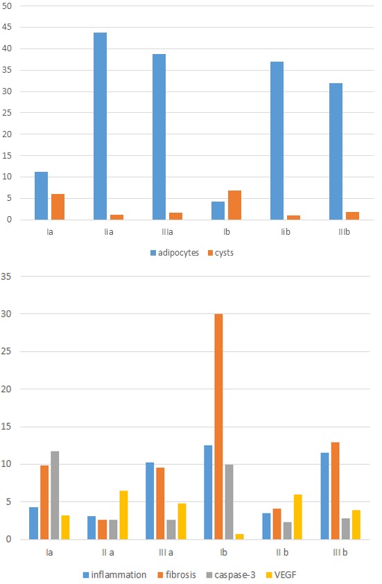
Flowchart 1: showing the mean number of normal adipocytes and fat cysts in the different subgroups.
Flowchart 2: showing the mean area percentage of inflammatory infiltration, fibrosis, and caspase-3 positive immune reaction and VEGF positive immune reaction in different subgroups.
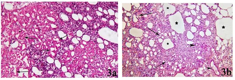
Figure 1: 1a: Photomicrograph of ASCs on day 4 of primary culture showing spindle (↑) and Star-shaped (↑↑) cells with vesicular nuclei and granular cytoplasm. Colonies started to appear. 1b: Photomicrograph of ASCs on day 6 of primary culture showing about 60-70% confluent fibroblast-like cells with vesicular nuclei. They are interconnected by cytoplasmic processes. 1c: Photomicrograph of ASCs on day 7 of primary culture showing 90-95% confluent cells. Whorly appearance of the colonies can be seen. 1d: Photomicrograph of ASCs on passage 3 showing star shaped cells with cytoplasmic processes, granular cytoplasm, and vesicular nuclei. Many nuclei show 2 or more nucleoli (↑). (Phase contrast microscopy x100).
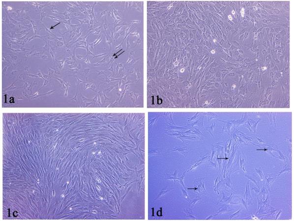
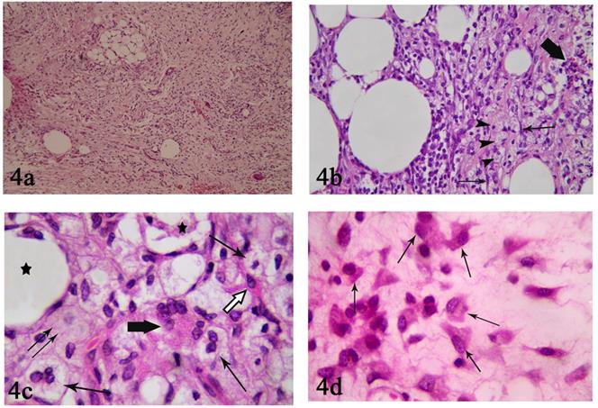
Figure 3: 3a: Photomicrograph of control group (I) showing numerous atrophied fat cells (↑). The atrophied cells contain peripheral acidophilic rim of cytoplasm and multiple fat vacuoles. Notice: intense cellular infiltrate. 3b: Photomicrograph of control group (I) showing few normal sized adipocytes, numerous small sized adipocytes (↑) and multiple large fat cysts (star). Notice: Severe inflammatory cellular infiltration, multiple blood vessels (arrow head). (H&E x100).
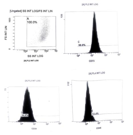
Figure 4: 4a: Photomicrograph of control group (I) showing a localized group of adipocytes and few fat cysts. Numerous inflammatory cells and blood vessels can also be seen. Notice: presence of large amount of connective tissue fibers. (H&E x100) 4b: Photomicrograph of control group (I) showing fat cysts surrounded by intense inflammatory infiltration. Spindle shaped fibroblasts can be seen (↑). Apoptotic cells and cellular debris are observed (thick arrow). Notice: numerous cells with foamy cytoplasm can be seen (arrow heads).(H&E x400). 4c: Photomicrograph of control group (I) showing adipocytes forming fat cyst containing cellular debris (star) and adipocytes with karyolitic nuclei (↑↑). Star-shaped preadipocyte with acidophilic cytoplasm and few fat droplets were also observed (white arrow). Notice! Presence of foam cells (↑) and multinucleated giant cells (thick arrow).(H&E x1000) 4d: Photomicrograph of control group (I) showing numerous spindle shaped cells with acidophilic cytoplasm (↑). Some of them contain small vacuoles. (H&E x1000).
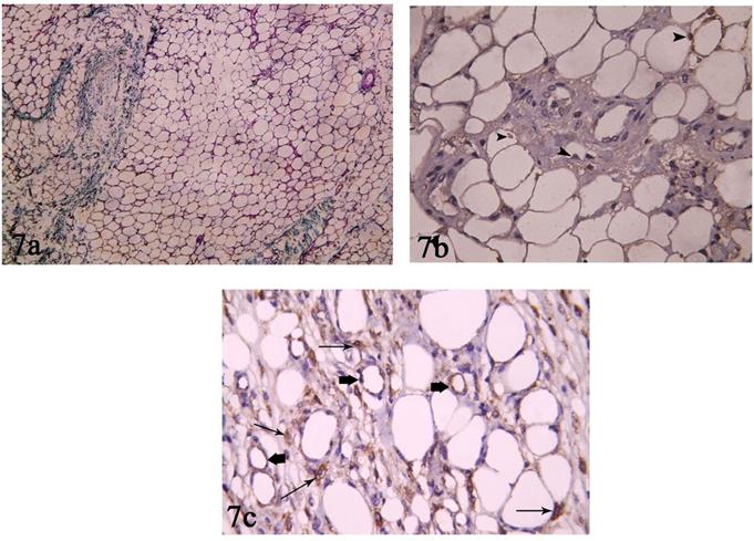
Figure 5: 5a: Photomicrograph of control group (I) showing large amount of collagen fibers surrounding a group of small normally sized adipocytes.(Masson’s Trichrome staining x100) 5b:Photomicrograph of control group (I) showing moderate number of caspase-3 positive adipocytes (arrow heads) and stromal cells (↑). (Caspase-3 immune histochemical staining x400) 5c: Photomicrograph of control group (I) showing weak positive VEGF immune reaction in few inflammatory and stromal cells (↑). Endothelial cells of few blood vessels were stained positive (arrow heads) while some others are negatively stained (star). (VEGF immune histochemical staining x400).
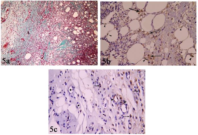
Figure 6: 6a: Photomicrograph of CAL group (II) showing many normal sized and shaped adipocytes with few numbers of degenerated fat cysts (star). Connective tissue septa (arrow heads) and blood vessels (↑) can be seen. (H&E x100) 6b: Photomicrograph of CAL group (II) showing multiple normal sized and shaped adipocytes, few fat cysts (triangles). The stromal vascular fraction contains few stromal cells (arrow heads) and blood vessels (star). (H&E x400).
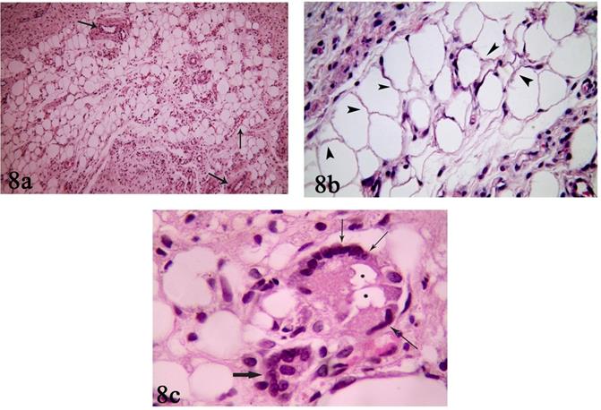
Figure 7: 7a: Photomicrograph of CAL group (II) showing few amount of collagen fibers surrounding normal sized and shaped adipocytes and blood vessels. (Masson’s Trichrome staining x100) 7b: Photomicrograph of CAL group (II) showing minimal caspase-3 positive reaction (arrow heads). (Caspase-3 immune histochemical staining x400) 7c: Photomicrograph of CAL group (II) showing moderate to severe positive VEGF reaction in reaction in blood vessels (thick arrow) and stromal cells (↑). (VEGF immune histochemical staining x400).
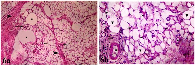
Figure 8: 8a: Photomicrograph of ALA group (III) showing normal sized and shaped adipocytes. Stromal fraction contains connective tissue fibers, inflammatory cells and many blood vessels (↑). (H&E x100) 8b: Photomicrograph of ALA group (III) showing adipocytes with irregular cell membranes (arrow heads). Connective tissue with stromal cells can also be seen.(H&E x400) 8c: Photomicrograph of ALA group (III) showing two multi nucleated giant cells (thin and thick arrows). One giant cell (thin arrow) contains fat droplets in its cytoplasm (stars). (H&E x1000).
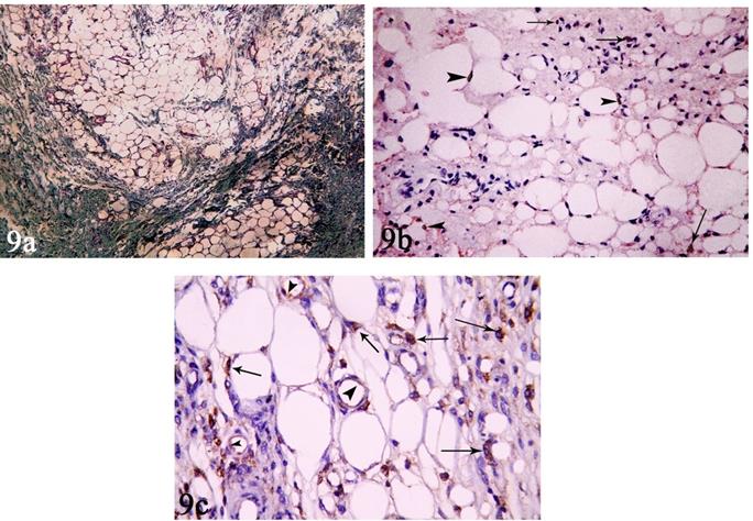
Figure 9: 9a: Photomicrograph of ALA group (III) showing moderate amount of collagen fibers surrounding areas of normal sized and shaped adipocytes forming septa.(Masson’s Trichrome staining x100) 9b:Photomicrograph of ALA group (III) showing mild mild caspase-3 positive immune reaction in some adipocytes (arrow head) and inflammatory cells (↑). (Caspase-3 immune histochemical staining x400) 9c: Photomicrograph of ALA group (III) showing moderate positive VEGF reaction in endothelium of blood vessels (arrow head) andstromal cells (↑). (VEGF immune histochemical staining x400).
Discussion
The current study was designed to compare the effect of ASCs versus ALA in autologous fat graft survival. Adipose tissues were formed of adipocytes and stromal vascular fraction. The stromal vascular fraction is a heterogeneous cell population. Some of them are blood derived and others are adipocytes derived. They included ASCs, progenitor cell, endothelial cell, resident monocytes, fibroblasts, and smooth muscle. In the present study, fat grafts were harvested by excision from inguinal pad of fat with subsequent physical fragmentation into sub millimeter units called micro fragmented adipose tissue (MAT). Excised fat had some advantages over aspirated fat; first, the yield of ASCs from excised fat is much more than the yield of ASCs from aspirated fat. Second, aspirated fat contained fewer vascular structures, especially large ones, compared with excised fat [7]. Blood vessels were considered as important for graft survival [11].
Keeping the microarchitecture of the fat intact by physical fragmentation maintain regenerative growth factors and cytokines. A previous study stated that enzymatic digestion could change the gene expression resulting in decrease of secretory activity of stromal vascular cells and their antigenic growth factors [12].
H&E stained sections of the control group (group I) showed few number of intact fat cells. Some adipocytes showed karyolitic nuclei.
Large Fat cysts could be detected. Some investigators declared that these cysts aroused from fusion of adipocytes after destruction of their cell membranes. Moreover, they recorded that fat cysts remained in the graft as a consequence of fat necrosis. These cysts were non-nucleated with impaired membranes [13].
Loss of adipocytes by necrosis and apoptosis was confirmed by significant increase in caspase-3 positive cells in group I of the present study.
In the present study, most of the fat cells revealed thick rim of acidophilic cytoplasm and multiple fat vacuoles. These atrophic changes were adaptive changes due to lack of vascularization [14].
Some areas of the grafts showed numerous spindle shaped cells with large vesicular nuclei with acidophilic cytoplasm containing minute fat vacuoles. Some researchers stated that fat cells might undergo dedifferentiation into preadipocytes by losing their fat content and transformed into fibroblast-like morphology [15]. Investigators declared that ASCs and preadipocytes could survive in ischemic tissues whereas mature adipocytes died easily [16].
Presence of small sized intact adipocytes was also observed. Previous studies stated that small intact adipocytes were regenerating cells resulted from activation of stem cells to restore the dead cells [17]. However, proliferated stem cells underwent apoptosis resulting in incomplete regeneration and fibrosis. They added that the mechanism of late apoptosis of ASCs was unknown [18].
Fat droplets released from fat cysts and necrotic adipocytes were phagocytized by macrophages resulting in formation of foam cells. Moreover, aggregation of macrophages around dead adipocytes leads to the formation of crown-like structure (multinucleated giant cell).
Another feature of control group was the extensive formation of connective tissue septa. In some specimens, these septa replaced most of the graft tissue. Many investigators explained the cause of fibrotic changes in the graft. A previous researcher stated that foam cells could up regulate lipid receptors with lipid accumulation. This resulted in change in macrophage gene expression from classically activated M1 into alternatively activated M2 which release proliferate mediators [18]. Hypoxia also is thought to enhance pro-inflammatory cytokine production in macrophages promoting an inflammatory shift of the adipose tissue macrophage phenotype [19].
Some scientists recorded that, PDGF A released few lymphocytes and eosinophil’s acted on perivascular adipocytes precursors (adventitial stromal cells and pericytes) and inhibited adipocyte differentiation, but stimulated myofibroblasts differentiation and fibrosis [20]. In addition, researchers stated that stressed adipocytes and hypoxia contributed to immune cell immigration and activation leading to aggravated adipose tissue fibrosis [21].
Researchers thought that stresses resulted after harvesting, early ischemic changes and nutrient deprivation resulted in apoptosis and cell death [18]. Moreover, adipocyte could undergo dedifferentiation and the loss of mature adipocytes may contribute to a significant loss in graft volume and decrease in normal adipocytes. In addition, clearance of adipocytes debris from extracellular matrix by aggregated macrophages also added to the decrease of adipocytes [15]. Being smaller in size than normal adipocytes, regenerating cells was neglected during image analysis contributing to the significant decrease of normal adipocytes in control group compared to the other groups.
Control group showed weak positive VEGF reaction. VEGF is the most important regulator of physiological angiogenesis during growth, healing and in response to hypoxia. VEGF-A is produced by endothelial cells, fibroblasts, smooth muscle cells, platelets, stem cells and macrophages [22, 23]. VEGF is up regulated by hypoxia- inducible transcription factor. However, VEGF alone may lead to leaky, unstable capillaries. Previous studies stated that PDGF can help stabilize blood vessels by recruiting mesenchymal progenitors forming mature vessels [24].
Histological evaluation of fat grafted from CAL group showed significant improvement of all histological parameters compared to control and ALA groups. Marked increase in number of normal size and shape adipocytes was observed. Also, significant decrease in number of fat cysts, inflammatory infiltration, fibrosis and apoptosis was present. Significant increase positive VEGF reaction in adipocytes, blood vessels, fibroblasts and inflammatory cells was observed Many previous studies demonstrated that the most important mechanism behind this improvement is that ASCs contributed to increased micro vascular density and accelerated neoangiogenesis by producing and releasing soluble antigenic factors such as vascular endothelial growth factor, hepatocyte growth factor, and insulin-like growth factor-1 [25]. The capacity of culture-expanded ASCs reported [6, 25] to differentiate into endothelial cells in vitro supports their contribution to neoangiogenesis. This was confirmed by the marked increase in positive VEGF reaction.
In addition, ASCs contributed to the increased number of normal adipocytes by direct adipocyte differentiation and preventing apoptosis by secretion of anti apoptotic cytokines [26] this was confirmed by the significant decrease of caspase-3 positive reaction.
Significant decrease of inflammatory infiltration was referred to the effect of ASCs in modulation of immune response by their paracrine effects. ASCs secrete prostaglandins E2 (PGE2), which alters the secretion profile of cytotoxic T cells by inhibiting the (tumor necrosis factor-α) TNF-α and (interferon-ɣ) INF-ɣ and by stimulating interleukin-10 secretion to modulate the immune inflammatory response [27].
Significant decrease of collagen fibers within the fat grafts of group II of the present study was detected. This was attributed to anti fibrolytic reagents secreted by ASCs [28, 29].
Histological study of the grafts of ALA group showed significant improvement of histological parameters compared to control group. Marked increase of normal adipocytes and VEGF and decrease of fat cysts, inflammatory infiltration, fibrosis and apoptosis were observed. However, some adipocytes showed irregular cell membranes.
The mechanism of ALA in improvement of graft survival might be due to its potent antioxidant and anti apoptotic activity [15, 30]. This was proved by the significant decrease of caspase-3 positive reaction. Antioxidant and anti apoptotic effects of ALA helped adipocytes to survive mechanical and hypoxic stresses after transplantation.
A study investigated the protective effect of ALA against oxidative stress, inflammation, and apoptosis. The anti oxidative activity of ALA was attributed to direct free radical scavenging effect or indirectly to the restoration of endogenous antioxidants (i.e. GSH, vitamin C, and vitamin E), metal binding activity, and regeneration of oxidized proteins [31].
Previous studies proved that ALA interacted with the membrane lipid bilayer and maintained cellular integrity [15, 32, 33]. This also contributed to its protective effect on stressed adipocytes.
Moreover, exogenous ALA had exhibited anti- inflammatory properties and it is now being pursued as a new therapy for inflammatory diseases [34]. A study demonstrated that ALA inhibited the (TNF-α)-induced inflammatory activation by blocking its signaling cascades in rheumatoid arthritis fibroblast-like synovial cells [35]. This explained the significant decrease of inflammatory infiltration within fat grafts compared to control group, and the decrease of fibrosis as a consequence.
Some adipocytes with irregular cell membrane were observed. This irregularity might be due to reducing the lipid content of adipocytes. A previous study stated that ALA had an important role in lipid metabolism. It prevented diabetes mellitus in obese diabetes-prone rats by reducing lipid accumulation in adipose and non- adipose tissues [36]. Other researchers stated that the lipolysis metabolic effect of ALA may be mediated by fibroblast growth factor-21.
Conclusion
Comparative study between CAL and ALA groups showed improvement in their histological parameters compared to control group. However, CAL gave better results.
References
-
Toyserkani NM, Quaade ML, Sorensen JA (2016) Cell- Assisted Lipotransfer: A Systematic Review of Its Efficacy. Aesthetic plastic surgery 40(2): 309-318.
-
Conde Green A, Wu I, Graham I, Chae JJ, Drachenberg CB, et al. (2013) Comparison of 3 techniques of fat grafting and cell-supplemented lipotransfer in athymic rats: a pilot study. Aesthetic surgery journal 33(5): 713-721.
-
Cansancao AL, Conde Green A, David JA, Vidigal RA (2019) Subcutaneous-Only Gluteal Fat Grafting: A Prospective Study of the Long-Term Results with Ultrasound Analysis. Plastic and reconstructive surgery 143(2): 447-451.
-
Jiang A, Li M, Duan W, Dong Y, Wang Y (2015) Improvement of the survival of human autologous fat transplantation by adipose-derived stem-cells- assisted lipotransfer combined with bFGF. The Scientific World Journal 2015: 968057.
-
Gassman AA, Lewis MS, Bradley JP, Lee JC (2015) Remote Ischemic Preconditioning Improves the Viability of Donor Lipoaspirate during Murine Fat Transfer. Plastic and reconstructive surgery 136(3): 495-502.
-
Zhu M, Zhou Z, Chen Y, Schreiber R, Ransom JT, et al. (2010) Supplementation of fat grafts with adipose- derived regenerative cells improves long-term graft retention. Annals of plastic surgery 64(2): 222-228.
-
Matsumoto D, Sato K, Gonda K, Takaki Y, Shigeura T, et al. (2006) Cell-assisted lipotransfer: supportive use of human adipose-derived cells for soft tissue augmentation with lipoinjection. Tissue engineering 12(12): 3375-3382.
-
Truong TT, Soh YM, Gardner DK (2016) Antioxidants improve mouse preimplantation embryo development and viability. Human reproduction 31(7): 445-1454.
-
Li K, Li F, Li J, Wang H, Zheng X, et al. (2017) Increased survival of human free fat grafts with varying densities of human adipose-derived stem cells and platelet-rich plasma. J Tissue Eng Regen Med 11(1): 209-219.
-
Ong SL, Vohra H, Zhang Y, Sutton M, Whitworth JA (2013) The effect of alpha-lipoic acid on mitochondrial superoxide and glucocorticoid-induced hypertension. Oxidative medicine and cellular longevity 2013: 517045.
-
Zannettino AC, Paton S, Arthur A, Khor F, Itescu S, et al. (2008) Multipotential human adipose-derived stromal stem cells exhibit a perivascular phenotype in vitro and in vivo. J cell physiol 214(2): 413-421.
-
Olenczak JB, Seaman SA, Lin KY, Pineros Fernandez A, Davis CE, et al. (2017) Effects of Collagenase Digestion and Stromal Vascular Fraction Supplementation on Volume Retention of Fat Grafts. Annals of plastic surgery 78(6S Suppl 5): S335-S342.
-
Smahel J (1989) Experimental implantation of adipose tissue fragments. Br J Plast Surg 42(2): 207- 211.
-
Wga (2013) Method of Preventing Fat Graft by Administering Fat-Derived Cells and Poloxamer P 188. United States Patent 8,512,695 B2.
-
Suga H, Eto H, Aoi N, Kato H, Araki J, et al. (2010) Adipose tissue remodeling under ischemia: death of adipocytes and activation of stem/progenitor cells. Plastic and reconstructive surgery 126(6): 1911- 1923.
-
Eto H, Kato H, Suga H, Aoi N, Doi K, et al. (2012) The fate of adipocytes after nonvascularized fat grafting: evidence of early death and replacement of adipocytes. Plast Reconstr Surg 129(5): 1081-1092.
-
Engin A (2017) Adipose Tissue Hypoxia in Obesity and Its Impact on Preadipocytes and Macrophages: Hypoxia Hypothesis. Advances in experimental medicine and biology 960: 305-326.
-
Snodgrass RG, Boss M, Zezina E, Weigert A, Dehne N, et al. (2016) Hypoxia Potentiates Palmitate-induced Pro-inflammatory Activation of Primary Human Macrophages. JBC 291(1): 413-424.
-
Trayhurn P, Wang B, Wood IS (2008) Hypoxia in adipose tissue: a basis for the dysregulation of tissue function in obesity?. Br J Nutr 100(2): 227-235.
-
Buechler C, Krautbauer S, Eisinger K (2015) Adipose tissue fibrosis. World J Diabetes 6(4): 548-553.
-
Al Sabti H (2007) Therapeutic angiogenesis in cardiovascular disease. J cardiothorac surg 2: 49.
-
Akhavani MA, Sivakumar B, Paleolog EM, Kang N (2008) Angiogenesis and plastic surgery. JPRAS 61(12): 1425-1437.
-
Deveza L, Choi J, Yang F (202) Therapeutic angiogenesis for treating cardiovascular diseases. Theranostics 2(8): 801-814.
-
Yamaguchi M, Matsumoto F, Bujo H, Shibasaki M, Takahashi K, et al. (2005) Revascularization determines volume retention and gene expression by fat grafts in mice. Exp Biol Med (Maywood) 230(10): 742-748.
-
Badimon L, Cubedo J (2017) Adipose tissue depots and inflammation: effects on plasticity and resident mesenchymal stem cell function. Cardiovascular research 113(9): 1064-1073.
-
Lindroos B, Suuronen R, Miettinen S (2011) The potential of adipose stem cells in regenerative medicine. Stem cell Rev Rep 7(2): 269-291.
-
Zografou A, Tsigris C, Papadopoulos O, Kavantzas N, Patsouris E, et al. (2011) Improvement of skin-graft survival after autologous transplantation of adipose- derived stem cells in rats. JPRAS 64(12): 1647-1656.
-
Klinger M, Marazzi M, Vigo D, Torre M (2008) Fat injection for cases of severe burn outcomes: a new perspective of scar remodeling and reduction. Aesthetic Plast Surg 32(3): 465-469.
-
Canepa P, Dal Lago A, De Leo C, Gallo M, Rizzo C, et al. (2018) Combined treatment with myo-inositol, alpha- lipoic acid, folic acid and vitamins significantly improves sperm parameters of sub-fertile men: a multi-centric study. European review for medical and pharmacological sciences. 22(20): 7078-7085.
-
Lebda MA, Sadek KM, Tohamy HG, Abouzed TK, Shukry M, et al. (2018) Potential role of alpha-lipoic acid and Ginkgo biloba against silver nanoparticles- induced neuronal apoptosis and blood-brain barrier impairments in rats. Life sciences 212: 251-260.
-
Kagan VE, Shvedova A, Serbinova E, Khan S, Swanson C, et al. (1992) Dihydrolipoic acid--a universal antioxidant both in the membrane and in the aqueous phase. Reduction of peroxyl, ascorbyl and chromanoxyl radicals. Biochem Pharmacol 44(8): 1637-1649.
-
Heitzer T, Finckh B, Albers S, Krohn K, Kohlschutter A, et al. (2001) Beneficial effects of alpha-lipoic acid and ascorbic acid on endothelium-dependent, nitric oxide-mediated vasodilation in diabetic patients: relation to parameters of oxidative stress. Free Radic Biol Med 31(1): 53-61.
-
Hwang JS, An JM, Cho H, Lee SH, Park JH, et al. (2015) A dopamine-alpha-lipoic acid hybridization compound and its acetylated form inhibit LPS- mediated inflammation. European journal of pharmacology 746: 41-49.
-
Lee CK, Lee EY, Kim YG, Mun SH, Moon HB, et al. (2008) Alpha-lipoic acid inhibits TNF-alpha induced NF-kappa B activation through blocking of MEKK1- MKK4-IKK signaling cascades. International immunopharmacology 8(2): 62-370.
-
Song KH, Lee WJ, Koh JM, Kim HS, Youn JY, et al. (2005) alpha-Lipoic acid prevents diabetes mellitus in diabetes-prone obese rats. Biochem Biophys Res Commun 326(1): 197-202.
-
Yi X, Pashaj A, Xia M, Moreau R (2013) Reversal of obesity-induced hypertriglyceridemia by (R)-alpha- lipoic acid in ZDF (fa/fa) rats. Biochemical and biophysical research communications 439(3): 390- 395.
- Research Progress of Induced Pluripotent Stem Cells and Their Clinical Application Prospects
- Nishan Al-Kamal is the Starting Point of A Feminist Scientist
- Current Concepts and Future Perspectives of Stem Cell Therapy in Peripheral Arterial Disease
- Stem Cell and Oxidative Stress-Inflammation Cycle
- Adipose Derived Mesenchymal Stem Cells Origin, Characteristics and Promises
- Mitochondria Targeted Antioxidants can Improve In Vitro Embryo Production in Buffalo