Preparation, Characterization and Application of Niosomes for the Delivery of Herbal Bioactive Agents: A Mini Review
The demands of herbal products including Nutraceuticals, cosmetics and drugs increased tremendously globally in recent times. Herbal compounds offer limited toxicity, high biocompatibility with numerous health benefits. The herbal drugs show many health benefits due to their presence of bioactive compounds in it. However, bioactive compounds suffer due to their poor solubility, poor bioavailability and stability. Niosomes, a non-ionic surfactant vesicle, can be a promising aspect to deliver the bioactive compound to enhance the bioavailability and stability. Niosomes have many advantages as it enables encapsulation of either hydrophilic and hydrophobic compounds. Preparation methods of niosomes are also simple and inexpensive. Moreover, encapsulating bioactive compounds in niosome not only improve the bioavailability but also enable it for sustained release or controlled release drug delivery. The review highlights various methods for niosome preparation, including thin-film hydration, reverse-phase evaporation, ether injection, microfluidization and bubble methods along with their advantages and disadvantages. Additionally, the characterization techniques, such as size determination, zeta potential measurement, and morphological evaluation, are also discussed.
Roy PKr*, Nongsiej B, Lalruatsangi H, Laldinchhana L and Lalhlenmawia H
Introduction
Compounds from natural sources are said to be safe and have minimal toxicity [1]. Natural compounds have multiple health benefits with high biocompatibility [2]. However, natural compounds face many challenges including poor solubility and bioavailability, stability which often leads to compromised bioactivity [3]. In the 21st century, pharmaceutical nanotechnology has achieved remarkable milestones in the development of novel drug delivery systems and presenting them with improved efficacy and safety. Among these, niosomes have risen as one of the most promising and versatile approaches for delivery of bioactive compounds [4].
Niosomes are vesicles similar to liposomes with non- ionic surfactant but come with several positive outcomes including stability, ease of preparation and low toxicity [5]. The reason behind the success of niosomal delivery is mainly due to the ability to deliver effectively many bioactive compounds, nutraceuticals, including therapeutic agents which suffer with their inherent poor solubility, instability and rapid clearance [6]. Niosomes are capable to encapsulate either hydrophilic and hydrophobic compounds and provide shields for degradation and upgrade its bioavailability and therapeutic effectiveness [7]. Additionally, niosomes improve the pharmacokinetic and pharmacodynamic properties of herbal bioactive agents [8]. Moreover, niosomes offer site specific delivery of herbal compounds which enhances the bio-efficacy and lowers the risk of toxicity [9]. Niosomes has been successfully applied for the delivery of bioactive compounds, including anti-inflammatory drugs, anticancer drugs, antioxidants etc [10, 12].
Overall, niosomes provide an attractive promising option for drug delivery for bioactive compounds due to their unique structure, versatility and biocompatibility. In this review article, we tried to provide an overview of different preparation methods of niosomes with their advantages and disadvantages, their characterization and application in different industrial aspects.
Structure and Components of Niosomes
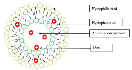
Figure 1 represents the structure of niosomes. Niosomes are the drug carrier for the novel drug delivery system where active pharmaceutical ingredients are encapsulated in a vesicle [13]. Niosomes are similar to liposomes and vesicles are bilayer usually made up from non-ionic surface-active agents, on the other hand liposomes use phospholipids instead of surfactant [14]. Niosomes can be unilamellar (single-layered) or multilamellar (multiple concentric layers). Basic structure of niosomes consists of Non-ionic surfactant, Cholesterol and optional lipids [15].
Non-Ionic Surfactant: Non-ionic surfactants such as Tween series (Tween 20, Tween 80 etc) and Span series e.g., Span 60; Span 80 etc are the primary components of niosomes [15]. The surfactant has hydrophilic head and lipophilic tail and is capable of self-assembling into a bilayer structure under aqueous environment. The concentration of the non- ionic surfactant may vary depending upon the application and type of surfactant used for niosomes [15]. Cholesterol: Cholesterol provides rigidity and stability to the niosomal membrane and prevents leakage of bilayer. Cholesterol decreases the fluidity of niosomal membranes, promotes formation of lipid rafts and regulates permeability and ensures membrane stability at different varying temperatures thereby preventing leakage of the encapsulated compounds [16]. Optional Lipids: In order to stabilize the physical characteristics, additional lipids may be added in the niosomal formulation. Depending upon the need, sterols or phospholipid may be added.
Methods of Preparation for Niosomes
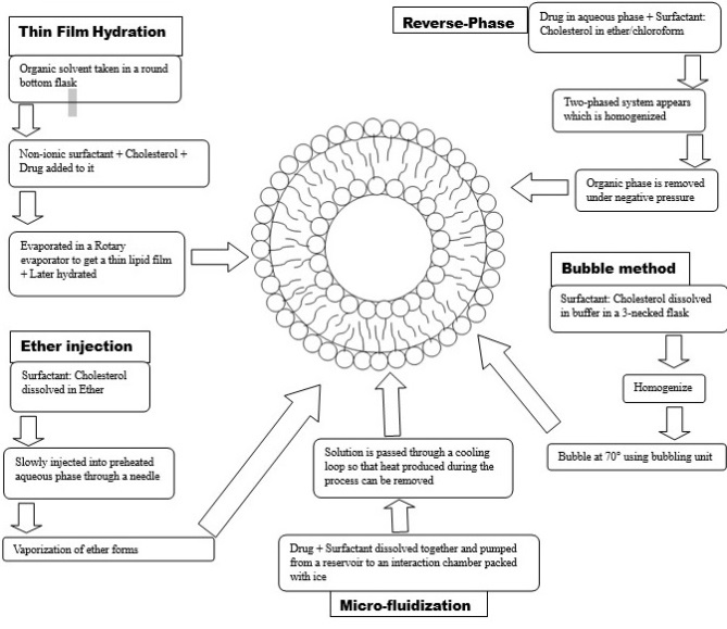
The method of preparation for niosomes has been demonstrated in Figure 2. The following methods are used for preparation of niosomes.
Thin Film Hydration Method: This method is the most commonly used method where vesicles are prepared using non-ionic surfactants. In this method, a thin film of non-ionic surfactant is produced by evaporating a solution of surfactant using suitable organic solvent in a rotary vacuum evaporator. This method prepares niosomes that are lipid-based vesicles and have the capability to encapsulate the herbal bioactive compounds. Here, a thin film of lipids is formed on the glass surface of a container by evaporating the solution of lipids.
Then, herbal extract or bioactive compounds are further added to blend with the film and hydrated with an aqueous phase containing distilled water. Hydration period may vary from 1-2 hours depending on the needs and to ensure complete formation of niosomes. The formed niosomes may be purified using centrifugation and the formed niosomes are further characterised [17]. Thin film hydration methods have several advantages and as well as Disadvantages [18]-Advantages are- a) The method is simple and cost-effective and requires basic laboratory instruments compared to other methods. b) High encapsulation capacity can be achieved; large amounts of drug can be entrapped within the vesicles. This method enables encapsulation of both hydrophilic and hydrophobic drugs.
c) Thin film hydration method enables wide ranges of molecules including small hydrophilic, hydrophobic, and amphiphilic compounds to encapsule thereby enhancing the versatility of drug delivery applications. d) This method can be easily scaled up from laboratory scale research to large scale industry. The formed niosomes possess good physicochemical stability during storage for long term use. e) Non-ionic surfactants used in the formation of niosomes are usually biocompatible, biodegradable and lowers the risk of adverse reaction. f) Thin film hydration methods allow formulation scientists to adjust the composition of lipid bilayer by varying type and composition. This enables customization of noisome size. g) Sustained release or prolonged drug release can be attained by the niosomes prepared using thin film hydration method thereby enhancing patient compliances. Despite thin film hydration method enjoys numbers of advantages, it has some limitations too- they are- a) Thin film hydration methods have several steps such as film formation, hydration and extrusion. They are complex and time consuming and require skilled operators for reproducibility of quality niosomes. b) There are chances of producing low entrapment efficiency of the drugs as in this technique only in the hydration phase active ingredients can be trapped. c) Uniform Size of niosomes are very difficult to obtain in this method. Size and homogeneity of the niosomes may affect their stability in drug performance d) The method is temperature and shear sensitive, therefore during processing of niosomes undesired aggregates may form which can create stability problems. e) Lipids used in formulation of niosomes may undergo oxidation or hydrolysis during thin film formation thereby affecting stability and shelf life of the final product. f) Surfactants and lipid used for the process is limited unlike liposomes, which may affect the optimization for the niosomal preparation g) There are chances that niosomes formed by thin film hydration method may leak during storage causing content loss and instability. Reverse-Phase Evaporation Method: This is one of the widely used methods for the preparation of niosomes. This method utilises water-in-oil emulsion which is formed as a result of mixing non-ionic surfactant with an organic solvent and water. Addition of cholesterol also enhances the rigidity and stability of the niosomes. Immiscible organic solvent is used to dissolve the herbal extract or bioactive compound and added to the surfactant solution containing aqueous phase. Both the phases are mixed vigorously to form the water -in- oil emulsion. The emulsion is then evaporated using a rotary Vacuum evaporator, where organic solvent evaporates and niosomes containing bioactive extract/compounds remain [19]. Once the solvent completely evaporates, aqueous phase containing buffer or distilled water is added to the content of lipid film. The system is allowed to hydrate with gentle stirring or by sonication. The niosomes are purified using ultrafiltration or dialysis to remove the untrapped compounds and further characterise them. Reverse phase evaporation method has several advantages and disadvantages over other methods [20] -Advantages are a) It is a very simple method and does not require any expensive equipment. It is cost effective and accessible to laboratories with limited laboratory resources. b) This method has the capability of loading high content of drugs/bioactive compounds thereby reducing the drug wastage. c) This method enables encapsulation of both hydrophilic as well as hydrophobic compounds providing versatility in drug delivery. d) Niosomes prepared by these methods are more stable compared to other methods making it enhanced shelf life. e) Controlled release drug delivery is possible. Limitations of Reverse-phase evaporation method are- a) The uses of organic solvent in this method may concern about toxicity as the removal residual of the organic solvent may be problematic. b) The process is time consuming compare to other methods. c) Producing narrow size distribution vesicles are problematic. d) There are chances of batch-to-batch variation in the size of niosomes. Ether Injection Method: The method uses non-ionic surfactant, a co-surfactant and an organic solvent which is injected into an aqueous phase with high pressure syringe and further sonicated to form niosomes. This method is most commonly applied to prepare niosomes where non- ionic surfactant-based vesicles encapsulate wide ranges of hydrophilic and hydrophobic bioactive compounds. Then the formed niosomes are purified by centrifugation or dialysis to eliminate non-encapsulated extract [21]. Ether injection method have several advantages and also some limitations [22] The method enjoys following advantages- a) It is very simple method and does not require sophisticated equipment’s making it cost effective. b) High efficiency of encapsulation of both hydrophilic as well as hydrophobic bioactive compounds may be achieved. c) Scalability to industrial level is possible. Limitations of Ether injection methods are a) Use of ether can impose potential risk of safety as it is highly inflammable. Proper handling and disposal methods are required to ensure safety. b) Despite the effort of removal of organic solvent, traces of organic solvent may cause harmful effects after ingestion. c) Niosomes produced by this method may have problem in long term stability. Microfluidization Method: Microfluidization is a technique used for the preparation of liposomes and niosomes. In this method a mixture of non-ionic surfactant containing bioactive compounds and cholesterol (for improving stability) are passed through a microfluidizer at high pressure to form niosomes. High pressure homogenization creates uniform dispersion of lipids or surfactant in the aqueous solution [23]. The resulting niosomes where herbal bioactive compounds have been encapsulated can be utilised for drug delivery and cosmetic formulations. Microfluidization methods enjoys the several advantages and also suffers from some limitations [24]. Advantages are as follows- a) Uniform size distribution of niosomes can be obtained using this technique. b) High encapsulation efficiency of drug can be achieved. c) Microfluidization enables easy scale up for commercial production of niosomes. d) Batch to batch variation can be reduced as microfluidization promotes uniformity in size of niosomes. e) Use of organic solvent can be avoided in this method, reducing the risk of toxicity to health and environment. Some of the limitations of microfluidization methods are a) Cost of the equipment is high compared to others methods, therefore may not affordable to every laboratory. b) Expert technician is required in order to run the machine. c) Microfluidization techniques generate heat during the process due to shear force applied, therefore it may cause stability issue with heat sensitive compounds. d) High shear forces are used in microfluidization, which may affect in the structure of niosomes e) In certain cases, microfluidization may cause particle aggregation which may reduce the encapsulation efficiency. Bubble Method: Bubble method uses aqueous solution where bioactive compound and non-ionic surfactant are mixed together, followed by bubbled with nitrogen gas. The formed bubbled suspension is then ultrasonicated for a few minutes to break down the bubbles and yield niosomes [25]. The formed niosomes are purified either by centrifugation or dialysis. The bubble offer certain advantages over some other methods and have limitations too [26]. Advantages are-a) The method is simple and cost effective. b) Without significant changes these methods offer scale up option. c) Lower energy consumption compared to other methods like sonication or microfluidization etc. d) Uniform size distribution can be achieved. e) Wide ranges of therapeutic agents can be encapsulated. Some limitations of Bubble methods are- a) For certain hydrophilic compounds high encapsulation efficiency cannot be achieved. b) Unable to have control over the size during formulation compared to other methods may leads to stability issue. c) Bubble methods takes longer duration to formulate niosomes compared to other methods, so large scale production may not be feasible in economic point of view. d) Bubbles methods can not be controlled over batch-to- batch variation. So, reproducibility is low compared to other methods.
Characterization of Niosomes
The characterization of niosomes containing herbal extract can be done through various methods, which are discussed below:
Size Analysis: a. Dynamic Light Scattering (DLS): Zeta sizer is one of the most popular instruments to study the size distribution of niosomes. It utilises a technique called Dynamic Light Scattering or photon correlation spectroscopy to determine the size distribution of niosomes. Usually, it measures the fluctuations in light scattering caused by the Brownian motion of the particles. DLS can provide information about the average size, size distribution, and polydispersity index of niosomes [27].
b. Transmission Electron Microscopy (TEM): TEM permits direct imaging of niosomes at high resolution. By taking images of niosomes, it determines the size by measuring dimensions of individual vesicles. TEM offers complete information about the morphology and size distribution of niosomes [27]. Shape Analysis: a. TEM and SEM: As stated earlier, TEM and SEM can offer the morphological information of niosomes. It examines the images; the shape of individual vesicles is determined. b. Atomic Force Microscopy (AFM): It is a high- resolution imaging technique used to analyse the surface morphology and shape of niosomes. It offers topographical information at the nanoscale and can reveal the three- dimensional structure of niosomes [27]. Fourier Transform Infrared (FTIR): FTIR is used to detect the incompatibility between the lipid, surfactant bioactive compounds or any other chemical used for the formulation development. FTIR spectra of all individual excipients are taken followed by a mixture of all the compounds used for the formulation. Any changes in the fingerprinting region of the active compound shows incompatibility [27, 28]. Thermal Analysis: In thermal analysis, Differential scanning calorimetry is used to detect the physical properties of a sample along with temperature against time. Initially, all individual compounds’ thermal properties are measured, followed by a mixture of all compounds. Any changes in thermal behaviour of the compound’s results in incompatibility [27, 28]. Entrapment Efficiency: Entrapment efficiency refers to how efficiently a compound or substance is entrapped during the process of encapsulation. The entrapment efficiency of niosome containing bioactive herbal extracts is measured by extracting the herbal extract from the niosomes with suitable techniques followed by measuring the concentration of compounds or extract concentration using UV-VIS spectroscopy or HPLC [27]. It is expressed as a percentage and can be calculated using the following formula: Entrapment Efficiency = (Amount of substance entrapped / Total amount of substance used) × 100 Drug Release Kinetics: Release kinetics from the niosomes refers to at which rate or extent a compound is released from the niosomes over the time. The release kinetics of a compound from niosomes can be determined by measuring the released content from the niosomes over the time period using UV-VIS spectroscopy or HPLC. After the determination of the rate constant is determined and checked for whether release kinetics is Zero order or first order or Higuchi’s square root of time kinetics or Hixson-Crowell cube root kinetics etc [27, 28]. Stability Studies: The stability of the niosomes containing the herbal extract can be evaluated by monitoring the size, shape, and entrapment efficiency of the vesicles over time under various storage conditions such as temperature, humidity, and light [27, 28].
Encapsulation of Bioactive Compounds
The primary goal of encapsulating bioactive compounds in niosomes is to improve the stability and delivery of these compounds in pharmaceutical, cosmeceuticals and biomedical applications. (Table 1) shows numbers of bioactive compounds and drugs that are encapsulated for different pharmacological activities. Bioactive compounds possess antioxidants, anti-inflammatory, anticancer etc and suffer from either solubility or stability issues [10, 11, 12]. By encapsulating these compounds pharmaceutical scientists can improve their stability and deliver them to alleviate disease or to promote health. While formulating proper choice of surfactant and lipid is important as these components are mainly responsible for release of the drugs and stability. In other words, we can say that by encapsulating bioactive compounds it is possible to not only enhance their stability and solubility, but also improve their bioavailability and sustained or target drug release can be achieved.
| Method of Preparation | Objective of the Study | Reference | |
|---|---|---|---|
| Calendula L.(Marigold) extract | Thin film hydration | cytotoxicity, wound healing and antioxidant activity | [29] |
| D-limonene | Thin film hydration | In-vitro cytotoxicity assay | [30] |
| Nerium oleander extracts | Thin film hydration | Antioxidant activity, cytotoxic activity. | [31] |
| Lawsone | Thin film hydration | Anticancer activity | [32] |
| Gymnema sylvestre extract | Thin film hydration | Antihyperglycemic activity | [33] |
| Curcumin | Thin film hydration | Anti-diabetic, anti-microbial, anti-cancer | [34] |
| Hedera Helix and Glycyrrhiza glabra extract | Thin film hydration | Breast cancer treatment | [35] |
| Lippia citriodora essential oil | Thin film hydration | Anti-microbial, anti-oxidant | [36] |
| Spermacoce hispida Extract | Thin film hydration | Antituberculosis Activity | [37] |
| Carum carvil seed extract | Thin film hydration | Anti-cancer activity | [38] |
| Spirulina platensis | Thin film hydration | Alzheimer’s disease treatment, Neuroprotective agent | [39] |
| Arbutin | Ultrasonic technique | Treatment of hyperpigmentation | [40] |
| Tradescantia pallida extract | Probe sonication technique | Anti-diabetic | [41] |
| Mupirocin with Bee honey and Curcumin | Ether injection method | Wound healing | [42] |
| Amphotericin B and Thymus essential oil | Thin film hydration | Anti-fungal | [43] |
| Lycopene | Thin film hydration method followed by bath sonication | Treatment of prostate cancer | [44] |
| Echinacea angustifolia Extract | Thin film hydration | Antibacterial activity | [45] |
| Propolis extract | Thin film hydration | Antimycobacterial | [46] |
| Aloe vera | Reverse-phase evaporation | Wound Healing | [47] |
| Hypericum perforatum extract | Reverse-phase evaporation | Transdermal drug delivery | [48] |
| Galangin | Reverse-phase evaporation | Antitumour activity | [49] |
| Rosmarinic acid | Reverse-phase evaporation | Acne Treatment | [50] |
| Withania somnifera Crude Extracts | Ether injection method | Transdermal delivery | [51] |
| Fumaria officinalis | Ether-injection method | Antidiabetic, Antineuropathic, Anti-inflammatory activity | [52] |
| Punica granatum extract | Ether-injection method | Optimalization for delivery | [53] |
| Turmeric oil | Microfluidization methods | Larvicidal activity | [54] |
Table 1: Different compounds encapsulated as niosomes for delivery of different purposes.
Application of Niosomes
A brief list of applications of niosomes are given in the Figure 3.
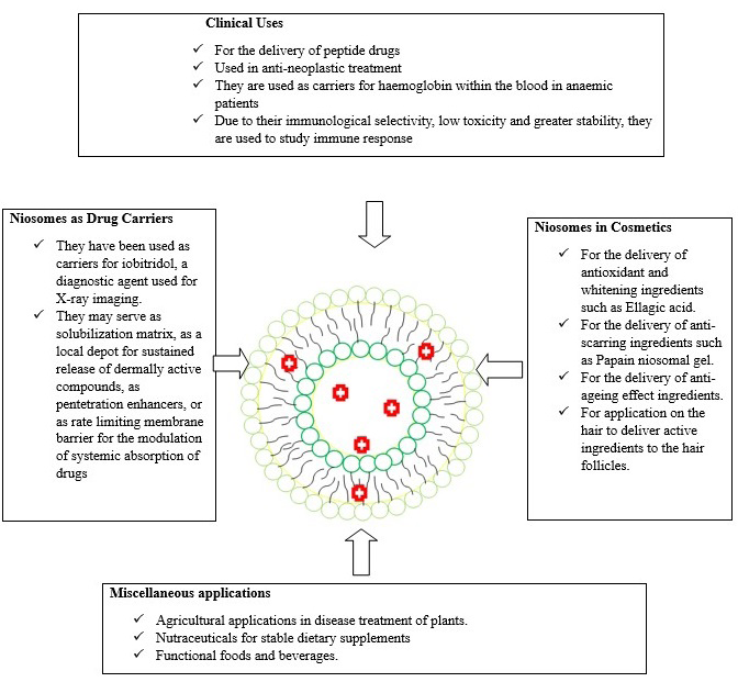
Drug Delivery: Niosomes have advantages over conventional techniques as it protects the compounds from biodegradation and hence increased absorption and bioavailability. It also offers controlled release drug delivery of the compounds and site-specific delivery of compounds thereby minimising toxicity [55]. Cosmetics: The demand of herbal cosmetics is tremendously high across the globe as they have several potential benefits to skin and hair. Niosomes can encapsulate the herbal bioactive compounds and are utilised in cosmetic formulations such as creams, lotions, and serums etc [56, 57]. Nutraceuticals: Entrapment of bioactive compounds used in nutraceuticals provided with stable dietary supplements. Niosomes protects the bioactive compounds from degradation in the digestive system and enhances its absorption and bioavailability [57, 58]. Agricultural Applications: Niosomes can serve as effective carriers for delivering herbal bioactive compounds. Herbal bioactive compounds are utilised for various agricultural purposes such as disease management etc. They can enhance the stability and controlled release of these compounds, allowing for targeted application and improved efficiency in agricultural practices [56, 57, 58, 59]. Functional Foods and Beverages: Niosomes can be used to encapsulate herbal bioactive compounds for nutraceuticals. Niosomes can protect the compounds from degradation and facilitate their controlled release during digestion, ensuring the desired bioactivity [58, 59].
Conclusion
In conclusion, this mini review article provides a brief overview of preparation of niosomes containing bioactive compounds, their characterization and application of niosomes in different industries. There is no doubt that niosomal drug delivery has emerged as one of the most promising delivery aspects for herbal bioactive compounds due its overall protection against bio-environment and enables enhanced solubility and bioavailability. Overall, niosomes offer great potential for delivery of herbal bioactive compounds with improved therapeutic effectiveness and patient compliance. As the research and development progress, it will be interesting to see the use of the niosomal delivery system of herbal bioactive compounds in the modern healthcare system.
Conflicts of Interests: The Authors declares no conflict of interest
References
-
Lourenço SC, Moldão Martins M, Alves VD (2019) Antioxidants of natural plant origins: From sources to food industry applications. Molecules 24(22): 4132.
-
Do NH, Truong Q T, Le PK, Ha AC (2022) Recent developments in chitosan hydrogels carrying natural bioactive compounds. Carbohydrate Polymers 294: 119726.
-
Pachuau L, Laldinchhana, Roy PK, Zothantluanga JH, Ray S, et al. (2021) Encapsulation of bioactive compound and its therapeutic potential. Bioactive natural products for pharmaceutical applications 140: 687-714.
-
Vieira IRS, Conte Junior C A (2022) Nano-delivery systems for food bioactive compounds in cancer: Prevention, therapy, and clinical applications. Critical Reviews in Food Science and Nutrition 8: 1-26.
-
Chen S, Hanning S, Falconer J, Locke M, Wen J (2019) Recent advances in non-ionic surfactant vesicles (niosomes): Fabrication, characterization, pharmaceutical and cosmetic applications. European journal of pharmaceutics and biopharmaceutics 144: 18-39.
-
Martins Gomes C, Souto EB, Silva AM (2022) Nanophytosomes: a novel approach for the delivery of herbal drugs. Systems of Nanovesicular Drug Delivery pp: 239-257.
-
Dehnad D, Emadzadeh B, Ghorani B, Rajabzadeh G, Kharazmi MS, et al. (2022) Nano-vesicular carriers for bioactive compounds and their applications in food formulations. Critical Reviews in Food Science and Nutrition pp: 1-20.
-
Wang G, Wang J, Wu W, Tony To SS, Zhao H, et al. (2015) Advances in lipid-based drug delivery: enhancing efficiency for hydrophobic drugs. Expert opinion on drug delivery 12(9): 1475-1499.
-
Martínez Ballesta M, Gil Izquierdo Á, García Viguera C, Domínguez Perles R (2018) Nanoparticles and controlled delivery for bioactive compounds: Outlining challenges for new smart-foods for health. Foods 7(5): 72.
-
Afereydoon S, Haghiralsadat F, Hamzian N, Shams A, Hemati M, et al. (2022) Multifunctional PEGylated niosomal nanoparticle-loaded herbal drugs as a novel nano-radiosensitizer and stimuli-sensitive nanocarrier for synergistic cancer therapy. Frontiers in Bioengineering and Biotechnology 10: 917368.
-
Leelarungrayub J, Manorsoi J, Manorsoi A (2017) Anti- inflammatory activity of niosomes entrapped with Plai oil (_Zingiber cassumunar Roxb_.) by therapeutic ultrasound in a rat model. International journal of nanomedicine 12: 2469-2476.
-
Machado ND, Gutiérrez G, Matos M, Fernández MA (2021) Preservation of the antioxidant capacity of resveratrol via encapsulation in niosomes. Foods 10(5): 988.
-
Kauslya A, Borawake PD, Shinde JV, Chavan RS (2021) Niosomes: a novel carrier drug delivery system. Journal of Drug Delivery and Therapeutics 11(1): 162-170.
-
Mishra V, Nayak P, Singh M, Sriram P, Suttee A (2020) Niosomes: Potential nanocarriers for drug delivery. J Pharm Clin Res 11(3): 389-394.
-
Ge X, Wei M, He S, Yuan WE (2019) Advances of non-ionic surfactant vesicles (niosomes) and their application in drug delivery. Pharmaceutics 11(2): 55.
-
Nakhaei P, Margiana R, Bokov DO, Abdelbasset WK, Jadidi Kouhbanani MA, et al. (2021) Liposomes: structure, biomedical applications, and stability parameters with emphasis on cholesterol. Frontiers in bioengineering and biotechnology 9: 705886.
-
Thabet Y, Elsabahy M, Eissa NG (2022) Methods for preparation of niosomes: A focus on thin-film hydration method. Methods 199: 9-15.
-
Kaur D, Kumar S (2018) Niosomes: present scenario and future aspects. Journal of drug delivery and therapeutics 8(5): 35-43.
-
Matos M, Pando D, Gutiérrez G (2019) Nanoencapsulation of food ingredients by niosomes. In Lipid-based nanostructures for food encapsulation purposes 2: 447- 481.
-
Mishra V, Nayak P, Singh M, Sriram P, Suttee A (2020) Niosomes: Potential nanocarriers for drug delivery. J Pharm Clin Res 11(3): 389-394.
-
Raut DJ, Shirode DS, Deokar SS, Saoji VV, Rajput GD, et al. (2023) Quercetin and Silymarin loaded Niosomal Formulation with Synergistic Effect on Hep G2 Cell Lines. Latin American Journal of Pharmacy 42(3): 347-354.
-
John CR, Sailaja AK (2023) A curcumin loaded niosomes as novel drug delivery system by ether injection method. Drug Discovery 17: e19dd1918.
-
Ag Seleci D, Maurer V, Stahl F, Scheper T, Garnweitner G (2019) Rapid microfluidic preparation of niosomes for targeted drug delivery. International journal of molecular sciences 20(19): 4696.
-
Moghtaderi M, Sedaghatnia K, Bourbour M, Fatemizadeh M, Salehi Moghaddam Z, et al. (2022) Niosomes: a novel targeted drug delivery system for cancer. Medical Oncology 39(12): 240.
-
Gharbavi M, Amani J, Kheiri Manjili H, Danafar H, Sharafi A (2018). Niosome: A Promising Nanocarrier for Natural Drug Delivery through Blood-Brain Barrier. Advances in Pharmacological Sciences pp: 1-15.
-
Masjedi M, Montahaei T (2021) An illustrated review on nonionic surfactant vesicles (niosomes) as an approach in modern drug delivery: Fabrication, characterization, pharmaceutical, and cosmetic applications. Journal of Drug Delivery Science and Technology 61: 102234.
-
Pando D, Gutiérrez G, Coca J, Pazos C (2013) Preparation and characterization of niosomes containing resveratrol. Journal of Food Engineering 117(2): 227-234.
-
Khan DH, Bashir S, Figueiredo P, Santos HA, Khan MI, et al. (2019) Process optimization of ecological probe sonication technique for production of rifampicin loaded niosomes. Journal of Drug Delivery Science and Technology 50: 27-33.
-
Un RN, Barlas FB, Yavuz M, Ag Seleci D, Seleci M, et al. (2015) Phyto-niosomes: in vitro assessment of the novel nanovesicles containing marigold extract. International Journal of Polymeric Materials and Polymeric Biomaterials 64(17): 927-937.
-
Hajizadeh MR, Maleki H, Barani M, Fahmidehkar MA, Mahmoodi M, et al. (2019) In vitro cytotoxicity assay of D-limonene niosomes: an efficient nano-carrier for enhancing solubility of plant-extracted agents. Research in pharmaceutical sciences 14(5): 448-458.
-
Gunes A, Guler E, Un RN, Demir B, Barlas FB, et al. (2017) Journal of Drug Delivery Science and Technology 37: 158-165.
-
Barani M, Mirzaei M, Torkzadeh Mahani M, Nematollahi MH (2018) Lawsone-loaded Niosome and its antitumor activity in MCF-7 breast Cancer cell line: a Nano-herbal treatment for Cancer. DARU Journal of Pharmaceutical Sciences 26(1): 11-17.
-
Kamble B, Talreja S, Gupta A, Patil D, Pathak D, et al. (2013) Development and biological evaluation of Gymnema sylvestre extract-loaded nonionic surfactant- based niosomes. Nanomedicine 8(8): 1295-1305.
-
Bashash M, Varidi M, Varshosaz J (2022) Composite Hydrogel-Embedded Sucrose Stearate Niosomes: Unique Curcumin Delivery System. Food and Bioprocess Technology 15(9): 2020-2034.
-
Akhlaghi M, Taebpour M, Lotfabadi NN, Naghib SM, Jalili N, et al. (2022) Synthesis and characterization of smart stimuli-responsive herbal drug-encapsulated nanoniosome particles for efficient treatment of breast cancer. Nanotechnology Reviews 11(1): 1364-1385.
-
Saleh A, Pirouzifard M, Khaledabad MA, Almasi H (2022) Optimization and Characterization of Lippia citriodora Essential Oil Loaded Niosomes: A Novel Plant-based Food Nano Preservative. Colloids and Surfaces A: Physicochemical and Engineering Aspects 650: 129480.
-
Anghore D, Kulkarni GT (2017) Development of novel nano niosomes as drug delivery system of spermacoce hispida extract and in vitro antituberculosis activity. Current Nanomaterials 2(1): 17-23.
-
Barani M, Mirzaei M, Torkzadeh Mahani M, Adeli Sardou M (2019) Evaluation of carum-loaded niosomes on breast cancer cells: Physicochemical properties, in vitro cytotoxicity, flow cytometric, DNA fragmentation and cell migration assay. Scientific reports 9(1): 7139.
-
Abdelghany AK, Gamal A, Abdel Wahab A, Abdel Razik ARH, El Samannoudy SI, et al. (2023) Evaluating the neuroprotective effect of Spirulina platensis–loaded niosomes against Alzheimer’s disease induced in rats. Drug Delivery and Translational Research.
-
Radmard A, Saeedi M, Morteza Semnani K, Hashemi SMH, Nokhodchi A (2021) An Eco-Friendly and Green Formulation in Lipid Nanotechnology for Delivery of a Hydrophilic Agent to the Skin in the Treatment and Management of Hyperpigmentation Complaints: Arbutin Niosome (Arbusome). Colloids and Surfaces B: Biointerfaces 201: 111616.
-
Imtiaz F, Islam M, Saeed H, Ahmed A, Asghar M, et al. (2023) Novel Phytoniosomes Formulation of Tradescantia Pallida Leaves Attenuates Diabetes more effectively than Pure Extract. Journal of Drug Delivery Science and Technology 83: 104399.
-
Theerdhala S, Harikrishnan N (2023) Mupirocin Niosomal Gel with Bee Honey & Curcumin as Nano-Drug Delivery in Wound Healing Applications. Current Trends in Biotechnology and Pharmacy 17(2): 885-900.
-
Rahimi F, Amoabediny G, Sabahi H, Zandieh Doulabi B (2022) Fungal Infected Adipose Stem Cells: The Effects of Novel Lipo-Niosome Nanoparticles Loaded with Amphotericin B and Thymus Essential Oil. Cell Journal (Yakhteh) 24(7): 391.
-
Kusdemir BC, Guldu OK, Kilcar AY, Medine EI (2023) Preparation and in Vitro Investigation of Prostate- Specific Membrane Antigen Targeted Lycopene Loaded Niosomes on Prostate Cancer Cells. International Journal of Pharmaceutics 640: 123013.
-
Moghtaderi M, Mirzaie A, Zabet N, Moammeri A, Mansoori Kermani A, et al. (2021) Enhanced Antibacterial Activity of Echinacea Angustifolia Extract Against Multidrug- Resistant Klebsiella Pneumoniae Through Niosome Encapsulation. Nanomaterials 11(6): 1573.
-
Sangboonruang S, Semakul N, Suriyaprom S, Kitidee K, Khantipongse J, et al. (2023) Nano-Delivery System of Ethanolic Extract of Propolis Targeting Mycobacterium Tuberculosis Via Aptamer-Modified-Niosomes. Nanomaterials 13(2): 269.
-
Dadashzadeh A, Imani R, Moghassemi S, Omidfar K, Abolfathi N, et al. (2020) Study of Hybrid Alginate/ Gelatin Hydrogel-Incorporated Niosomal Aloe Vera Capable of Sustained Release of Aloe Vera as Potential Skin Wound Dressing. Polymer Bulletin 77: 387-403.
-
Ali M, Abdel Motaal A, Ahmed MA, Alsayari A, El Gazayerly ON (2018) An in Vivo Study of Hypericum Perforatum in a Niosomal Topical Drug Delivery System. Drug Delivery 25(1): 417-425.
-
Sabry S, Okda T, Hasan A (2021) Formulation, Characterization, and Evaluation of the Anti-Tumor Activity of Nanosized Galangin Loaded Niosomes on Chemically Induced Hepatocellular Carcinoma in Rats. Journal of Drug Delivery Science and Technology 61: 102163.
-
Budhiraja A, Dhingra G (2015) Development and Characterization of a Novel Antiacne Niosomal Gel of Rosmarinic Acid. Drug Delivery 22(6): 723-730.
-
Chinembiri TN, Gerber M, Du Plessis LH, Du Preez JL, Hamman JH, et al. (2017) Topical Delivery of Withania Somnifera Crude Extracts in Niosomes and Solid Lipid Nanoparticles. Pharmacognosy Magazine 13(3): S663.
-
Raafat KM, El Zahaby SA (2020) Niosomes of Active Fumaria Officinalis Phytochemicals: Antidiabetic, Antineuropathic, Anti-Inflammatory, and Possible Mechanisms of Action. Chinese Medicine 15: 1-22.
-
Priya H, Singh H (2012) Formulation and Evaluation of Niosomes Containing Punicalagin From Peels of Punica Granatum. Journal of Drug Delivery and Therapeutics 2(6).
-
Asaithambi K, Muthukumar J, Chandrasekaran R, Ekambaram N, Roopan M (2020) Synthesis And Characterization of Turmeric Oil Loaded Non-Ionic Surfactant Vesicles (Niosomes) and its Enhanced Larvicidal Activity Against Mosquito Vectors. Biocatalysis and Agricultural Biotechnology 29: 101737.
-
Sahin NO (2007) Niosomes as Nanocarrier Systems. Nanomaterials and Nanosystems for Biomedical Applications pp: 67-81.
-
Tripathi PK, Choudary SK, Srivastva A, Singh DP, Chandra V (2012) Niosomes: an Study on Noval Drug Delivery System-A Review. Inter J Pharmac Res Develop 3: 100- 106.
-
Chen S, Hanning S, Falconer J, Locke M, Wen J (2019) Recent Advances in Non-Ionic Surfactant Vesicles (Niosomes): Fabrication, Characterization, Pharmaceutical and Cosmetic Applications. European Journal of Pharmaceutics and Biopharmaceutics 144: 18-39.
-
Tavano L, Muzzalupo R, Picci N, De Cindio B (2014) Co- Encapsulation of Antioxidants into Niosomal Carriers: Gastrointestinal Release Studies for Nutraceutical Applications. Colloids and Surfaces B: Biointerfaces 114: 82-88.
-
Chen S, Hanning S, Falconer J, Locke M, Wen J (2019) Recent Advances in Non-Ionic Surfactant Vesicles (Niosomes): Fabrication, Characterization, Pharmaceutical and Cosmetic Applications. European Journal of Pharmaceutics and Biopharmaceutics 144: 18-39.
- Solution-Processed Chiral Perovskites for Biomedical Applications
- Nanotechnology in Health Chemistry and Medicine: Current Challenges and Future Directions
- Human Exposure to Micro- and Nanoplastics: Pathways, Toxicity, and Intervention Strategies
- Exosome Nanomedicine for Cancer Therapy
- Micro and Nanoplastics–Plastisphere, Biotoxicity, Impact on Human Health, and Mitigation Strategies
- Process Validation of Cefixime Powder for Suspension Dosage Form, 50 mL