A Study on the Phytochemical, Antioxidant and Antimicrobial Activities of Ficus glumosa Delile (Moraceae)
Phytochemical, Antioxidant and Antimicrobial studies of the ethanolic leaf and stem extracts of African rock fig (Ficus glumosa) were carried out to determine its potency as a medicinal plant. The presence of these phytochemicals (alkaloid, phenol, flavonoid, sterol, tannin, saponin, lycopene, beta carotene and ascorbic acid) was determined using standard techniques. Antioxidant activity of leaf and stem extracts was determined by reducing power capacity. The antimicrobial assay of leaf and stem extracts was carried out at different concentrations against some selected human pathogens (Staphylococcus aureus, Salmonella typhi, Escherichia coli, Candida albicans and Aspergillus niger) using disc diffusion method. Analysis of variance was employed in data analysis. Qualitative phytochemical screening revealed the presence of alkaloid, phenol, flavonoid, sterol, tannin and saponin. Antioxidant studies indicated that the extract had antioxidant potentials and the effectiveness of the extract increases progressively as the concentration increases. Antimicrobial studies indicated that the ethanolic leaf and stem extracts of Ficus glumosa inhibited the growth of the pathogen but at varied levels. The extract showed higher inhibition against the fungal strains than the bacterial strains. Inhibitory effect of the leaf extract was significantly higher than those of the stem extract. Data obtained from this research showed that the plant possessed antioxidant and antimicrobial potentials and could be used in treatment of microbial infections especially fungal infections and also used to protect cells against free radical damages.
Introduction
The discovery and use of medicinal plants in ethno- medicine practices dates back to prehistoric times. Medicinal plants are plants that have medicinal properties that can treat various ailments [1]. Unexploited are these plants, which are a rich source of biologically active compounds (phytochemicals) that are crucial for human health [2]. In non-industrialized societies, they are commonly employed because they are easily accessible and cheaper than modern medications [3].
Ficus glumosa (African rock or mountain fig), a species of Moraceae family, is a well-known plant that is widely employed in traditional medicine because of its medicinal and economic properties. It is present in most of tropical Africa, with an extension to western Saudi Arabia in the east and South Africa in the south [3]. F. glumosa is an erect shrub or small tree that can reach a height of 10 m. The outer bark flakes, and the inner bark emits sticky white latex. The leafy twigs are glabrous or white hairy. Seed and cuttings are the means by which it can be propagated. Throughout recorded history, F. glumosa herbal remedies have been widely utilized by diverse cultures and continue to be the primary method of therapeutic medical care [4]. In
central, eastern, and southern African the bark extract of F. glumosa is used for tanning hides. F. glumosa demonstrated high hypoglycemic actions in alloxan induced diabetic mice [4]. It is chewed like chewing-gum in Uganda. Young leaves are eaten as a vegetable in Ghana. It is employed in the treatment of diabetes and skin conditions in Senegal and Cameroon [5]. In Zimbabwe latex is applied in the eyes against sore eye Adeniji, et al. In Central African Republic, a bark decoction is used as a mouthwash to prevent toothaches, and in Nigeria, powdered bark combined with latex is used to seal cavities [6]. The plant is utilized in traditional medicine, which may be demonstrated by the effectiveness of a methanol extract from the leaves against diarrhoea [7].
Many investigations have been carried out using plant extracts to screen for antioxidant and antimicrobial properties against a variety of microorganisms and to find novel compounds with these properties [8, 9, 10, 11]. Antioxidants are secondary metabolites, vitamins and other substances that shield cells from damage caused by free radicals. Studies conducted both in vivo and in vitro have demonstrated that antioxidants aid in preventing the damage caused by free radicals, which is linked to heart disease and cancer. The majority of fruits and vegetables are rich in antioxidants, but culinary and medicinal plants can also have significant antioxidant content [12].
An antimicrobial is a substance that either destroys or stops the growth of microorganisms. Antimicrobial drugs are categorized based on the primary germ they target. Additionally, they might be categorized based on their roles [12]. Microbicidal antibiotics kill bacteria, while micro- biostatic antibiotics only stop them from growing.
Expanding the quest for novel antioxidants and antimicrobials derived from natural sources is becoming more critical. Despite the species many benefits, insufficient scientific data has been available to determine if it can restrict the growth of microbes, and how it shields cells from the damaging effects of free radicals, particularly in South Eastern Nigeria. Thus, the objectives of this research were to evaluate the phytochemical, antioxidant and antimicrobial potentials of the leaves and stem extracts of Ficus glumosa as a medicinal plant.
Materials and Methods
Study Area
The experiments were carried out at Special Research Center Department of Plant Science and Biotechnology, University of Nigeria Nsukka, Enugu State.
Procurement and Identification of the Plant
The species Ficus glumosa was collected between the months of April-May 2022 from Nsukka, Enugu State. The species was identified by a taxonomist in the Department of Plant Science and Biotechnology, University of Nigeria Nsukka, Enugu State. The voucher specimen was deposited at the herbarium of Department of Plant Science and Biotechnology, University of Nigeria Nsukka, Enugu State.
Preparation of Plant Sample
The leaves and stem of Ficus glumosa were cut into bits with knife and were dried at room temperature to remove all moisture. The sample was ground in a mortar with a pestle, and then blended into fine powder.
Materials used for Experiments
The materials and instruments used for the study included plant specimen (F. glumosa) Mechanical blender, Whatman filter papers No. 42, Conical flask, Beakers, Syringes, Glass funnel, Oven, Pipettes, Volumetric flasks, Measuring cylinders, Burettes, Petri dish, Rotatory evaporation, Spatula, Electronic scale, Bursen burner, Test tube, Aluminium foil, Cotton wools. Chemicals include ethanol, potassium ferricyanide, Butylated Hydroxyanisole, sodium phosphate, Trichloroacetic acid, Ciocalteu’s phenol reagent, ferric chloride, deionised water, sodium carbonate, 2,6-dichlorophenolindophenol.
Extraction of Plant Material
• Ethanolic Extraction Ethanolic extract of the sample was prepared by soaking 20g of the powdered sample of the leaf and stem in 200ml of 70% ethanol. This was placed in a shaker for one hour after which it was allowed to stand for 24hours at room temperature. This was then filtered using Whatman paper and the filtrate evaporated at 80°C to dryness and the weight of the extracts noted and used to calculate the percentage yield of the extract. The stem and leaf extract was reconstituted in 70% ethanol at a concentration of 10mg/ml and stored at 4°C in a refrigerator till it is require for analysis.
Qualitative Phytochemical Analysis
Qualitative tests were conducted to determine the determine the presence or absence of phytochemicals such as tannin, saponin, flavonoid, alkaloid, sterols and phenols in the parts (leave and stem) of Ficus glumosa • Test for Saponins Presence or absence of saponin in each sample was determined by the forth test and emulsion test of Harbone JB [13]. The froth test involved the use of aqueous extract of each sample mixed with 3ml of distilled water in a test tube. The mixture was shaken very well and observed for the presence of a steady froth which was taken as positive for the test tube following the froth test. The mixture was shaken well and observed for the presence of a stable emulsion. The formation of a stable emulsion indicates the presence of saponin.
• Test for Tannin Presence or absence of tannin in each of the samples was tested using the method described by Harbone JB [13]. An ethanolic extract of each sample was obtained by adding into 100ml of water 5g of each sample. The mixture was left for 3 hours and filtered. Two milliliters of the filtrate was measured into test tube, and two drops of dilute ferric chloride [FeCl2] solution was added. The formation of dark precipitate indicates the presence of tannin.
• Test for Flavonoids The test was investigated using acid-alkaline test method. Two milliliters of the ethanolic extract of each sample was weighed into a test tube and few drops of bench sodium hydroxide (NaOH) added. The formation of yellow colouration was recorded as a positive test. In another experiment, three drops of concentrated hydrogen tetraoxosulphate VI (H2SO4) were added to the yellow solution. The yellow solution was colourless. This indicated the presence of flavonoids.
• Test for Alkaloids Presence or absence of alkaloids in each of the samples was investigated using Wagner’s reagent. Two grams of each sample was measured into separate boiling tubes, and 2ml of 5% ethanoic acid solution was added. The mixture was boiled in a water bath for 5 minutes, cooled and filtered through Whatman filter paper. Two milliliters of each sample filtrate was dispensed into a test tube, and three drops of Wagner’s reagent were added and observed. The formation of orange colouration indicated the presence of alkaloids.
Quantitative Phytochemical Determinations
• Determination of Alkaloid This was done by the alkaline precipitation gravimetric method described by Harbone JB [13]. A measured weight of the sample was dispersed in 10% acetic acid solution in ethanol to form a ratio of 1:10 (10%). The mixture was allowed to stand for 4h at 28°C. It was later filtered via Whatman No 42 grade of filter paper. The filtrate was concentrated to one quarter of its original volume by evaporation and treated with drop wise addition of conc. aqueous NH4OH until the alkaloid was precipitated. The alkaloid precipitated was received in a weighed filter paper, washed with 1% ammonia solution dried in the oven at 80°C. Alkaloid content was calculated and expressed as a percentage of the weight of sample analyzed.
• Determination of Flavonoid This was determined according to the method of Harbone JB [13]. Five grams of the sample were boiled in 50 ml of 2 m HCl solution for 30min under reflux. It was allowed to cool and then filtered through Whatman No 42 filter paper. A measured volume of the extract was treated with equal volume of ethyl acetate starting with drop.
The flavonoid precipitated was recovered by filtration using weighed filter paper. The resulting weight difference gave the weight of flavonoid in the sample.
• Determination of Saponin According to the method outlined by Obadoni BO, et al. [14], 10ml of each extract was transferred in 250 ml separator funnels and washed with 20 ml diethyl ether. Two layers were separated in each- the aqueous layer and the ether layer. The aqueous layers were recovered and the ether layers discarded. The purification process was repeated. 60 ml of n-butanol was added into the extracts and the extracts washed twice with 10ml of 5% aqueous NaCl. The remaining solutions were heated over a water bath. After evaporation, the samples were dried in already weighed beakers in an oven to constant weights. The final weights were obtained and the saponin calculated in percentages.
• Determination of Tannin Tannin content was determined by the Folin-Denis colorimetric method described by Kirk II, et al. [15]. Five grams of the sample was dispersed in 50mls of distilled water and shaken. The mixture was allowed to stand for 30min at 28°C before it was filtered through Whatman No. 42 grade of filter paper. Two millilitres of the extract was dispersed into a 50ml volumetric flask. Similarly 2ml standard tannin solution (tannic acid) and 2ml of distilled water were put in separate volumetric flasks to serve as standard and reagent was added to each of the flask and the 2.5ml of saturated Na2CO3 solution added. The content of each flask was made up to 50mls with distilled water and allowed to incubate at 28°C for 90min. Their respective absorbance was measured in a spectrophotometer at 260nm using the reagent blank to calibrate the instrument at zero.
Antioxidant Assay
• Power Capacity Assay The reducing power was determined according to the method of Barros L, et al. [16]. This method is based on the principle of increase in the absorbance of the reaction mixture.2.5ml of various concentration of ethanol extract of the leaf and stem of F. glumosa was mixed with 2.5ml of 0.2M sodium phosphate buffer (pH 6.6) and 2.5ml of 1% potassium ferricyanide. The mixture was incubated at 50oC for 20mins. 2.5ml 0f 10% Trichloroacetic acid was added and the mixture centrifuged at 1000rpm for 8min. the upper layer (5ml) was mixed with 5ml of deionised water followed by the addition of 1ml of 0.1% ferric chloride. The absorbance was measured at 700nM. The graph of absorbance at 700nM against the extract concentrations was plotted. Butylated Hydroxyanisole (BHA) was used as a standard antioxidant.
Determination of Major Antioxidant Phytochemicals
• Determination of Phenol The total phenol content of the samples was determined using the method of Barros L, et al. [16]. The extract solution (1 ml) was mixed with Folin and Ciocalteu’s phenol reagent (1 ml). After 3 min, saturated sodium carbonate solution (1 ml) was added to the mixture and adjusted to 10 ml with distilled water. The reaction will be kept in the dark for 90 min, after which the absorbance was measured at 725 nm (UV-Visible spectrophotometer). Gallic acid was used to make standard curve (0.01- 0.4 mM; Y = 2.8557X-0.0021; R2 = 0.9999) and the results was expressed as mg of gallic acid equivalents (GAEs) per g of extract.
• Determination of Flavonoids The flavonoid content was determined by the use of a slightly modified colorimetry method of Barros L, et al. [16]. A 0.5 ml aliquot of appropriately (2 mg/2 ml) diluted sample solution was mixed with 2 ml of distilled water and subsequently with 0.15 ml of 5 % NaNO2 solution. After 6 min, 0.15 ml of 10% AlCl3 solution was added and allowed to stand for 6 min, and then 2 ml of 4% NaOH solution was added to the mixture. Immediately, water was added to bring the final volume to 5 ml, and then the mixture was thoroughly mixed and allowed to stand for another 15 min. Absorbance of the mixture was determined at 510 nm versus water blank with reference standard prepared with catechin concentrations. The analyses were performed in triplicate. The results were expressed as mg Catechin equivalents per 100g of sample (mg CE/100 g).
• Determination of β-carotene and Lycopene These were determined according to the method of Barros L, et al. [16], 0.1g of dried ethanol extract was vigorously shaken with acetone-hexane mixture (4:6, 10ml) for 1 minute and filtered through Whatman filter paper. The absorbance of the filtrate was measured at 453, 505, and 663nm. Contents of β-carotene and lycopene were calculated using the equations:
Lycopene (g/100ml)= -0.0458A663+0.372A505- 0.0806A453 β-Carotene (g/100ml) = 0.216A663-0.304A505+0.452A453 • Determination of Ascorbic Acid Ascorbic acid content of the leaf and stem of ficus glumosa was determined according to Klein BP, et al. [17]. 20mg of powdered samples were extracted with 10ml of 1% metaphosphoric acid. It was allowed to stand for 45 min at laboratory temperature after which it was filtered through Whatman filter paper. 1ml of the filterate was mixed with 9ml of 50µM 2,6-dichlorophenolindophenol sodium salt hydrate and the absorbance was measured at 515nm using a UV-Vis spectrophotometer after 30min. Ascorbic acid content was calculated from the calibration curve of authentic L-ascorbic acid and the result expressed as mg ascorbic acid equivalent per gram of the dried sample (mgAE/g) .
Determination of Antimicrobial Activity
• Test Organisms The following microorganisms; (Escherichia coil, Staphylococcus aureus, Salmonella typhii, Aspegillus niger, Candida albicans ) were collected based on their clinical and pharmacological importance.
• Sources of Test Organism The pure cultures of the micro-organism were obtained from the Department of microbiology at University of Nigeria Nsukka, Enugu State.
• Sample/Extract Preparation The ethanolic extract of the plant was prepared by soaking the powdered sample of the leaf and stem in 100ml of ethanol. The concentration of the extract was determined by adding (150, 100, 75 and 50) mg/ml in 100 ml of ethanol. The whole set up was left for 24 hours at room temperature and thereafter filtered using Whatman filter Paper. The extract was then concentrated to 50 ml of the extract and stored in a tight container in a refrigerator at 40°C until it is required for analysis.
• Antimicrobial Susceptibility Testing Antimicrobial activity of the leaf and stem of the F. glumosa ethanol extracts was determined using a modified disc diffusion method [18]. Both bacteria/fungi were grown in nutritional bath before use. The extracts were tested using 10 mm sterilized filter paper discs. The plates were allowed to stand at 4°C for 2 hours before incubation with the test microbial agents. The Plates were later observed for the zones of inhibition. The effect of the extract on bacteria/ fungi Pathogens are compared with those of the standard antibiotics ampicillin and fungabacter for bacteria and fungi as standard control.
• Statistical Analysis The results were analyzed using ANOVA. The Duncan’s multiple range test significance was used to test the difference among treatments All analyses were carried out at 5% level of significance.
Results
The Result of the study were shown in Tables 1-6, Figures 1-3 and Plate 1
Phytochemical Result
Qualitative Phytochemical Analysis of Ficus glumosa
| Phytochemicals | Leaf | Stem | ||
|---|---|---|---|---|
| Saponin | + | + | ||
| Tannin | + | + | ||
| Flavonoid | + | + | ||
| Alkaloid | + | + | ||
| Phenol | + | + | ||
| Sterol | + | + | ||
| Betacarotene | + | + | ||
| Lycopene | + | + |
Table 2: ** Qualitative Phytochemical Analysis of the Leaf and Stem Extracts of _Ficus glumosa_. Qualitative phytochemical result
Keys: Positive (+ve) = presence Negative (-ve) = absence Table 1: Qualitative Phytochemical Analysis of the Leaf and Stem Extracts of Ficus glumosa. Qualitative phytochemical result (Table 1) showed that, the phytochemicals were present in all the parts tested except Ascorbic acid that was absent in the leaf.
Antioxidant Result
| Phytochemicals (mg/100g) | Leaf | Stem |
|---|---|---|
| Total Phenol | 4619.23±0.13 | 3273.08±0.658 |
| Flavonoid | 919.26±0.52 | 608.19±0.032 |
| Beta carotene | 73.77±0.15 | 2.47±0.056 |
| Lycopene | 16.40±0.053 | 7.19±0.311 |
| Ascorbic acid | - | 2.47±0.049 |
Table 1: ** Antioxidant phytochemical Composition.
Results are in mean ± Standard deviation. Table 2: Antioxidant phytochemical Composition.
The result of antioxidant phytochemical showed varying quantities of the phytochemicals in the leaf and stem extracts of Ficus glumosa. Result indicated that the ethanolic extract of the leaf of Ficus glumosa contained higher compostion of all the phytochemicals assayed than the stem except in ascorbic acid that was absent in the leaf (Table 2).
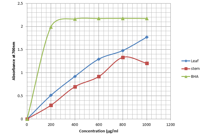
Reducing power capacity of the ethanol extract of the leaf and stem of F. glumosa compared with that of butylated hydroxyanisole (BHA), a standard antioxidant. The concentration of the samples that gave optical density of 0.5 at 700nm were 370µg/ml for the leaf and 420µg/ml for the stem while BHA had 50.15µg/ml. the lower the OD0.5, the better the reducing capacity indicating that the plant possess good antioxidant potential (Figure 1).
• Effect of concentration on the antioxidant activity of ethanolic leaf and stem extract of Ficus glumosa
Antimicrobial Result
| Inhibition Zone of Pathogen (mm)* | |||||
|---|---|---|---|---|---|
| Microbes | Staphylococcus Aureus | Salmonella typhi | Escherichiacoli | Candida albicans | Aspergillus niger |
| Control | 12.71±0.014c | 8.68±0.035b | 9.46±0.085c | 14.78±0.028c | 15.32±0.03c |
| Stem | 1.20±0.000a | - | 0.64 ±0.014a | 4.32±0.028a | 5.50±0.000a |
| Leaf | 1.82±0.000b | 0.92±0.000a | 1.67±0.050b | 5.53±0.106b | 6.78±0.035b |
| P-value | ** | ** | ** | ** | ** |
Table 3: Antimicrobial activity of the Leaf and stem extract of Ficus glumosa at 50 mg/ml Concentration.
Results are in mean ± standard deviation *Columns with the same superscript are not significantly different**P<0.05 Table 3: Antimicrobial activity of the Leaf and stem extract of Ficus glumosa at 50 mg/ml Concentration.
The result of the antimicrobial activity of leaf and stem extracts of F. glumosa at 50 mg/ml concentration indicated that the leaf extract showed significantly higher inhibitory affect against all pathogens than the stem extract. However, in comparison with the control, the inhibition of the pathogens is significantly higher in the control than in plant extract (Table 3).
| Inhibition Zone of Pathogen (mm)* | |||||
|---|---|---|---|---|---|
| Microbes | Staphylococcus aureus | Salmonella typhi | Escherichia coli | Candida albicans | Aspergillus niger |
| Control | 15.61±0.014c | 11.53±0.11c | 12.72±0.03c | 18.31±0.01c | 19.46±0.085c |
| Stem | 6.43±0.035b | 4.71±0.014 | 4.78 ±0.035a | 7.44±0.085a | 7.71±0.127b |
| Leaf | 5.29±0.014a | 6.78±0.035b | 5.83±0.035b | 8.68±0.035b | 9.57±0.042b |
| P-value | ** | ** | ** | ** | ** |
Table 4: Antimicrobial activity of the Leaf and stem Extract of Ficus glumosa at 75 mg/ml Concentration.
Results are in mean ± standard deviation*Columns with the same superscript are not significantly different **P<0.05 Table 4: Antimicrobial activity of the Leaf and stem Extract of Ficus glumosa at 75 mg/ml Concentration.
The result of the antimicrobial activity of the leaf and stem extracts of F. glumosa at 75mg/ml concentration indicated that the leaf extract showed a significantly higher inhibitory effect against the pathogens than the stem The stem showed only significantly higher activity against Staphylococcus aureus than the leaf extract. However, In comparison with the control, the inhibition of the pathogens is significantly higher in the control than in plant extract (Table 4).
| Inhibition Zone of Pathogen (mm)* | |||||
|---|---|---|---|---|---|
| Microbes | Staphylococcus aureus | Salmonella typhi | Escherichia coli | Candida albicans | Aspergillus niger |
| Control | 18.48±0.177c | 15.32±0.03c | 17.54±0.09c | 21.77±0.09c | 23.61±0.014c |
| Stem | 7.83±0.035a | 6.61±0.127a | 6.77 ±0.099a | 9.48±0.035a | 9.56±0.290a |
| Leaf | 8.38±0.106b | 9.28±0.035b | 8.18±0.021b | 11.74±0.00b | 12.71±0.127b |
| P-value | ** | ** | ** | ** | ** |
Table 5: Antimicrobial activity of the Leaf and stem Extract of Ficus glumosa at 100 mg/ml Concentration.
Results are in mean ± standard deviation *Column with the same superscript is not significantly different **P<0.05 Table 5: Antimicrobial activity of the Leaf and stem Extract of Ficus glumosa at 100 mg/ml Concentration.
The result of the antimicrobial activity of the leaf and stem extracts of F. glumosa at 100mg/ml concentration indicated that the leaf extract showed a significantly higher inhibitory effect against all pathogens than the stem extract.
However, In comparison with the control, the inhibition of the pathogens is significantly higher in the control than in plant extract (Table 5).
| Inhibition Zone of Pathogen (mm)* | |||||
|---|---|---|---|---|---|
| Treatment | Staphylococcus aureus | Salmonella typhi | Escherichia coli | Candida albicans | Aspergillus niger |
| Control | 12.60±0.000c | 19.52±0.03c | 20.48±0.04c | 23.72±0.017c | 24.46±0.226c |
| Stem | 9.53±0.318a | 8.66±0.57a | 8.52±0.255a | 11.56±0.08a | 11.67±0.099a |
| Leaf | 11.56±0.085b | 12.45±0.07b | 10.78±0.04b | 12.68±0.11b | 13.63±0.247b |
| P-value | ** | ** | ** | ** | ** |
Table 6: Antimicrobial activity of the Leaf and stem Extract of Ficus glumosa at 150 mg/ml Concentration.
Results are in mean ± standard deviation *Column with the same superscript is not significantly different**P<0.05 Table 6: Antimicrobial activity of the Leaf and stem Extract of Ficus glumosa at 150 mg/ml Concentration.
The result of the antimicrobial activity of leaf and stem extract of F. glumosa at 150mg/ml concentration indicated that the leaf extract showed a significantly higher inhibitory effect against all pathogens than the stem extract. However, In comparison with the control, the inhibition of the pathogens is significantly higher in the control than plant extract (Table 6).
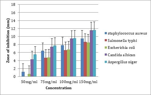
The figure revealed that the inhibitory activity of the stem extract increased with concentration. The stem extract gave the highest inhibition of Aspergillus niger and Candida albican in all concentrations and lowest inhibition of Salmonella typhii and Escherichia coli (Figure 2).
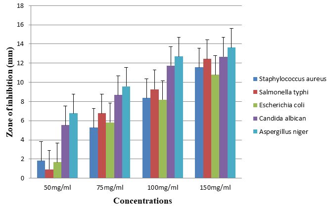
The figure revealed that the inhibitory activity of the leaf extract increased with concentration. The leaf extract gave the highest inhibition of Aspergillus niger in all concentrations and lowest inhibition of Escherichia coli (Figure 3).
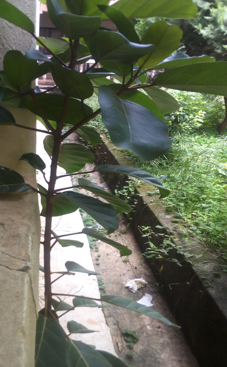
Plate 1: Ficus glumosa in its natural habitat.
Discussion
Phytochemical screening revealed that the ethanolic leaf and stem extracts of Ficus glumosa contain the phytochemical assayed (alkaloid, tannin, Saponin, sterol, flavonoid, phenol, beta carotene, lycopene) but in diverse proportions (Table 1). It was observed that depending on the plant variety, the chemical contents of herbal extracts can differ significantly. These phytochemicals are known to have therapeutic qualities [19]. These compounds validate the F. glumosa plant’s application as a therapeutic herb. According to Agate G, et al. [20] flavonoids have protective effects including anti- inflammatory, anti-oxidant, biological activities of flavonoids include action against allergies, inflammation, free radicals, and hepatoxins. They serve in illness management and defense against herbivores. Many alkaloids are known to have effect on the central nervous system and act as a painkiller. Similarly, Saponins which are a special class of glycosides have been found to possess antifungal activity [21]. Saponins have been reported to have wide range of pharmacological and medicinal activities. Many dicotyledonous plants are said to contain bitter-tasting triterpene glycosides called saponins. Tannin is a potential metal ion chelator, proton precipitating agents and biological antioxidant [22]. The primary properties of tannins, which are found naturally in plants, are their ability to bind and precipitate protein and to significantly impact the nutritional content of a wide variety of foods. Tannin serves as a plant defense mechanism against viruses, herbivores, and unfavorable environmental circumstances [23]. Sterols have been used in medicine to treat variety of conditions ranging from endocrine hormonal alteration to coronary insufficiency. They are applied to treat skin diseases. Phenols neutralize free radicals acting as primary antioxidant [24]. Because of their antibacterial action, they are the main active component of disinfectants and antiseptics.
The result of the antioxidant activity of the leaf and stem extracts of Ficus glumosa as determined by using the Reducing Power Capacity indicated that Ficus glumosa leaf and stem extracts have antioxidant potentials (Table 2). The antioxidant effects of the extracts increases as the concentration increases (Figure 1). Bioactive compounds activities are a measure of contribution of diet as sources of antioxidant for maintenance of health. Free radical damage to cells is prevented by antioxidants, which are phytochemicals, vitamins, and other minerals. It has been recognized that flavonoid show antioxidant activity and their effects on human nutrition and health are considerable [25]. The reducing properties are generally associated with the presence of reductants, which have been shown to exert antioxidant action by donating a hydrogen atom or by removing free radical. The antioxidant activity amongst them has attributed to their flavonoid, phenolic content, beta carotene and lycopene [26]. The reason behind their antioxidant activity is their capacity to delocalize unpaired electrons by resonant stabilization.
Moreover from the study, Leaf extract of Ficus glumosa showed significant higher composition in the antioxidant phytochemical of Total Phenol, Flavonoid, Beta carotene and lycopene and therefore could serve as better source of these compounds for medicinal purpose than the stem extract. The stem extract only showed significantly higher composition of Ascorbic acid and could therefore play an important role in protein thiol group protection against oxidation (Table 2).
The antimicrobial study carried out showed that Ficus glumosa extracts all showed inhibitory effects on pathogens at varied level (Tables 3-6). This could be attributed to the presence of phytochemical compounds (Alkaloid, Flavonoid, Tannin, Sterol, Saponin and phenol) in the extracts (Tables 3-6). These phytochemicals are known to have medicinal properties. This finding supports the use of F. glumosa extracts for other socioeconomic goals as well as the treatment of a variety of illnesses and maladies. However, the leaf extract showed higher inhibitory activity against the pathogen than the stem extract According to Hassan HS, et al. [27], this could be attributed to presence of higher bioactive ingredients or phytochemicals in leaf extracts than stem extract. Furthermore, the sensitivity and susceptibility of the pathogen to the plant extracts varies. In particular, the fungal strains were highly sensitive and susceptible to the plant extracts than the bacterial strains. This difference according to Ogu GI, et al. [21] could be due to the fact that gram positive bacteria such as Escherichia coli develop resistant to inhibition caused by plant extract [28, 29, 30].
Conclusion
This study showed that the plant extracts are rich source of naturally occurring antioxidant and possess bioactive substances that have antimicrobial activities against some human pathogens. The leaf of Ficus glumosa showed more inhibition against the tested pathogenic organisms than the stem. The data obtained from the study indicated that the plant possessed antibacterial and antifungal potentials especially antifungal. It could also be used to protect cells against free radical damages. Thus, its uses in ethno- medicines are justified.
Competing Interests
Authors have declared that no competing interests exist.
References
-
Gershenzon J, Ullah C (2022) Plants protect themselves from herbivores by optimizing the distribution of chemical defenses. Proc National Academy of Science 119(4): e2120277119.
-
Arbonnier M (2004) Trees, Shrubs and Lianas of West African Dry Zones. CIRAD, Margraf Publishers Gmbh, MNHHHN, Paris, France, pp: 573.
-
Ukwubile CA (2010) Pharmacognostic, Antibacterial and Toxicity Investigations of Ficus abutlilifolia Miq (Moraceae). International Journal of Biological Sciences 2(8): 98-111.
-
Onoja OS, Omeh NY, Ezeja IM, Odo CE, Elendu SD (2014) Subacute antidiabetic and in vivo antioxidant effects of methanolic extract of _Ficus glumosa_ stem bark on alloxan-induced hyperglycaemic rats. Comparative Clinical Pathology 23: 1689-1695.
-
Madubunyi II, Onoja SO, Asuzu IU (2012) In vitro antioxidant and in vivo antidiabetic potential of the methanolic extract of _Ficus glumosa_ Del (Moraceae) stem bark in alloxan-induced diabetic mice. Comparative Clinical Pathology 21: 389-394.
-
De Boer HJ, Kool A, Brokerg A, Mziray WR, Hedberg I, et al. (2005) Antifungal and antibacterial activity of some herbal remedies of Tanzania. Journal of Ethnopharmacology 96(3): 461-469.
-
Tanko Y, Aladey O, Ahmed MK, Mohammed A, Musa KY (2012) The effect of methanol leaves extract of _Ficus_ _glumosa_ on gastrointestinal motility and on castor oil induced diarrhea in laboratory animals. J. Nat. Prod. Plant 2(3): 360-367.
-
Chah KF, CA Eze, CE Emuelosi, CO Esimone (2006) Antibacterial and wound healing properties of methanolic extracts of some Nigerian medicinal plants. Journal of Ethnopharmacology 104(2): 164-167.
-
Nair R, S Chanda (2006) Activity of some medicinal plants against certain pathogenic bacterial strains. Indian Journal of Pharmacology 38(2): 142-144.
-
Parekh J, S Chanda (2007) In vitro antimicrobial activity of Trapa natans L. fruit rind extracted in different solvents. African Journal of Biotechnology 6(6): 766- 770.
-
Ilodibia CV, Ezeaja IJ, Akachukwu EE, Chukwuma MU, Egboka TP, et al. (2015) Phytochemical Screening and Antimicrobial Effects of Aqueous and Ethanol Leaf and Stem Extracts of Gongronema latifolium Benth. Research Journal of Botany 10(2): 50-60.
-
Mahapatra A, Mishra S, Basak UC, Panda PC (2012) Nutrients analysis of some selected wild edible fruits of deciduos forests of India. An explosive study towards Non-conventional Bio-nutrition. Advance Journal of Food Science Technology 4(1): 15-21.
-
Harborne JB (1973) Phytochemical methods. A Guide to Modern Technique in Plant Analysis. Chapman and Hall, New York, USA, pp: 600.
-
Obadoni BO, Ochuko PO (2001) Phytochemical Studies and Comparative Efficacy of the Crude Extract of some Homeostatic Plants in Edo and Delta States of Nigeria. Global Journal of Pure and Applied Science 8(2): 203- 208.
-
Kirk II, Sawyer R (1998) Frait Pearson Chemical Analysis of Food. In: 8th (Edn.), Longman Scientific and Technical, Edinburgh, USA, pp: 211-212.
-
Barros L, Ferreira MJ, Queiros B, Ferreira IC, et al. (2007) Total phenols, ascorbic acid, á-carotene and lycopene in Portuguese wild edible mushrooms and their antioxidant activities. Food Chemistry 103(2): 413-419.
-
Klein BP, Perry AK (1982) Ascorbic acid and vitamin A activity in selected vegetables from different geographical areas of the United States. Journal of Food Science 47(3): 941-945.
-
Rajesh K, Harsha R, Mohammed GA, Hareesh AR, Thammanna G, et al. (2010) Antimicrobial activity of ethanol extract of leaf and flower of Spathodea campanulata. Research Journal of Pharmaceutical, Biological and Chemical Sciences 1(3): 691-693.
-
Ilodibia CV, Okoli BE (2016) Anatomical and Phytochemical Studies on Various Parts of Morinda lucida Benth. (Rubiaceae). International Journal of Life Sciences 5(2): 100-106.
-
Agate G, Azzarello E, Tattini M, Pollastri S (2012) Flavonoids as Antioxidants in Plants, Location and Functional Significance. Plant Science 196(4): 67-76.
-
Ogu GI, Tanimowo WO, Nwachukwu PU, Igere A (2012) Antimicrobial and phytochemicals root extracts of Cyathuta prostate (L) Blume against some pathogens. Journal of Intercultural Enthopharmacology 1(1): 35-43.
-
Okonkwo SI (2009) Isolation and characterization of tannin metabolites in _Spondias mombin_ I (Linn) (Anacardiaceae). Nat Applied Sci J 10: 21-29.
-
Heming WRW (1989) Chemistry and significance of condensed tannins. New York: Academic Press, USA, pp: 134.
-
Gulçin I, Berashvili D, Gepdiremen A (2005) Antiradical and antioxidant activity of total anthocyanins from Perilla pankinensis decne. Journal of Ethnopharmacology 101(3): 287-293.
-
Ewim JA, Oliveria D, Lapa AJ (2010) Pharmacological evaluation of the anti-inflammatory activity of a citrus bioflavonoid, hesperidin, and the isoflavonoids. Journal of Pharmacy and Pharmacology 46(2): 118-122.
-
Ilodibia CV, Okoli BE, Okeke CU (2017) Studies on Antimicrobial and Antioxidant Activities of Morinda lucida Benth. International Journal of Life Sciences 6(3): 168-175.
-
Hassan HS, Sule MI, Usman MA, Ibrahim A (2009) Preliminary phytochemical and antimicrobial screening of the stem bark extracts of Bauhinia rufescens using some selected pathogens. Bayero Journal of Pure and Applied Science 2(2): 53-55.
-
Adeniyi BA, Ayepola OO, Adu FD (2015) The antiviral activity of leaves of _Eucalyptus camaldulensis_ (dehn) and _Eucalyptus torelliana_ (_R. muell_). Pakistan Journal of Pharmaceutical Sciences 28(5): 1773-1776.
-
Iwu MW, Duncan AR, Okunji CO (1999) New antimicrobials of plant origin. In: Janick (Ed.), Journal of Education Perspective of New Crops and New Uses, ASHS Press, Alexandria, VA, USA, 2: 457-462.
-
Odunbaku OA, Ilusanya OA, Akasoro KS (2008) Antibacterial activity of ethanol leaf extracts of Ficus exasperata on Escherichia coli and Staphylococcus albus. Scientific Research Essay 3(11): 562-564.
- Evaluation of Comparative Morphological and Phytochemical Studies on the Seeds Extracts of Cocos nucifera (L.) and Elaeis guineensis Jacq. (Arecaceae)
- Aborted Spores in Argentine Ferns
- Ficus middletonii Chantaras. – A New Distributional Record For Central India
- Paclobutrazol (PBZ) and its Metabolic Function in Agriculture: A Review
- Decalepis arayalpathra: Ethnobotany, Scientific Interventions and Prospects
- Effect of Different Cultural Treatments on Branching and Yield of Groundnut (Arachis hypogea L.)