A Comprehensive Review on the Prevalence of Bruxism with Stress and Aggression
Bruxism is a type of neuro-psychic disease that influences the activity of the mandible and distorts multiple structures of the face and mouth muscles. This study goes through the complicated relationship between bruxism, stress, and aggression. The aim is to interpret the extensiveness of these three factors and evaluate the scientific perception of this. Psychological discomfort may manifest subtly as bruxism. A person having Bruxism is a spontaneous activity with the problem of clenching and grinding of teeth. It is a pervasive oral parafunctional activity. It accumulates growing recognition because of its complex connection with several serious psychological disorders. Bruxism is a type of neuro-psychic disease that influences the activity of the mandible and distorts multiple structures of the face and mouth muscles. According to previous studies, most bruxism is the result of suppressed stress which can occur due to lifestyle, and career pressure. People with severe anger issues can show signs of bruxism as a result of tension build-up in their oral cavity. As it is studied by various research, people show stages of bruxism in various stages of life depending on the situation they are struggling with. To shed light on the intricate interactions between stress, aggression, and oral habits, this review investigates the prevalence of bruxism in forensic contexts. We examine the body of research to clarify the possible applications of bruxism in forensic contexts, highlighting its importance in comprehending psychological profiles and behavioural patterns.
Abbreviations
TMJ: Temporomandibular Joint; TG: Tooth Grinding; TMD: Temporomandibular Disorder; CBA: Clinically Based Assessment; ASA: Average Stage of Attrition; PTSD: Post- Traumatic Stress Disorder.
Introduction
Bruxism is a condition that involves grinding, gnashing, and clenching of teeth that often occurs involuntarily. It usually happens during sleep but can occur when a person is awake. In those cases, it is wakefulness or daytime bruxism. It can affect both children and adults. s and anxiety are considered to be the foremost causes associated with teeth grinding. People may grind their teeth as an unconscious response to emotional stress or fear. Stress on a psychological level can be the factor of building up tension in the muscles, especially jaw muscles. Stress becomes a factor in bruxism in those cases. Bruxism is frequently linked to movement abnormalities that are related to sleep, as well as other sleep disorders. Anxiety and stress, which are frequently, associated with mental health issues, can also appear when you’re sleeping and exacerbate episodes of bruxism.
Emotional discomfort may manifest subtly as bruxism. People may grind their teeth while they sleep as a physical sign of tension or emotional upheaval. Bruxism might act as a survival mechanism for an individual to release stress. Headaches, temporomandibular joint (TMJ) issues, and jaw pain can all result from bruxism. Bruxism can lead to tooth deterioration, chipping, and sensitivity over time. Many kids outgrow their bruxism habit, which is rather frequent in them. The prevalence rates of it can last into adulthood and range from 8% to 31%, according to estimates.
Masticatory Muscles
The main muscles used in mastication or chewing food are the masseter, medial, lateral, lateral, and temporalis muscles. The mandible, or jaw, is moved by the four primary masticatory muscles attached to its rami. The five basic mandibular movements involved in chewing are protrusion, retraction, elevation, and side-to-side movement. The platysma muscle, which is located in the neck, helps to depress the mandible when it encounters resistance. This muscle is considered to be superficial in nature [1]. The Temporalis Muscle: The vertically oriented anterior fibers, obliquely oriented mid fibers, and more horizontally oriented posterior fibers make up the fan-shaped temporalis muscle. The temporalis muscle originates from the inferior temporal line of the skull and the temporal fossa. Its posterior fibers retract the mandible [2]. Medial Pterygoid Thick and rectangular, the medial pterygoid muscle has two heads: superficial and deep. The maxillary tuberosity of the inferior maxilla is the origin of the superficial head of the medial pterygoid. The medial pterygoid’s deep head is bigger than its superficial head. The medial surface of the lateral pterygoid plate of the sphenoid bone is where the deep head originates. It also assists the lateral pterygoid muscle with a side-to-side mandibular motion to help grind food [3].
Lateral Pterygoid
The principal muscle of the inferior temporal fossa is the lateral pterygoid muscle. There are two sections to the lateral pterygoid: the upper and lower heads. The inferior temporal surface of the greater wing of the sphenoid bone is the source of the upper head of the lateral pterygoid muscle. The lateral aspect of the lateral pterygoid plate of the sphenoid bone is where the lower head originates.
Masseter
The three layers of the masseter muscle are superficial, deep, and intermediate. It has a rectangular shape. The inferior zygomatic arch and the anterior two-thirds of the zygomatic arch, which connect to the zygomatic bone’s posterior aspect, are the masseter muscle’s source. The mandibular ramus and coronoid process of the mandible are inserted by a tendon formed by the inferior convergence of the masseter muscle fibers.
Pathophysiology of Bruxism
Dentists are extremely concerned about tooth grinding (TG) because of the following consequences: tooth decay, fractures in dental rehabilitation or restorations, worsening of temporomandibular disorders, or induction of a temporal tension headache and grinding sounds that could keep family members or life partners awake at night.
Patients who suffer from sleep bruxism, a condition in which a person grinds or clenches their teeth during sleep, tend to have higher levels of anxiety and a stronger focus on tasks than the general population [4]. Regarding risk factors, Sleep Bruxism is thought to be slightly more likely in individuals who smoke [5].
Definition of Bruxism According to the American Academy of Orofacial Pain
Bruxism is “diurnal or nocturnal parafunctional activity including clenching, bracing, gnashing, and grinding the teeth” [6].
According to the American Sleep Disorder Association
Bruxism is “tooth grinding or clenching during sleep plus one of the following: Tooth wear, sounds or jaw muscle discomfort in the absence of medical disorder” [7].
The most common definition of bruxism is the non- functional tooth grinding or gnashing. The normal function may include habitual clenching without causing harm to the teeth, muscles, temporomandibular joints, or supporting structures, even though braces involve excessive forces and movements during static or dynamic tooth contact. A restricted investigation of the etiology, diagnosis, and treatment of bruxism is not allowed in studying the condition because definitions can encompass abnormal and normal clenching.
Pathophysiology of bruxism depends on the two main factors (Table 1).
| Exogenous or Peripheral Activity: | Endogenous Activity |
|---|---|
| Stress-anxiety | Personality (e.g., anxious) |
| Environmental influences (jaw-clenching reactions, tongue habits) | Genetic (no proven transmission |
| Occlusal interferences (controversy) | Neurochemicals (e.g., dopamine, noradrenaline, serotonin); see text. |
| Medication (e.g., L-dopa, neuroleptics, amphetamine, SSRI*) | Neurological disorders (e.g., Parkinson’s, Meige syndrome/oral tardive dyskinesia, RBD, olivopontocerebellar atrophy, cerebellar hemorrhage) |
| Substance abuse (e.g., cocaine, alcohol) | Psychiatric-related disorders (e.g., dementia, mental retardation, tics/ Tourette’s syndrome) |
| Sleep disorders (e.g., PLMS, apnea, RBD) |
Table 1: Pathophysiology of Sleep Bruxism.
The current international consensus defines bruxism as an activity of the masticatory muscles, being characterized as two different, non-exclusive entities, according to their manifestation in the circadian cycle: awake bruxism, and sleep bruxism. These are phenomena regulated by the central nervous system, of multifactorial origin, with peripheral factors (anatomical or occlusal) playing a secondary role [8, 9].
Prevalence of Bruxism
The prevalence of bruxism is as high as 30% of the population [10]. In some cases, it can be pathological, resulting in issues such as damaged teeth and orofacial pain, and it is one of the potential risk factors for the development of temporomandibular disorders (TMD), even though it is regarded as a behavior rather than a disease. Furthermore, several studies have discovered biological markers associated with psychological changes, such as elevated cortisol, and catecholamines. Additionally, certain studies have found a link between bruxism and stressful activities. In this vein, studies have shown that during spontaneous awake bruxism episodes, participants with muscular TMD exposed to relaxing music experienced less muscular strain, whereas stressful music increased it. A recent study also discovered that, compared to the non-bruxism group, people with definitive awake bruxism showed increased muscle activity in response to negative-valence videos and texts, particularly those about pain.
The process of tooth wear that happens when teeth come into contact with one another while doing tasks like chewing is referred to as teeth attrition. Over time, the tooth’s surface may change as a result of this normal wear and tear. Several things can lead to attrition, such as chronic teeth clenching and grinding (bruxism) and regular chewing and grinding. The degree of tooth attrition varies from person to person and is influenced by personal habits, dental hygiene, and diet. Excessive attrition can occasionally result in issues like tooth sensitivity, altered bite patterns, or even structural damage to the teeth. Environmentally, chronic exposure to dust and dirt can also cause increased wear in humans [11, 12].
| Area Affected | Sign and Symptoms |
|---|---|
| Teeth | Flattened, chipped, cracked, or loose teeth. |
| Worn tooth enamel, exposing the inner layers of the tooth. | |
| Tooth pain or sensitivity. | |
| Jaw | Soreness in the muscle. |
| Tightness in the jaw | |
| Tiredness of jaw muscles. | |
| Head and face | Headache |
| Facial pain |
Table 2: List of signs and symptoms.
Clinical Manifestation
Bruxism can result in tooth wear or cracks, dental restorations breaking, implants failing, muscle hypertrophy, jaw pain and/or fatigue, headaches, toothaches, disrupted sleep patterns, and a general decline in quality of life. Dental wear is the first clinical sign of bruxism, but it does not indicate if the condition is current or caused by previous lesions. Age, gender, occlusion, enamel hardness, diet, acidic, isotonic, or carbonic beverages, salivary flow, and digestive disorders (such as gastroesophageal reflux) significantly impact dental wear variability [13].
Symptoms
- Symptoms of teeth Table 2 grinding include a distinct sound that can wake up a sleeping partner and TMJ pain.
- Soreness in the neck and masticatory muscles. Headache, especially in the morning when the patient wakes up in the temporal zone.
- Susceptible teeth.
- Too much movement between the teeth.
- Poor quality of sleep, fatigue
Signs
- Unusual dental wear.
- Dimples on the tongue.
- A Linea alba in the bite plane.
- Gum recession.
- Mandibularies or torus maxillaries is present.
- Increased muscle activity (as recorded by the polysomnography).
- Masseter muscle hypertrophy.
- Reduced salivary secretion.
- Teeth or fillings break.
- A reduction in the ability to open the mouth.
Clinical Evaluation of Bruxism
If left untreated, bruxism (abnormal, nonfunctional tooth contact) can lead to orofacial pain and dentition breakdown. Early detection and treatment are therefore regarded as critically important. Several treatment options must be used to identify and manage the multifactorial causes of bruxism. The diagnosis of bruxism can be traced by taking a good case history and evaluating the tooth mobility, tooth wear, and other clinical findings of TMJ [14].
Questionnaires: History Taking Part
A questionnaire is the quickest and easiest way to assess something. The main disadvantage of this assessment method is that it is subjective. Because bruxism episodes can occur with or without noise, most children and adults may be unaware that they are doing it.
Extraoral Examination
The Clinically Based Assessment (CBA) includes domains on Joints and Muscles, Intra- and Extra-Oral Tissues, and Teeth and Restorations, based on information collected by an examiner [15]. For this examination, facial asymmetry, masseter, and temporalis muscle should be inspected. Assessment of the range of mandibular movement (maximum mouth opening, lateral movement) also holds an important role in this examination.
Maximum mouth opening should be measured and checked for the presence of pain while doing the task, participants were asked to open their mouths as wide as possible and asked whether they were feeling any kind of pain or not.
Temporomandibular Joint
Palpation of pretragus area is done for the examination of TMJ. In this process, the participant was asked to open their mouth. The joint of the mandible is felt when the participant opens their mouth-palpation of the masseter at its attachments to the zygomatic arch and angle of the mandible. Through palpation, condylar movement can be easily felt. By asking them to close their jaw again condyle movement can be felt against our finger. TMJ can also be assessed by the palpation through anterior wall of the external auditory meatus.
The tenderness during this process can be alarming and a sign of articular inflammation. Participants were asked about any pain they were feeling during this process. It was also designed to examine whether the pain elevated or not during the whole process.
2 types of joint sound should be looked for during this examination. Those are: Clicking Sound: Single explosive noise of short duration. The timing of the click is also important. It should be noted whether the clicking sound is heard while opening their jaw. Crepitus: Continuous grating noise. This type of noise can only be felt with the stethoscope. The main cause of this is the articular surfaces of the joint being worn.
Examination of Muscles
The most frequent complaint of TMD patients is likely related to functional disorders of the masticatory muscles. The broad category known as “masticatory muscle disorder” is typically where they are categorized. Like in any pathologic condition, there are two main signs to look out for: • Pain is undoubtedly the most prevalent symptom among patients with masticatory muscle disorders; it can vary from mild soreness to severe discomfort. Dysfunction: contraction or stretching exacerbates pain in muscles whose tissues have been damaged by excessive use. Because of this, the patient limits their range of motion to keep themselves comfortable without exacerbating their pain. According to clinical opinion, this is incapacity to open the mouth widely.
Temporalis Muscle: It is recommended to palpate the temporalis muscle’s anterior, middle, and posterior regions. it is visible when a person firmly clenches their teeth which helps in palpation.
Multiple Masseter Muscle: areas are palpated with the temporalis muscle. This muscle can be easily observed when a person forcibly closes their jaw. Palpation of masseter muscle can be done with the thumb and index finger.
The Medial Pterygoid
Manually inserting one finger orally in the lingual vestibule in the retromolar region and one finger externally at the medial aspect of the mandible’s angle. One can palpate the anterior part of the insertion by inserting a finger at a 45-degree angle into the patient’s mouth near the base of their relaxed tongue. It’s possible to palpate the inferior and posterior parts of the insertion extra orally with the other hand. By turning the index finger upward and pressing it against the muscle until it is close to its origin on the tuberosity, one can feel the muscle’s body.
The Lateral Pterygoid
Muscle is palpated. Place the little or index finger medially to the coronoid process and lateral to the maxillary tuberosity to palpate the muscle. It is possible to detect pain when the finger presses upward and inward. The examining finger can reach the lateral pterygoid intraorally. Through this process, any type of muscle spasm and tenderness can be examined for presence in those areas. The extraoral examination should be done under the supervision of a certified dentist.
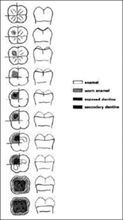
Figure1: Li and Ji’s Staging Chart.
Forensic Importance of Bruxism
Forensic bruxism research is significant for several reasons, including its capacity to shed light on a subject’s behaviour, mental health, and possible involvement in legal issues. The following particular arguments emphasize how crucial it is to investigate bruxism from a forensic angle:
Clinical Examination or Intraoral Examination
The attrition which is a probable cause due to bruxism needs to be examined to understand the severity or level of it. Oral examination needs to be done to check the attrition of the teeth. The modern Li and Ji method is applied to calculating the average surface attrition.
Li and Ji Method
Li and Ji introduced a new clinical method that uses permanent first and second molars to estimate age. The attrition values of each molar cusp are recorded using the Average Stage of Attrition (ASA) chart (stages 0–9)),[refer to figure 1], and an average is subsequently calculated. All cusps of maxillary and mandibular first and second molars were used, and their average attrition levels were used. They developed a new graduation standard and employed it to calculate age with a 4.53-year maximum error [16] (Figure 1).
Behavioural Markers
Potential for Stress and Aggression: Bruxism can be caused by stress or aggression, and understanding the psychological aspects associated with it can improve the assessment of one’s behaviour. Bruxism during wakefulness (clenching) is likely to be a result of emotional tension or psychosocial disorders that force the subject to respond with a prolonged contraction of his/her masticatory muscles [17].
Assessments by Forensic Psychiatrists
Mental State Assessments: If stress-related behaviours are relevant to the case, forensic psychiatric evaluations may take bruxism into account when evaluating an individual’s mental state. Bruxism has been associated with emotional tension, psychosomatic disorders, hostility, aggressiveness, apprehension, and a tendency to worry, and also with psychiatric disorders such as schizophrenia [18]. Relation to Emotional Distress: Bruxism and emotional distress may be associated, and the existence of this relationship may be taken into account when assessing a person’s mental health in court. Clinically oriented-studies have shown that some symptoms related to anxiety disorders have a significantly higher prevalence in bruxers than in non- bruxers [19]. Expert Testimony Dental and Psychological Experts: In court proceedings, forensic psychologists and dentists may offer their expert opinions. They can talk about the possible connections between aggression, stress, and bruxism as well as provide insight into the person’s mental health. The hypothesis that wake-clenching is strictly related to depression, or maybe an expression of a depressed mood, is fascinating and found some support in the psychiatric literature suggesting that bipolar patients are characterized by disturbances in the central neurotransmitter system which may also be involved in the etiology of bruxism [20]. Mitigating Factors: When bruxism is associated with stress, it can be offered as a mitigating factor, giving an account of the person’s emotional condition at the time of a particular incident.
Dental Records as Proof
Dental History: A person’s stress levels and oral health history can be inferred from the documentation of their bruxism in their dental records. Bruxism increases the risk of oral complications such as dental attrition and masticatory muscle and TMJ pain [21]. Long-term Patterns: Analyzing dental records for long- term bruxism patterns may reveal a history of ongoing stress or psychological discomfort.
Comprehending Aggressive Conduct
Aggression as a Manifestation: One of the outward signs of aggressive tendencies could be bruxism when combined with aggression. This knowledge could be useful in situations where there has been aggressive or violent behaviour.
Psychological Autopsies
Post-mortem Exams: Psychological autopsies may be performed in cases of death. Reconstructing the person’s mental state and actions before the incident can be aided by an understanding of bruxism.
Implications for Treatment
Rehabilitation Programs: For those engaged in legal proceedings, rehabilitation programs may take into account the knowledge of bruxism and its associations with aggression and stress. One aspect of the rehabilitation process might involve addressing underlying psychological issues. Diurnal bruxism can be managed by considering interventions such as habit modification, relaxation therapy, and biofeedback [22]. In patients with sleep bruxism, which does not appear to be impacted by psychological or psychosocial factors, appropriate intervention might include appliance therapy [23].
Considering Public Safety
Risk Assessment: When aggressive behavior is a concern, knowing the interactions between bruxism, stress, and aggression can help with risk assessments and public safety considerations.
In conclusion, researching bruxism from a forensic standpoint can improve our comprehension of the psychological elements that could influence a person’s behavior. This knowledge will be beneficial to legal practitioners, mental health specialists, and other parties with an interest in the legal system.
Bruxism in Stress and Aggression
Many researchers have concluded that bruxism is related to certain personality traits such as aggression, emotional suppressions, and stress. Anxiety and neuroticism personality factors have been seen in individuals with bruxism. This study is also focused on the establishment of a relationship between stress and bruxism and how other factors such as lifestyle, and professional factors can cause it. The patient’s struggle with himself and the environment around him is manifested in the occlusal syndrome. Bruxism is an outward manifestation of stress. People encounter stressful and conflictual situations daily. The body’s physiological reactions are thought to be brought on by stress. The variations are specific to each patient. Everybody responds differently to stressful circumstances. The most frequent cause for a patient to seek medical attention is pain, which manifests when the muscles and temporomandibular joint structures are unable to adjust to the environment. It is common opinions that sleep bruxism, and bruxism in general, is related to stress. This belief is typical of patients, who usually report an increase in their night-time teeth grinding during stressful life periods, as well as of clinicians, who often attribute a patient’s bruxing behavior to an increase in stress. This theory is based on some early case series that reported a relationship between stressful daily events and an increase in nocturnal masseter muscle activity [24, 25, 26, 27]. Excessive stress sensitivity was found to be the most significant difference between women with bruxism and the control group in research studies. It is stressed that a person’s anxious personality and mindset toward success are key factors in the development of bruxism. Prolonged stress modifies the sensitivity of the muscle’s receptors, and when these receptors are overstimulated, the masseter muscles contract more forcefully and uncontrollably. In addition to dealing with issues of a personal and familial nature, modern women also have responsibilities related to their work. It makes sense that it has an impact on her health, including her teeth. Numerous studies have also demonstrated the link between stress and bruxism, which lowers university students’ quality of life.
There have been more studies recently on the connection between bruxism and stress in college students. The university environment, with its commitment, transitional nature, and challenges, can be a time when students either learn to deal with stress or remain unaware of it or become more vulnerable to its negative effects. Stress is increasingly considered an initiating, predisposing, and perpetuating factor for bruxism, although their implicit relationship remains unclear [28]. The second category includes psychological elements like anxiety, character traits, and sensitivity to stress. Compared to individuals without bruxism, patients with bruxism, both adults and children, appear to perform better on measures measuring the severity of mental illnesses, anxiety, and stress. Additionally, there are scientific reports that suggest a link between the symptoms of depression and bruxism. In addition to being linked to painful temporomandibular disorder (TMD), post- traumatic stress disorder (PTSD) may also play a role in the development of both awake bruxism and sleep bruxism. The type of traumatic event can have an impact on the likelihood of painful temporomandibular disorder, awake bruxism, and sleep bruxism in patients with severe PTSD.
Legal Admissibility of Bruxism
The admissibility of bruxism (teeth grinding or clenching) in court is determined by the circumstances surrounding its consideration. The following are some possible situations and legal considerations where bruxism may be relevant:
In a malpractice case, expert testimony is frequently needed to determine the relevant standard of care and show how the defendant violated it [29]. Often bruxism misconceptions as medical malpractices, in those cases expert testimony is the important part.
Sullivan MF, et al. [29] is the case where expert testimony was needed to prove the fact that bruxism is not associated with malpractices. Because bruxism can impact the outcome of specific dental procedures, it is important to take it into account when evaluating a patient’s dental history and treatment plan [30]. Expert testimony is frequently needed in malpractice cases to establish the relevant standard of care and show how the defendant’s actions violated it. There are, however, some exceptions to this general rule, such as in cases where the lack of professional care is so obvious that a layperson would be able to recognize it [31]. As demonstrated by sertraline [antidepressant] induced bruxism, bruxism can also be linked to specific drugs [32]. This emphasizes how important it is for medical personnel to be informed about possible drug side effects and to keep an eye out for them in their patients.
Treatment
The management of bruxism encompasses various therapeutic approaches, including the following: Detection of Bite Force: Takeuchi, et al. [33] created the intra-splint force detector (ISFD), a recording tool for sleep bruxism that measures the force generated by tooth contact on an intra-oral appliance. A thin, deformation-sensitive piezoelectric film embedded 1-2 mm below the appliance’s occlusal surface is used to detect the force.
Splints and Mouth Guards: These devices are specifically designed to mitigate teeth damage caused by clenching and grinding by maintaining separation between the teeth. They are custom-fitted over either the upper or lower teeth and can be fabricated using soft materials or hard acrylic. In instances of advanced dental degradation, it may be necessary for a healthcare professional to undertake restorative measures to address a chipped or worn tooth, thereby ameliorating the compromised chewing function and effecting a reshaping of the dental crown [34].
Relaxing Technique: Stress and depression are recognized as the underlying causes of bruxism. It is crucial to alleviate stress through meditation and therapy, as this has the potential to reduce the symptoms of bruxism. In severe cases, seeking psychological assistance becomes imperative.
Medication: Medication that inhibits muscle function by
impeding the release of acetylcholine at the neuromuscular junction, such as botulinum toxin, diminishes the occurrence of bruxism. This effect is especially pronounced in severe cases concomitant with conditions such as autism, amphetamine abuse, coma, and brain damage [35]. In cases of severe bruxism, low doses of the dopamine D1/ D2 receptor agonist pergolide were administered to reduce sleep bruxism activity [36]. Huynh N, et al. [35] discovered that propranolol, a non-selective adrenergic beta-blocker, did not affect sleep bruxism. However, two cases of bruxism caused by antipsychotics responded well to this medication [37].
Biofeedback: The theory behind biofeedback is that individuals who grind their teeth (bruxers) can change their behaviour by becoming aware of their maladaptive jaw muscle activity through a process called “aversive conditioning.” This method has been used to treat both sleep bruxism and bruxism during wakefulness. Patients can be trained to control their jaw muscle activity while they are awake by receiving visual or auditory feedback from surface electromyography. As for sleep bruxism, feedback can be provided through auditory, electrical, vibratory, or taste stimuli [38].
Additional Aids: To alleviate bruxism symptoms, it’s essential to address the psychological issues underlying the condition, such as depression or anxiety. Engaging in regular exercise, maintaining a balanced diet, and ensuring an adequate amount of sleep can effectively reduce stress and help alleviate the symptoms associated with bruxism. These lifestyle changes can positively impact overall well-being and contribute to managing bruxism.
Conclusion
This review article endeavours to unravel the connection between stress and aggression with bruxism, providing a more comprehensive understanding of the condition. Based on the study findings, it can be inferred that awake bruxism is commonly linked to aggression. However, stress and anxiety are primarily linked to sleep bruxism. It is possible that stress and aggression can cause bruxism in some individuals, leading to visible stages of attrition. Because stress can cause increased muscle tension and clenching, which can contribute to the development of bruxism, it explains the pathophysiological mechanism by which stress influences the presence of bruxism [38]. This study can be used to gain a preliminary understanding of bruxism and increase student awareness of the condition. This study can help in creating a summary of how bruxism relates to stress and aggression. Stress and aggression can be gas lighted as the tendency of bruxism in a person. Those with obsessive, domineering, or violent personalities are more likely to become bruxists.
Stress at work and erratic schedules can make bruxism worse. Although the precise mechanisms causing the condition are still somewhat unknown, researchers generally agree that stress plays a significant role in the onset and maintenance of bruxism. In conclusion, this study will help in understanding the dynamicity of bruxism in determining personality traits, anxiety, and psychological health.
Ethics Approval and Consent to Participate
The given manuscript is a review article. Therefore, there is no requirement for ethical approval or participation consent.
Consent for Publication
Since the given article manuscript is a review article, consent is not required for publication. The author gives full consent for the publication of the article on approval.
Availability of Data and Material
All required data about the manuscript has been already provided. There is no supplemental data.
Competing Interests
The author(s) declare that there are no competing interests in the article.
Funding
The given manuscript is a review article and is an institutional contribution. There are no funding sources.
Authors’ Contributions
Both the authors have participated in the conception and design, analysis and interpretation of the data, drafting the article or revising it critically for important intellectual content, and approval of the final version.
References
-
Alomar X, Medrano J, Cabratosa J, Clavero JA, Lorente M, et al. (2007) Anatomy of the Temporomandibular Joint. Semin Ultrasound CT MR 28(3): 170-183.
-
Arsenina OI, Komarova AV, Popova NV, Popova AV, Egorova DO (2020) Elimination of Discoordination of the Masticatory Muscles Works in Patients with Muscular- Articular Dysfunction of the Temporomandibular Joint by using Elastocorrector Appliance. Stomatologiia 99(2): 61-65.
-
Li GW, Liu CK, Liu P, Deng TG, Li JL, et al. (2020) Anatomical Study of Rat Trigeminal Motor Nucleus- Lateral Pterygoid Muscle Projection Pathway. Zhonghua Kou Qiang Yi Xue Za Zhi 55(4): 259-263.
-
Por CH, Watson L, Doucette D, Dolovich L (1996) Sertraline Associated Bruxism. Can J Clin Pharmacol 3: 123-125.
-
Lavigne GJ, Lobbezoo F, Rompre PH, Nielsen TA, Montplaisir J (1997) Cigarette Smoking as a Risk Factor or an Exacerbating Factor for Restless Legs Syndrome and Sleep Bruxism. Sleep 20(4): 290-293.
-
Okeson JP (1996) The American Academy of Orofacial Pain Orofacial Pain Guidelines for Assessment Diagnosis and Management. Quintessence Publishing Co Inc Chicago 113-184.
-
American Academy of Sleep Medicine (2005) International Classification of Sleep Disorders. Diagnostic and Coding Manual pp: 148-152.
-
Lavigne GJ, Kato T, Kolta A, Sessle BJ (2003) Neurobiological Mechanisms Involved in Sleep Bruxism. Crit Rev Oral Biol Med 14(1): 30-46.
-
Kato T, Thie N, Montplaisir J, Lavigne GJ (2001b) Bruxism and Orofacial Movements during Sleep. Dent Clin North Am 45(4): 657-684.
-
Anguita AEO, Sanchez TS, Goni XAS, Gonzalez MG, Farinas FA, et al. (2022) Awake and Sleep Bruxism Prevalence and their Associated Psychological Factors in First-Year University Students A Pre Mid-Post COVID-19 Pandemic Comparison. Int J Environ Res Public Health 20(3): 2452.
-
Melo G, Duarte J, Pauletto P, Porporatti AL, Barbosa JS, et al. (2019) Bruxism an Umbrella Review of Systematic Reviews. J Oral Rehabil 46(7): 666-690.
-
Goldstein RE, Curtis JW, Farley BA, Siranli S, Clark WA (2018) Abfraction Abrasion Attrition and Erosion. Ronald E Goldsteins Esthetics in Dentistry 692-719.
-
h t t p s : / / w w w. n i d c r. n i h . g o v / h e a l t h - i n f o / bruxism#overview
-
Aizpurua JLDL, Alonso E, Touche-Arbizu R, Jimenez JM (2011) Sleep Bruxism Conceptual Review and Update. Med Oral Patol Oral Cir Bucal 16(2): 231-238.
-
Hema K, Ashwin P, Bharathi P, Mallikarjun D (2018) Diagnosis and Treatment of Bruxism: Concepts from Past to Present. International Journal of Applied Dental Sciences 4(1): 290-295.
-
Manfredini D, Ahlberg J, Aarab G, Bender S, Bracci A, et al. (2023) Standardised Tool for the Assessment of Bruxism. J Oral Rehabil 51(1): 29-58.
-
Miles AEW (1962) Assessment of the Ages of a Population of Anglo-Saxons from their Dentitions. Proceedings of the Royal Society of Medicine 55(10): 881-886.
-
Prabu D, Sindhu R, Dhamodhar D, Bharathwaj VV, Manipal S, et al. (2022) Examining the Relationship Between Physiological Job Stress and Sleep Bruxism Among Working and Non-Working Women in North Chennai Tamilnadu. International Journal of Dental and Clinical Study 3(2): 08-16.
-
Kampe T, Edman G, Bader G, Tagdae T, Karlsson S (1997) Personality Traits in a Group of Subjects with Long‐ Standing Bruxing Behaviour. J Oral Rehabil 24(8): 588- 593.
-
Manfredini D, Basso D, Arboretti R, Nardini LG (2009) Association between Magnetic Resonance Signs of Temporomandibular Joint Effusion and Disk Displacement. Oral Surg Oral Med Oral Pathol Oral Radiol Endod 107(2): 266-271.
-
Lobbezoo F, Soucy JP, Montplaisir JY, Lavigne GJ (1996) Striatal D2 Receptor Binding in Sleep Bruxism a Controlled Study with Iodine 123 Iodobenzamide and Single Photon Emission Computed Tomography. J Dent Res 75(10): 1804-1810.
-
Raphael KG, Santiago V, Lobbezoo F (2016) Is Bruxism a Disorder or a Behaviour? Rethinking the International Consensus on Defining and Grading of Bruxism. J Oral Rehabil 43(10): 791-798.
-
Lal SJ, Sankari A, Weber KK (2024) Bruxism Management. In: StatPearls [Internet]. Treasure Island (FL) StatPearls Publishing.
-
Manfredini D, Lobbezoo F (2009) Role of Psychosocial Factors in the Etiology of Bruxism. J Orofac pain 23(2): 153-166.
-
Rugh JD, Solberg WK (1975) Electromyographic Studies of Bruxist Behaviour before and during Treatment. J Calif Dent Assoc 3(9): 56-59.
-
Clark GT, Rugh JD, Handelman SL (1980) Nocturnal Masseter Muscle Activity and Urinary Catecholamine Levels in Bruxers. J Dent Res 59(10): 1571-1576.
-
Rugh JD (1981) Psychological Stress in Orofacial Neuromuscular Problems. Int Dent J 31(3): 202-205.
-
Rugh JD, Harlan J (1988) Nocturnal Bruxism and Temporomandibular Disorders. Adv Neurol 49: 329-341.
-
Hartmann F, Cucchi G (2013) Stress and Orality New Data about Teeth Clenching & Outcomes Migraine Fibromyalgia Fatigue. Springer Science & Business Media.
-
Sullivan MF, Durham NC (1983) 417 Mich. 398, 338 N.W.2d 181.
-
Albrahim S, Albobali Y, Elzain M (2020) A Challenging Case of Sertraline-Induced Bruxism. J Med Case Rep Case Series 1(2).
-
Takeuchi H, Ikeda T, Clark GT (2001) A Piezoelectric Film-Based Intrasplint Detection Method for Bruxism. J Prosthet Dent 86(2): 195-202.
-
Shetty S, Pitti V, Babu CLS, Kumar GPS, Deepthi BC (2010) Bruxism A Literature Review. J Indian Prosthodont Soc 10(3): 141-148.
-
Monroy PG, Fonseca MAD (2006) The Use of Botulinum Toxin‐a in the Treatment of Severe Bruxism in a Patient with Autism a Case Report. Spec Care Dentist 26(1): 37- 39.
-
Huynh N, Lavigne GJ, Lanfranchi PA, Montplaisir JY, Champlain JD (2006) The Effect of 2 Sympatholytic Medications Propranolol and Clonidine on Sleep Bruxism Experimental Randomized Controlled Studies. Sleep 29(3): 307-316.
-
Cherasia M, Parks L (1986) Suggestions for Use of Behavioral Measures in Treating Bruxism. Psychol Rep 58(3): 719-722.
-
Fluerasu MI, Bocsan IC, Țig A, Iacob SM, Popa D, et al. (2021) The Epidemiology of Bruxism in Relation to Psychological Factors. Int J Environ Res Public Health 19(2): 691.
-
Miles AE (1962) Assessment of the Ages of a Population of Anglo-Saxons from their Dentitions. Proc R Soc Med 55(10): 881-886.
- Diagnosis and Management of Mental Nerve Paresthesia Secondary to Apical Periodontitis of Mandibular Second Premolar: A CBCT Based Case Report
- A Randomized, Double Blinded Clinical Trial to Compare the Effect of Oral Premedication (Diclofenac Potassium or Dexamethasone) on Post-Operative Pain Following Pulpectomy
- Modified Lip Repositioning Technique for the Management of Excessive Gingival Display
- Integral Role of Non-Dental Providers and Fluoride Dissemination
- Root Canal Treatment Rate in Deciduous Teeth Among 6-Year- Olds in the Era of Discontinuing Water Fluoridation - Historical Cohort Study
- The Impact of the Notch1 on the Migratory Capacity and the Expression of E-Cadherin and CyclinD1 in Ameloblastoma Cells