Effect of L-Tocopherol on Morphological Reformations of Rat’s Pineal Gland under the Long-Term Impact of Heavy Metal Salts
The article presents the results of the study on morphological rearrangements of structural changes in the rat pineal gland under the influence of heavy metal salts and correction with L-tocopherol during 60 days. General morphological and statistical research methods were used (histological, morphometric, immunohistochemical and statistical methods). The article presents the results of the study on morphological rearrangements of structural changes in the rat pineal gland under the influence of heavy metal salts and correction with L-tocopherol during 60 days. General morphological and statistical research methods were used (histological, morphometric and statistic methods). The protective effect of L-tocopherol caused a number of compensatory-adaptive processes in the pineal gland aimed at neutralizing the effects of stressors on the organ. Under the influence of α-tocopherol acetate, the tension on the part of the synthetic apparatus of pinealocytes decreases compared to the indicators of animals that did not use the corrector drug. The 60-days protective period of L-tocopherol is insufficient to completely neutralize the heavy metals impact on the organ: the glial response activated, the vascular area increased, vascular wall’s permeability impaired, edema was formed and the connective tissue component grew, indicating dysfunctional reorganization of the gland’s secretory activity. The above morphological changes in the pineal gland’s structural components negatively affect the processes of hormones evacuation into the blood, the course of the general adaptation syndrome and homeostasis restoration in the organ.
Introduction
The United Nations Environment Program (UNEP), together with partner organizations, conducted a preliminary monitoring of the environmental situation in Ukraine and analyzed the impact of hostilities on the environment. Thousands of cases of air, water, land pollution and ecosystem degradation have already been discovered and recorded, which poses a threat not only to Ukraine, but also to neighboring countries. According to UNEP, the vast majority of regions of the country suffered as a result of hostilities. In addition, the range of destruction is quite wide: there have been incidents at nuclear power plants and facilities, in energy infrastructure, including oil storage tankers, oil refineries, drilling platforms and gas facilities, distribution pipelines, mines, industrial facilities and enterprises for processing agricultural products.
As a result, the level of air, ground and surface water pollution has significantly increased. Damage to the infrastructure of water supply, sewage treatment plants, industrial facilities leads to the leakage of toxic, sometimes chemical, substances into the environment [1]. The development of the pathology of individual organs and systems undoubtedly depends on adverse environmental factors. Particular attention of researchers attracts heavy metal salts. Rapid urbanization causes a significant anthropogenic and techno genic load on the environment by increasing the number of chemical elements in the air, soil, water resources, living organisms, and plants [2, 3, 4]. According to the literature, heavy metals include about 40 chemical elements with an atomic mass of more than 50 units: Fe, Zn, Pb, Hg, Mn, Cu, Cr, Co, V, Mo, Cd, Sn, Ni, Bi and others [5]. It contributes to impairment of metabolic reactions, damage to membranes, reduced cell antioxidant protection, impaired protein and nuclear acids synthesis, adversely affects physiological activity of living bodies [6, 7]. Today, an important environmental problem of some northern regions of Ukraine is the accumulation of heavy metal salts (zinc, chromium, lead, manganese, copper and iron) in the soil, water and air, which is observed in various combinations depending on the region and causes adverse effects on population’s health. Such a negative impact determines the development and course of oncological pathology, disorders of the body homeostasis and morphological transformations in various tissues. The endocrine system together with the immune and nervous systems maintains homeostasis in the body. The central link, in particular the pineal gland, is involved in triggering a stress response, limiting its further development preventing adverse effects on the body [5, 8]. As of today, the various external and internal factors impact on the pineal gland: antipsychotic therapy and neuroleptics [9], electromagnetic radiation [10], light and radiation in the experiment [11], fluorine [12], and the degree of pineal gland calcification in the aged people [13] has been extensively studied.
The purpose of the study was to elucidate the morphological rearrangements of the structural components changes in the pineal gland of mature rats after the long- term influence of heavy metal salts and correction with L-tocopherol.
Material and Methods
The experiment was performed on 12 white mature male rats weighing 200-250g at the age of 7-8 months, which were divided into 2 groups (group 1 control and group 1 experimental). Animals of both groups were kept in normal vivarium conditions, where equal conditions of management, nutrition, proper care and natural lighting (day/night) with a constant ambient temperature (20-22°) were maintained. The animals had free access to drinking water. Animals of the experimental group were simulated microelementosis by adding to drinking water a mixture of heavy metal salts for 60 days: zinc (ZnSO4 7H2O) - 5mg/L, copper (CuSO4 5H2O) - 1mg/L, iron (FeSO4) - 10mg/L, manganese (MnSO4 5H2O) - 0.1mg/L, lead (Pb (NO3) 2) - 0.1mg/L and chromium (K2Cr2O7) - 0.1mg/L and received L-tocopherol corrective drug (9.1mg/kg of 10% oral oil solution). Dose recalculation for animals was taken into account. Selection and calculation of the drug dose was carried out based on the mean therapeutic daily dose for adults, which is 100mg per day (30 drops of 10% solution). The dose calculation for rats was performed taking into account the recommendations of R.S. Rybolovlev and Yu.R. Rybolovlev according to the formula: dose for rats = r × Dose for humans/R, where r is the coefficient of species endurance for rats, r=3.62, R is the coefficient of species endurance for humans, R=0.57. The selected concentration of salts in the mixture was due to the presence of similar concentrations of these salts in the soil and drinking water of some regions of Ukraine. Animals were kept and manipulated in compliance with national and international norms on bioethics. Groups of experimental animals were sacrificed after previous thiopental anesthesia (at the dose of 30-40mg/10g body weight) on the 30th day of the experiment (Minutes No. 8 of 17.11.2020 of the Bioethics Commission of Sumy State University). The subject of the study was the pineal gland of experimental and control animals. To study morphological changes of the pineal gland’s structural components, the usual methods of microanatomical (histological) study were used (hematoxylin-eosin staining, staining by Einarson). To determine the morphofunctional features of the pineal gland’s pinealocytes the following morphometric parameters were used: large and small diameters of cells and cell nuclei (μm), cross-sectional area of pinealocytes and their nuclei (μm²), cytoplasm’s area (μm²), nuclear-cytoplasmic ratio, pinealocytes’ optic density of the nucleus and cytoplasm (RU), medium diameter of the caryon. In order to study the ratio of pinealocytes and glial elements in the pineal gland, the absolute number of pinealocytes, astrocytic glia and gliocyto-neuronal index were determined. To determine the morphological rearrangements in the pineal gland’s vascular bed, the area of the vessels (μm²) was determined.
To calculate the level of expression of receptors for antibodies, used a semi-quantitative method. Determination of the expression of the heat shock protein marker 90 (Hsp90α) was performed using an antibody panel “Thermo scientific”, USA: rabbit polyclonal antibodies to the Hsp90α protein with a titer of 1:200 according to the manufacturer’s recommendations. Evaluation of Hsp90α marker expression was performed by the number of stained nuclei and cytoplasm of gland cells. The result was expressed as a percentage and evaluated on a scale in the case of a positive reaction: low positive (1 point), moderately positive (2 points) and strongly positive (3 points) reaction, taking into account the number of cells and the intensity of their color. The number of HSP90-positive cells was counted in a grid by experimenters in various random fields of view of the pineal gland (at least 10 fields from each control and experimental animal). Immune histochemical diagnosis of the KI-67 proliferation marker was performed on de- paraffin sections with a thickness of 4-5 microns, with the previous demo-masking of the antigen in citrate buffer (pH 6.0) under microwave conditions for 10 minutes. Rabbit monoclonal antibody clone SP6 for the determination of KI- 67 (USA) was used for the immunohistochemical reaction. The evaluation of KI-67 proliferation marker expression was performed on the number of the stained cells nuclei of the gland and stroma. Microscopically it was determined the brown color of nuclei of the glandular epithelium and cells of the stroma. The Ki-67 expression was evaluated by counting the number of stained nuclei per 100 cells in 3 fields of view, the result was expressed as a percentage and evaluated according to the scale adopted: 1) 0-20% - low proliferative activity, 2) 21-50% - moderate proliferative activity, 3) 51- 100% - high proliferative activity. General morphological and morphometric analysis was performed using a light optical microscope “Leica DM 500” with lenses x4, x10, x40, oculars 7, 10. Photo documentation of the results was performed with a digital video camera “Leica DM IC C50 HD Camera” (Leica Microsystems, Germany, 2010). Statistical processing of the obtained data was performed by parametric method of variation statistic using the software package STATISTIKA v.10 (“StatSoft Inc.”, USA). Data are presented as the mean (X) ± standard deviation (SD), using the Student’s t test. The error probability of less than 5% (p≤0.05) was considered sufficient.
Results of the Study
The 60-day exposure to the complex of heavy metal salts on the body caused deepening of morphological changes in all structural components of the pineal gland compared to the previous, 30-day period of the experiment. In the future, negative morphological changes in the pineal gland had a non-specific polymorphic character, which was expressed in an increase in the area of blood vessels, the initial stages of a violation of the rheological properties of blood, an active glial reaction, swelling of the parenchyma, an increase in the value and swelling of the connective tissue component of the gland. The gland was dominated by pinealocytes with signs of indoleamine production, with hypertrophied nuclei, the optical density of which decreased compared to the parameters of control animals. The main pathogenetic mechanisms of the influence of heavy metal salts on the organ have been established: change in the vascular lumen area, blood rheological properties impairment, tissue hypoxia, nucleus hypertrophy and change in their optical density.
The protective effect of vitamin E (α-tocopherol acetate) on the pineal gland’s structures at 60 days of exposure to a combination of heavy metal salts was studied. In this case, the pineal gland of the experimental animals retained its anatomical structure, had an oval shape, but the linear parameters changed (Table 1).
| Index | Groups of laboratory animals | |
|---|---|---|
| Rats of the control group | Rats of the experimental group, n=6 | |
| Large diameter of pinealocyte nuclei, μm | 3.83±0.19 | 4.67±0.58 |
| Small diameter of pinealocyte nuclei, μm | 2.73±0.44 | 3.12±0.68 |
| Cross-sectional area of pinealocyte nuclei, μm 2 | 7.27±1.24 | 18.37±2.56** |
| Large diameter of pinealocyte bodies, μm | 6.02±0.93 | 7.84±0.28 |
| Small diameter of pinealocyte bodies, μm | 4.37±0.38 | 3.81±0.59 |
| Cross-sectional area of pinealocyte bodies,μm | 23.04±1.83 | 45.19±1.12*** |
| Area of the pinealocyte cytoplasm, μm 2 | 15.77±0.39 | 26.82±1.36*** |
| Nuclear cytoplasmic ratio | 1:0.46±0.22 | 1:0.68±1.05 |
| Medium diameter of the caryon | 3.23±0.37 | 3.82±0.97 |
| Optical density of the nucleus, RU | 110.14±1.06 | 98.16±1.73*** |
| Optical density of cytoplasm, RU | 132.64±1.31 | 118.04±2.82*** |
| Vessels area, μm2 | 61.27±0.67 | 117.07±0.63*** |
| Absolute number of pinealocytes | 113.72±1.53 | 75.46±1.21*** |
| Absolute number of astrocytic glia cells | 42.25±1.48 | 88.32±2.59*** |
| Glyocyto-neuronal index | 0.37±0.12 | 1.17±1.71 |
Table 1: Results of the pineal gland structural components’ morphometric study in sexually mature rats under the heavy metal salt
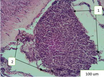
The length of the gland increased by 3.7% (1.165±0.07 mm; Р >0.05), the width was reliably increased by 1.2% (0.826±0.091 mm; Р >0.05) compared to the intact animals (Figure 1).
A reactive-compensatory reaction of the gland’s stromal component an increase in the number of fibroblasts, as well as qualitative changes - an increase in the area of connective tissue in the intertrabecular spaces and a significant thickening of the capsule - were observed. The capsule thickening by 2.5 times (4.46±0.47µm; P<0.01, t = 3.4).
A slight decrease in vascular perfusion was observed, but a violation of the rheological properties of blood (manifestations of stasis, sludge) remained. The vessels of the capsule and sub capsular zone remained dilated, in some fields of vision full of blood, in others with signs of emptying; the wall of the supplying arteries and arterioles was thickened. Some vessels of the capsule had an increase in the permeability of the vascular wall of the supplying vessels with the release of formed blood elements into the extravascular space and diffuse perspiration by blood cells of the gland parenchyma. The area of vascular lumen significantly increased by 1.9 times (p˂0.0001, t=43.65) compared to the control animals (Figure 2).
An active glial reaction, both local and diffuse, was preserved in the peripheral areas of the pineal gland, the degree of expressiveness of which practically did not differ from the indicators of animals that did not use the corrector drug. Astrocytes formed foci of glial complexes in the form of perivascular growths of various sizes. The nuclei of astrocytes were small, oval-shaped, hyper chromic (Figure 2).
Thus, the absolute number of pinealocytes decreased compared to the control animals by 33.6% (p˂0.001, t=19.61), and the absolute number of astrocytic glia cells, in contrast, increased by 2.1 times (p˂0.001, t=15.44). The glyocyto-neuronal index increased and exceeded the indices of control animals by 3.14 times (p>0.05).
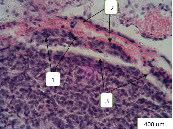
Figure 2: Morphological rearrangements of the pineal gland’s structural components under the condition of 60-days exposure to heavy metal salts and correction with α-tocopherol acetate: 1 - reactive glial response; 2 - aggregation and sludge of erythrocytes; 3 - increase in the severity of the gland’s trabecular connective tissue component. Hematoxylin-eosin staining.
During the study of the pineal parenchyma of experimental animals, a slight sponginess of the parenchyma and intertrabecular spaces, a slight violation of the cytoarchitectonics of cellular trabeculae was observed. As in the epiphysis of experimental animals that did not use the corrector drug, in the preparations stained with Einarson’s hallocyanin, pinealocytes with signs of indoleamine production prevailed. At the same time, dark pinealocytes were located on the periphery of the gland, and light ones, on the contrary, in the center. Light-colored pinealocytes had a distinctive regrowth form, the oval nuclei of such cells were enlarged in size, occupying almost the entire cytoplasm. The chromatin network of the nuclei was lightened and had diffuse chromatin. Hyperchromic nucleoli were contoured in some of the nuclei against the background of lightened karyoplasm. Dark pinealocytes had an elongated shape, with elongated, often deformed, hyperchromic homogeneous nuclei, without nucleoli. The preparations also contained a sufficient number of cells with chromatin margination, its shallow and coarse condensation in the form of blocks of various sizes, which were diffusely located in the illuminated nucleus.
There were changes in the structure of pinealocytes. At the same time, the index of pinealocyte body’s large diameter was increased by 30% (p>0.05), and the index of small diameter, on the contrary, decreased by 13% (p>0.05) compared to the control animals. The cross-sectional area of the cytoplasm and pinealocyte bodies grew by 70% (p˂0.001, t=6.34), by 1.96 times (p˂0.001, t=10.32), respectively, compared to the control animals. Thus, the pinealocytes nuclei large diameter index grew by 22% (p >0.05), and the index of their small diameter - by 14.3% (p>0.05) compared to the indices of the control animals. The cross-sectional area of pinealocyte nuclei was increased by 2.5 times (p˂0.01, t=3.9) compared to the control animals. The optical density of the nuclei and cytoplasm in pinealocytes decreased by 10.9% (Р˂0,001, t=5,9) and 11% (p<0.001, t=4,7), respectively, compared to the control animals, which probably indicated an improvement in the synthetic activity of pinealocytes when using the L-tocopherol corrector drug. This indicator, however, increased relative to the optical density of nuclei and cytoplasm of pinealocytes of experimental animals that did not use the corrector drug. The nuclear-cytoplasmic ratio increased to 1:0.68±1.05 compared to the control animals. The medium diameter of the caryon increased by 18.2% (P>0.05) compared to the parameters of the control animals.
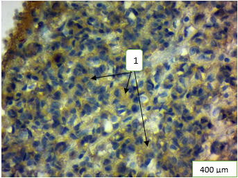
A weakly positive expression level of Ki-67 was established in 14.7 ± 0.53% (Р < 0.05) of pineal cells in comparison with indicators in control animals. The intensity of staining of cell nuclei was assessed as moderate (++). A slight increase in the expression level of Hsp90α was observed in the cytoplasm of 51-64% of pinealocytes in comparison with the indicators of experimental animals that did not use the corrector drug and control animals. A moderately positive level of expression of this type of heat shock protein was established (Figure 3).
Discussion
The protective effect of α-tocopherol acetate on the state of the pineal gland’s structural components in mature rats at 60-days exposure to a combination of heavy metal salts was determined. At the same time, the linear parameters of the organ increased relative to the indicators of the control animals.
The protective effect of α-tocopherol acetate was, first of all, to activate the reactive glial response, to increase the pronouncement of the gland’s stromal component, to improve the secretory activity of pinealocytes, however, the production of indoleamines by pinealocytes continues. A reactive compensatory response of the gland’s stromal component was observed. Thus, in contrast to animals that received only salts of heavy metals, in the pineal gland of animals with tocopherol correction significantly increased not only the thickness of the capsule, but there was an increase in the pronouncement of the trabecular connective tissue component, as well as thickening of the vessel wall. This can be explained by the activation of fibroblasts under the action of hypoxia, because the effect of α-tocopherol acetate at this term of the experiment is still insufficient to fully restore the condition of the vascular wall and the rheological properties of blood. Blood vessels were preserved, but it slightly decreased compared to the group of animals that did not use the corrector drug. Phenomena of stasis, coagulation of erythrocytes and increased permeability of the vascular wall were detected in the vessel lumen. This, in its turn, prevented the complete evacuation of hormones through the vascular wall into the blood and affected the course of the general adaptation syndrome. An increase in the permeability of the vascular wall led to the development of edema in the parenchyma of the gland and diffuse permeation of the parenchyma by plasma. On the part of pinealocytes, there was an improvement in their secretory activity was noted in comparison with the group of animals that did not use the corrector drug. Pinealocytes with cytological features of indole amine production predominated in the specimens [10]. Under the influence of α-tocopherol acetate, the tension on the part of the synthetic apparatus of pinealocytes decreases. The condition of the nucleus and nucleoreticulum was significantly improved, the area of the nucleus, medium diameter of the caryon, the optical density of the nuclei decreased compared to the indices of animals, receiving only heavy metal salts, which can be considered a positive protective effect of α-tocopherol acetate on the cell status. However, the presence of cells with nuclei with the phenomenon of chromatin margination in the preparations of the pineal gland indicates pre-apoptotic rearrangements of part of the cells.
The results of immunehistochemical studies indicate an increase in the proliferative activity of some cells. In the cytoplasm of most cells, a slight increase in the expression level of Hsp90α was detected, which is certainly a positive sign of the protective effect of α-tocopherol acetate on the state of cells.
The neuroglia reaction in response to the damaging agent is also activated, especially in the peripheral subcapsular areas of the gland. The formed perivascular astroglial complexes according to Hubina-Vakulik GI, et al. [14] may indirectly indicate more intense processes of pineal cell apoptosis in these animals, as evidenced by a reliable decrease in the number of pinealocytes compared to the control animals’ indices. In addition, the increase in the number of glial elements in the pineal gland certainly have a certain compensatory-adaptive value, especially in the transfer of RNA, amino acids, growth factors to pinealocytes and controlling water-ion homeostasis in the gland [15, 16]. According to the authors, such proliferates perform a barrier function, preventing the penetration of heavy metals into the parenchyma of the gland. It is impossible to overestimate the contribution of astrocytes in the protection of the gland’s parenchyma from the oxidative stress through the synthesis of hydrogen sulfide (H2S). This gas gliotransmitter has synaptic modulator and neuroprotective properties, protecting the pineal parenchyma from the oxidative stress [16].
Conclusion
The protective effect of L-tocopherol caused a number of compensatory-adaptive processes in the pineal gland aimed at neutralizing the effects of stressors on the organ. Under the influence of α-tocopherol acetate, the tension on the part of the synthetic apparatus of pinealocytes decreases compared to the indicators of animals that did not use the corrector drug.
However, the 60-days protective period of L-tocopherol is insufficient to completely neutralize the heavy metals impact on the organ: the glial response activated, the vascular area increased, vascular wall’s permeability impaired, edema was formed and the connective tissue component grew, indicating dysfunctional reorganization of the gland’s secretory activity. The above morphological changes in the pineal gland’s structural components negatively affect the processes of hormones evacuation into the blood, the course of the general adaptation syndrome and homeostasis restoration in the organ. The studies of the pineal gland, of course, expand the range of knowledge about the participation of this central neuroendocrine system’s organ in the body’s adaptive response to the combination of heavy metal salts and encourage researchers to seek for new drugs to correct morphological changes in the organ.
The work is a fragment of the research project “Modern views on the morphogenesis of general pathological processes”, state registration No. 0119U100887 (02.2019- 02.2024).
References
-
Demyanik D (2022) The war has significantly worsened the state of Ukraine’s environment.
-
Li X, Song J, Lin T, Dixon J, Zhang G, et al. (2016) Urbanization and health in China, thinking at the national, local and individual levels. Environmental health: a global access science source. Environ Health 15(1): 1-32.
-
Pollack L, Ondrasekm MR, Calisi R (2017) Urban health and ecology: the promise of an avian biomonitoring tool. Current Zoology 63(2): 205-212.
-
Yan X, Liu M, Zhong J, Guo J, Wu W (2018) How Human Activities Affect Heavy Metal Contamination of Soil and Sediment in a Long-Term Reclaimed Area of the Liaohe River Delta. North China 10(2): 338.
-
Hryntsova NB, Timakova OO, Romanyuk AM (2020) Morphofunctional reconstructions of the epiphysal- parathyroide axis structural components of rats in the period of readaptation after prolonged exposure to heavy metals. Problems of Endocrine Pathology 74(4): 106-114.
-
Bernstein C, Bernstein H (2015) Epigenetic reduction of DNA repair in progression to gastrointestinal cancer. World Journal Gastrointestinal Oncology 7(5): 30-46.
-
Wan D, Han Z, Yang J, Yang G, Liu X (2016) Heavy Metal Pollution in Settled Dust Associated with Different Urban Functional Areas in a Heavily Air-Polluted City in North China. International Journal of Environmental Research Public Health 13(11): 1119.
-
Romanyuk AM, Hryntsova NB, Karpenko LI, Kiptenko LI, Ustyansky OO, et al. (2019) The long-term effect of the complex of heavy metal salts on the morpho functional changes in the structural components of the intermediate lobe of the mature rat’s pituitary gland-the female. Problems of Endocrine Pathology 68(2): 98-103.
-
Volkov VP (2014) Functional morphology of the pineal gland during antipsychotic therapy. Universum: Medical and Pharmacological Electron 9 (10).
-
Bondarenko AA, Gubyna-Vakulyk GI, Gevorgyan AR (2013) The pineal gland and the hypothalamic-pituitary- thyroid system: age-related and chronobiological aspects. Institute of Endocrine Pathology Kharkiv, Ukraine, pp: 262.
-
Pshichenko VV (2014) Morpho functional features of the pineal gland of rats during simulation of round-the- clock lighting and acute stress. Kuban Scientific Medical Journal 1(143): 150-154.
-
Chlubek D, Sikora M (2020) Fluoride and Pineal Gland. Applied Science 10(8): 1-10.
-
Starchenko II, Grinko RM, Shkodina AD (2021) The Degree of Pineal Gland Calcification in the Aged People is Associated with Changes in the Internal Structure. Journal of International Dental and Medical Research 14(2): 841-844.
-
Hubina-Vakulik GI (2006) An attempt to summarize the results of histopathological examination of the pineal gland. Bukovyna Medical Bulletin 10(4): 34-36.
-
Drozdova GA, Samygulina AF, Nurgalieva EA, Bayburina GA, Sorokin AA (2017) Posthypoxic reaction of astroglial cells of the visual cortex in an experiment. Kazan Medical Journal 98 (6): 984-988.
-
Goryainov SA, Protsky SV, Okhotyn VY (2013) About the role of astroglia in the brain in normal and pathological conditions. Analysis of clinical and experimental neurology 7(1): 45-51.
- Shaping Healthy Futures: Pediatric Endocrine Breakthroughs of 2025
- Precision Medicine in Obesity: Customizing Treatment for 2025
- The Thyroid Revolution: How 2025 is Redefining Hormone Health
- Editorial- Targeting Immunometabolism for Generating Innovative Therapies for Cancer
- Current Knowledge of Chickenpox
- Correlation of Preinjection Values of Gonadotropins and Estradiol Level with Clinical and Radiologic Evidence of Sufficient Pubertal Suppression in Girls with Central Precocious Puberty