The Challenges of the Design-Driven Biotech Resources and Unique Translational Tools to Manage Cardiac Self-Renewal and Regeneration to be utilized in Clinical Practice
The approaches securing cardiac regeneration in post-infarction period are not available to be practiced. The key problem is the identity of cells is born to generate functionally active cardiac myocytes replenishing those being lost during ischemia. Over the last years, stem cells (SCs) have been a promise for the cure of several diseases, including heart failures, not only due to their plasticity but also acting as paracrine modulators and influencing the affected tissue. Human SC-based therapy derivatives are extremely attractive for therapeutic development because they have direct pharmacologic utility in clinical applications, unlike any other adult cells. Moreover, SC derived paracrine factors are to suppress inflammation and apoptosis, whilst stimulating angiogenesis, and amplifying the proliferation and differentiation of resident cardiac SCs (CSCs). SC therapies are thus viable alternatives to canonical treatments with substantial therapeutic potential; market opportunities are huge as multiple product candidates are expected to be approved over the coming decade. In this review, we accumulated recent data in the field of cardiac regeneration after MI using the latest resources of personalized and precision medicine (PPM), and considered promising ways of targeting pharmacotherapy and regenerative medicine in the field of cardiac rehabilitation.
Introduction
The ultimate goal for regenerative medicine is to channel multipotent human cells with high proliferative capacity into specified differentiation programs within the body. Meanwhile, the idea of extending the lifetime of the human heart has been fueled by a series of major advances in transplantation and drug therapies.
Nevertheless, myocardial infarction is characterized by the irreversible loss of cardiac myocytes (CMs) because of their ischemic necrosis. Therefore, the need to re-establish the structural and functional features of native heart tissue represents a major challenge for the field of cardiac tissue engineering. And current therapies do not address the underlying pathophysiology of ischemic conditions, namely, the progressive loss of functionally active CMs. The notion of regenerating lost myocardium via cell based therapies remains highly appealing.
SC-based therapy has been considered as a promising option in the treatment of ischemic heart disease. Although SC administration resulted in the temporary improvement of myocardial contractility in the majority of studies, the formation of new CMs within the injured myocardium has not been conclusively demonstrated [1, 2, 3].
And then, the focus of research and design driven applications in this field have since shifted to SC derived paracrine factors, including cytokines, growth factors, mRNA, and miRNA. Notably, both mRNA and miRNA can enter into the extracellular space either in soluble form or packed into membrane vesicles. SC derived paracrine factors have been shown to sup-press apoptosis, stimulate angiogenesis, and amplify the proliferation and differentiation of resident CSCs [4]. Such features have led to exosomes being considered as potential drug candidates affording myocardial regeneration. The search for chemical signals capable of stimulating cardiomyogenesis is ongoing despite continuous debates regarding the ability of mature CMs to divide or dedifferentiate, trans-differentiation of other cells into CMs, and the ability of CSCs to differentiate into CMs.
The recent identification of adult SCs, including both CSCs/progenitor cells, bone mar-row SCs (BMSCs) and others Figure 1, has triggered an explosive interest in using those cells for physiologically relevant cardiomyogenesis.
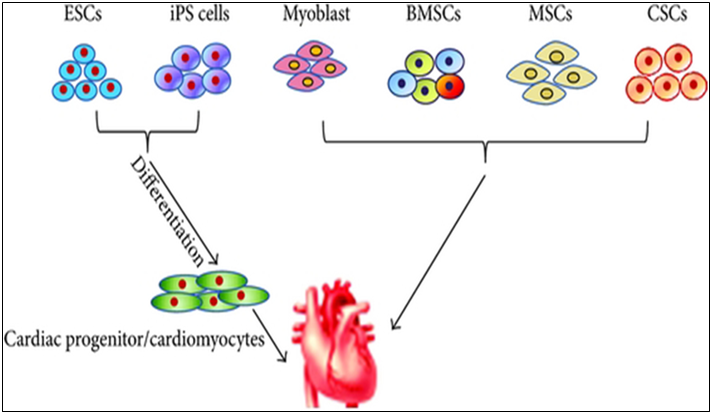
Source: Afjeh-Dana, Elham & Naserzadeh, Parvaneh & Moradi, Elham & Hosseini, Nasrin & Seifalian, Alexander & Ashtari, Behnaz. (2022). Stem Cell Differentiation into Cardiomyocytes: Current Methods and Emerging Approaches. Stem Cell Reviews and Reports 18: 1-27 HSCs, hematopoietic stem cell; iPSCs, induced pluripotent stem cells; BMSC, bone marrow stem cells; MSC, mesenchymal stem cell; CSC, cardiac stem cell; CM, cardiac cell.
Meanwhile, the heart is an extremely complex organ, and the techniques influencing its regeneration depend on many variables of nontrivial character. In addition, the efficacy of cell based therapies is somewhat limited by their poor longterm viability, homing, and engraftment to the myocardium. In response, a range of novel SC based technologies are in development to provide additional cellular modalities, bringing CTs a step closer to the clinic. Enthusiasm for cardiac regeneration via cell therapy has further been fueled by the many encouraging reports in both animals and human studies. Further intensive research in basic science, biodesign driven translational applications and clinical arenas are needed to make this next great frontier in cardiovascular regenerative medicine a reality.
However, SC based therapy has been considered to be a promising option in the treatment of ischemic heart disease (Figure 2).
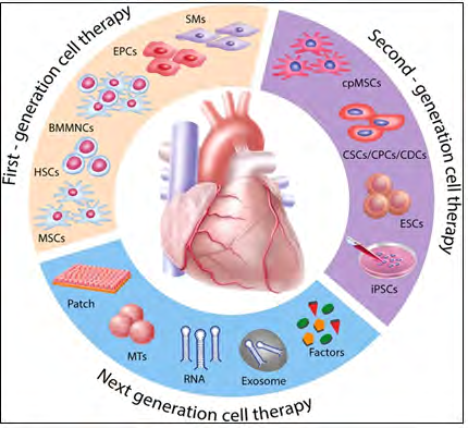
Figure 2: Evolution of translational cell driven cardiac regenerative therapies First generation cell types such as SMs, BMMNCs, HSCs, EPCs, and MSCs demonstrated feasibility and safety with, however, heterogeneous outcomes and limited efficacy in the clinical setting. In order to better match the target organ, second-generation cell therapies propose the use of cpMSCs, CSCs/CPCs, and CDCs, and pluripotent stem cells such as ESCs and iPSCs. Next-generation therapies for cardiac repair are directed toward cell enhancement (e.g., biomaterials, 3D cell constructs, cytokines, miRNAs) and cell free concepts (e.g., growth factors, noncoding RNAs, extracellular vesicles, and direct reprograming SMs, skeletal myoblasts; BMMNCs, bone marrow mononuclear cells; HSCs, hematopoietic stem cell; EPCs, endothelial progenitor cell; MSCs, mesenchymal stem cells; cpMSCs, chorionic plate derived mesenchymal stem cells; CSCs/CPCs, cardiac stem cell: cardiac progenitor cell ration; CDCs, cardiac dendritic cell; ESCs, embryonic stem cells; iPSCs, induced pluripotent stem cell; RNA, ribonucleic acid.
Source: Cambria, E., Pasqualini, F.S., Wolint, P. et al. Translational cardiac stem cell therapy: advancing from first-generation to next-generation cell types. npj Regen Med 2, 17 (2017). https://doi.org/10.1038/s41536-017- 0024-1.
Regarding the current status of SC therapies for patients with myocardial infarction and post infarction complications is critically being discussed now, describing:
- The current status of clinical trials of human pluripotent SCs (hPSCs) compared with clinical trials of human adult or fetal SCs;
- The gap between basics and design driven translational application of human SCs;
- The use of biomaterials in clinical and preclinical studies of SCs;
- Trends in design driven bioengineering to promote SC therapies for patients with myocardial infarction [1, 5].
SC-based therapies represent a possible paradigm shift for cardiac repair. However, most of the first-generation approaches displayed heterogeneous clinical outcomes regarding efficacy. Stemming from the desire to closely match the target organ, second generation cell types were introduced and rapidly moved from bench to bedside. Unfortunately, debates remain around the benefit of SC therapy, optimal trial design parameters, and the ideal cell type [6, 7, 8, 9, 10, 11, 12, 13, 14, 15, 16, 17, 18, 19, 20, 21, 22].
Having the above mentioned summarized and assessed, we might mention that SC driven therapy holds potential to tackle myocardial infarction and heart failure. But the key issues such as the cell type, cell number, delivery route, timing, follow-up periods, and endpoints remain un- solved. Meanwhile, the field has rapidly evolved to address in particular the ideal cell type and does require to get the problems be surmounted to secure the full potential of cell therapy be realized in the near future to come.
Toward Cardiac Regeneration: Combination of PSC-Based Therapies and Design Driven Bioengineering Strategies
At present, the approaches aimed at increasing myocardial regeneration after infarction are not available. The dispute over the existence of cardiac progenitors in the adult heart is still unsolved; on the same trail, the ability of immature cardiac progenitor cells to engraft with pre- existing CMs in vivo has not been irrefutably determined. Due to their unique capacity to pro-duce functional CMs, a wide variety of cells have been evaluated for therapeutic delivery, including BMCs, MSCs, and endogenous CSCs as the most promising sources for cardiac regenerative medicine application. Thus, the key question now is the identity of cells capable of producing functional CMs, replenishing those lost during ischemia.
But despite the encouraging results from these and other in vivo studies on the potential beneficial effects of PSC based cell therapy for heart failure, preclinical experiments, using CMs derived from human PSCs, have given controversial results [23].
Design-driven tissue engineering strategies are probably a relatively more suitable option. And given the limited regenerative capacity of the heart, the design-driven bioengineered scaffolds and tissue engineering approaches are thus ideal for cardiovascular regenerative medicine applications. The main goal is to create a cardiac graft which can be implanted and restore the functionality of the myocardium without major side effects. To this aim, tissue engineering in-tends to recreate the microenvironment of the cardiac tissue, in terms of cell composition, stiffness, geometry, physical and electrical stimuli and, extracellular matrix, and in order to generate constructs with an actual translational value, keeping in mind the native characteristics of the cardiac tissue [24]. And although SC therapy is a promising treatment for myocardial infarction, the minimal functional improvements observed clinically limit its widespread application. Where a need exists to maximize the therapeutic potential of those SCs by first understanding what factors within the infarct microenvironment affect their ability to regenerate the necrotic tissue, and to define a novel mechanism by which the extracellular environment of the infarction regulates the therapeutic potential of MSCs.
With respect to the developments within the biomaterial scaffold space, although they offer an effective solution to myocardium cell retention and cell number, the transplant process is generally more invasive, and challenges with effective electromechanical integration still remain. Meanwhile, the scaffolds could be modified or implemented in combination with molecules that activate the recruited cells (e.g., statins) and amplify their therapeutic potential. In conclusion, it is likely that no single approach to SC membrane reengineering will provide the “magic bullet” for cardiac cell therapies, and that the next generation of therapies will likely utilize combinations of these technologies to fully harness the therapeutic potential of transplanted SCs.
However, regardless of the employed strategy, generation of tissue-like engineered structures suitable for regenerative cardiology applications needs further considerations, specifically in relation to some aspects that may be critical for their efficacy and potential undesired effects in vivo.
Cardiac SCs (CSCs) as Promising Cell Types for Cardiac Regeneration
Myocardial infarction results in an irreversible loss of CMs due to aging or pathophysiological conditions, which is generally considered irreversible, and can lead to lethal conditions with subsequent adverse remodeling and heart failure. But human PSCs, including ESCs and iPSCs, can selfrenew while maintaining their pluripotency to differentiate into all cell types, including CMs. Furthermore, identifying new sources for CMs and promoting their formation rep-resents a goal of cardiac biology and regenerative medicine.
Meanwhile, because of the lack of strong evidence supporting the existence of resident CSCs, efforts are focused on how to mobilize and promote those few CMs that have potential to proliferate in the hearts. Whereas endogenous CMs can proliferate at a low frequency, methods that promote CM proliferation may be a vital research field in the future. The aforementioned pathways could be manipulated, optimized, and synergized to achieve better functional improvement after cardiac injury. In addition to inducing CM proliferation, other alternative approaches could be considered (Figure 3).
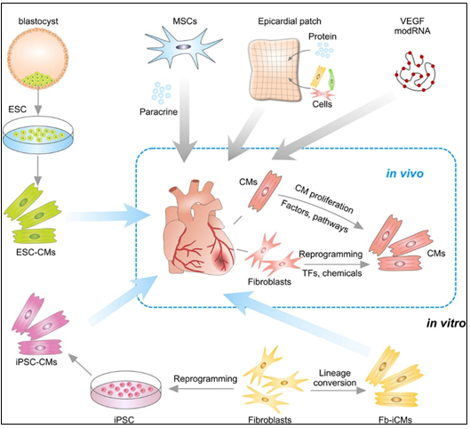
Figure 3: Strategies for treating cardiac repair and regeneration Transplantation of embryonic stem cell (ESC) derived CMs or iPSC derived CMs into myocardial infarction heart. CMs could also be reprogrammed from Fb in vitro or by transcriptional factors (TFs) or chemicals in vivo. Transplantation of MSCs promotes neovascularization and cardiomyocyte survival through paracrine mechanism. Epicardial patch containing growth factors or cardiac cells restores heart function after myocardial infarction. Administration of modified RNA (modRNA) of paracrine factors promotes heart function and drives heart progenitor cell fate. CMs could be promoted to proliferation by regulation of factors or signaling pathways.
Fb, fibroblasts; ESC, embryonic stem cell; CMs, cardiac myocytes; TF, transcriptional factor; iPSC, induced pluripotent stem cell; MSCs, mesenchymal stem cells; modRNA, modified RNA.
Source: Lingjuan He, Ngoc B. Nguyen, Reza Ardehali and Bin Zhou. Heart Regeneration by Endogenous Stem Cells and Cardiomyocyte Proliferation. Controversy, Fallacy, and Progress. Circulation. 2020; 142: 275-291.
In situ reprogramming of fibroblasts to cardiomyocytes by overexpression of specific transcriptional factors represents a promising direction for future study and translational applications to be implemented into clinical practice [25].
Recent studies have also suggested that small molecules can induce reprogramming of fibroblasts to CMs in vivo, which offers a dual pharmaceutical approach to regenerate CMs and reduce scar formation [26].
Human ESCs or iPSCs derived cardiac progenitors and CMs have been successfully transplanted and confirmed the survival of transplanted cells within the host myocardium and an improvement in cardiac function after injury. Furthermore, dual SC therapy, such as human iPSC derived CMs and MSCs, or human ESC-derived epicardium and CMs, synergistically improves cardiac function and augments vascularization in the injured myocardium. Moreover, re- cent advances in engineered epicardial patch containing multiple cardiac cell types are improved heart function and post-infarction neovascularization [27, 28, 29].
In this context, CSCs are considered to be very promising cell types for cardiac regeneration. And within the past decade, among CMs many types of putative CSCs have been reported to regenerate the injured myocardium by differentiating into new CMs. Some of these CSCs have been translated from bench to bed with reported therapeutic effectiveness. However, recent basic research studies on stem cell tracing have begun to question their fundamental biology and mechanisms of action, raising serious concerns over the myogenic potential of CSCs [30].
In particular, c-Kit+ CSCs represent a promising candidate for cardiac-specific SC lineages, which is likely heterogeneous in nature [31]. Moreover, resident CSCs are self-renewing and can give rise to CMs. Their multi-potentiality allows them to differentiate along the three main cardiac lineages: myocytes, endothelial cells, and smooth muscle cells. After their injection in the ischemic heart, the formation of the above-mentioned cell types contributes to the regeneration of myocardium and the improvement of its contractility.
But the approaches securing cardiac regeneration in post-infarction period are not available to be practiced. And the key problem is the identity of cells be born to generate functionally active cardiac myocytes replenishing those being lost during ischemia [32, 33].
Meanwhile, resident CSCs (rCSCs) may be a crucial source to initiate and prompt myocardial self-renewal and regeneration. And along with the latter, endogenous cardiomyogenic SCs (CMSCs) might be better choice for cardiac repair. Ideally, those cells should be directly stimulated in situ, avoiding extraction, purification, culture and reinjection. It is therefore of uttermost importance to understand the identity and function of the cells that constitute the natural environment of cardiac progenitors and support their quiescence, self-renewal and activation [34].
CSC therapy holds great potential to prompt myocardial regeneration in patients with is-chemic heart disease. The selection of the most suitable cell type is pivotal for its successful application. Therefore, different next generation cell types are currently under investigation for the treatment of the diseased myocardium [35].
Targeted Management of Regenerative Cardiomyogenesis
Drug (including SC driven) development continues to move in the direction of Personalized and Precision Medicine (PPM), where ideally the most effective therapy or treatment is determined by the genetic makeup of the patient and thus by a spectrum of biomarkers to be targeted by the therapeutics (including those being based on SCs). Regarding PPM based cardiology practice and the innovations made in the field of SC driven technologies, the focus in the last years has been moved towards a concept of the new wave of cardiac myocyte (CM) formation via a scenario of dedifferentiation and proliferation of mature CMs. The observation that CSCs can be developed inside a pool of immature cardiac cells by formation of “cell-in-cell structures” (CICSs) has enabled us to conclude that CICSs being encapsulated are implicated into mammalian cardiomyogenesis over the entire lifespan [2, 36, 37] (Figures 4A,4B).
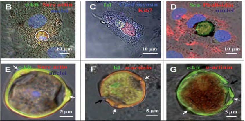
Figure 4A: The CSCs inside CMCs and the formation of CSC- containing CICSs in the cultures obtained from newborn and 20- and 40-day-old rats.
(A) Experimental design. The cells were plated and cultured for up to 30 days, followed by immunostaining or time-lapse microscopy. (B–G) Immunocytochemistry.
The nuclei of the cells have been stained with Hoechst. Transmitted light and fluorescent images are merged. (B) c-kitC CSC inside a CM obtained from a newborn rat (day in vitro). (C) Isl1C CSC inside a CM obtained from a newborn rat (day in vitro). As documented by the expression of Ki67, both the CSC and the host cell exhibit proliferative ability. (D) ScaC CSC encapsulated between the nuclei of the host cell (20-day-old rat, day in vitro). (E) A mature c-kitC CSC containing CICS with a prominent coating (“capsule”) with 3 pores (white arrows, 40 day old rat, day in vitro 6). Optical sectioning shows the host cell nucleus (blue) just above the CICS. (F-G) The CICS capsule in detail. (F) Erosion of the Isl1C CSC-containing CICS capsule (black arrow) obtained from a 40-day-old rat, day in vitro. The pores are also visualized (white arrows). The capsule interior is positive for sarcomeric a actinin, also observed in (G). (G) Erosion of the c-kitC CSC containing CICS capsule (black arrow) obtained from a newborn rat, day in vitro 20. The pores are seen (white arrows). CMs, cardiac myocytes; CSC, cardiac stem cell; CICSs, cell-in- cell structures”
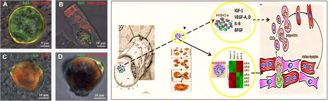
Figure 4B: Cell-in-cell structures (CICSs) identified in the suspension of freshly isolated myocardial cells (ex vivo) of 20- and 40-day-old rats.
Transmitted light and fluorescent images are merged. (A and B) Isl1C CSCs inside cardiomyocytes of 20-day-old rats (Isl1, green), a-Sarcomeric actin, red). (C) c-kitC CICS. (40-day-old rat, c-kit, green; Ki67, red). (D) c-kitC CICS. (40-day-old rat, c-kit, green; a-Sarcomeric actin, red).
It had been demonstrated before that new CMs are generated through formation of CSC derived transitory amplifying cells (TACs) either in the CM colonies or in a process of intracellular development of CICSs being encapsulated.
The data obtained, accumulated and interpreted, provide the basis for the suggestion that CSC mediated regenerative cardiomyogenesis is initiated in the myocardium immediately after the onset of myocardial ischemia. We can only assume that both encapsulated and nonencapsulated CICSs with poorly differentiated TACs inside are undergoing early rupture due to greater sensitivity to hypoxia and pH reduction at the acute stage of ischemia. If this is true, the key proximal question is the estimation of the survival rate of TACs released from encapsulated and non-encapsulated CICSs in the damaged area. In the case of their massive death, the myocardium is deprived of the significant number of TACs, which are in principle able to replace lost mature CMs.
Earlier on syngeneic animals were shown that a local laser apoptotic effect on tissues caused an intensive transition of mesenchymal BMSCs from the bloodstream to the zone of programmed cell death (Figure 5).
Apoptotic bodies (ApB) of cardiomyocytes combine the functions of CSC and HSCs in the areas of myocardial regeneration, and contain a complex of molecules, carriers of “epigenomic memory” about the tissue belonging of a dead cell. When ApB enters the CSC via endocytosis, a specific set of long and short noncoding RNAs express genes that determine the direction of differentiation of resident myocardial stem cells. It can be assumed that this hypothesis is true not only for the heart, but also for other organs and tissues [38, 39].
Those data for the first time suggest that the development of CSCs inside the encapsulated CICSs is important for cardiac self-renewal and maintenance of CSC based pool. At the same time, the development of CSCs inside a population of mature CMs is resulting in the formation of pre-cardiac myocytes, which are able to substitute for irreversibly injured CMs, representing the major mechanism of myocardial regeneration. And that, in turn, would open up a green light to secure the targeted management of regenerative cardiomyogenesis.
One of the main findings is the identification of a new type of CSC containing CICSs in heart samples obtained in the early post-infarction period. Due to the opinion, the above mentioned characteristics of two types of CICSs, that is, encapsulated vs. non-encapsulated CICSs, are responsible for the differences in maturity of TACs which develop inside them. Of note, the greater extent of TAC maturity was observed in non-encapsulated CICSs, which might have been accounted for by the higher level of differentiation of host cell and easier availability of humoral factors diffusing to the membrane-enveloped CSCs. In contrast, less differentiated TACs were released from encapsulated CICSs, which is paralleled by lower differentiation status of the host cell and poor diffusion of paracrine factors stimulating differentiation through the thick capsule. Formation of new CMs through proliferation and differentiation of CSCs within colonies is blocked after infarction and restored only 10–14 days after, the cardiomyogenesis in the early postinfarction period occurs by using population of TACs, which develop inside cardiac cells of different levels of maturity with formation of encapsulated and non-encapsulated CICSs. Therefore, improved understanding of molecular mechanisms giving rise to colonies and CICSs, may contribute to development of new pharmacological therapies targeting the process of cardiomyogenesis [2, 3, 4]. The results from those research and observation provide direct evidence for the replicative division of encapsulated SCs, followed by their partial cardiomyogenic differentiation. The latter is substantiated by the release of multiple TACs following the capsule rupture.
In conclusion, functional CSCs can reside not only exterior to but also within CMs. The presence of CSC-derived colonies, CICSs and TACs in myocardium proved the previous hypothesis about two pathways that generate new CMs in adult heart. Moreover, we suggest that TACs play a central role in self-renewal of myocardium throughout the lifetime of mammals [40].
The Prospects for the Introduction of Biodesign Inspired Drugs for Cardiac Regeneration
SC-driven translational applications have made significant progress in the field of cardiovascular regenerative medicine over the past few decades. Based on the new mechanisms and unique phenomenon, we are developing improvement strategies to boost the potency of SC repair and to generate the “next generation” of SC based and regulatory biomolecules-based (bimodal) therapeutics. Moreover, our strategies should aim at more personalized SC therapies in which individual disease parameters influence the selection of optimal cell type, dosage and delivery approach. And encouraging preclinical and clinical studies as one and solid entity reporting significant SC mediated cardiac regeneration would rapidly pave the way for clinical translation. Thus, a desire to discover innovative SC based technologies of the next step generation would encourage governments and companies to focus directly on regenerative medicine as a future potential economy and social insurance booster.
In reality, the global (worldwide) SC therapy market is still in an early stage. And the developing translational pipelines for rising applications will build the competition among merchants amid the conjecture time frame. Consequently, the focus of research in the field has since shifted to SC derived paracrine factors, including mRNA, and miRNA, which are entering into the extracellular space either in soluble form or packed into membrane vesicles. The search for chemical signals capable of stimulating cardiomyogenesis is ongoing despite continuous debates regarding the ability of mature cardiac myocytes to divide or dedifferentiate, transdifferentiation of other cells into cardiac myocytes, and the ability of CSCs to differentiate into cardiac myocytes. Future research is aimed at identifying novel cell candidates capable of differentiating into cardiac myocytes. The observation that CSCs can undergo intracellular development with the formation of “cell-in-cell structure” and subsequent release of transitory amplifying cells with the capacity to differentiate into cardiac myocytes may provide clues for stimulating regenerative cardiomyogenesis.
The worldwide SC therapy market is still in an early stage. And the developing translational pipelines for rising applications in the above-mentioned microecosystem will build the competition among merchants amid the conjecture time frame. And thus partnering between SC-related researchers, cell biodesigners and bioengineers, cardiac clinicians and surgeons, business and regulatory bodies and government can help ensure an optimal development program that leverages the Academia and industry experience and FDA’s new and evolving toolkit to speed our way to getting new tools into the innovative SC markets. We guess that the above mentioned would illustrate a unique Micro Ecosystem to be used for SC treatment in the near future to come, and tp provide a unique platform for dialogue and collaboration among thought leaders and stakeholders in government, academia, biopharma, foundations, and disease and patient advocacy with an interest in improving the system of healthcare delivery on one hand and drug discovery, development, and translation, on the other one, whilst educating the policy community about issues where biomedical science and policy intersect.
The Discussion and Conclusions
Cardiac aging manifests as functional tissue degeneration that leads to heart failure. Adult cardiac stem/progenitor cell senescence has been accordingly associated with processes encompassing both age related decline in cardiac tissue repair and cardiac dysfunction and disease. For instance, due to the limited regenerative capacity of cardiac myocytes, the infarction and its resulting heart failure have become the number one killer of human health in the senescent population. In addition, the idea that the heart is capable of regeneration has raised the possibility that cell-based therapies may provide such an alternative to conventional treatments, including cell-driven manipulations in elderly people, since cells having the potential to generate cardiac myocytes and vascular cells, have been identified in both the adult heart and peripheral tissues. And thus the stem cell therapy has exhibited broad application prospects in basic and clinical research on cardiovascular disease because of its plasticity, self-renewal and multidirectional differentiation potential [41, 42, 43, 44, 45]. The further understanding of the reversibility of the senescence phenotype will help to develop novel rational SC-driven therapeutic strategies [44, 45, 46, 47, 48, 49].
PPM, as we already mentioned, incorporates genetic variability, lifestyle, ageing and micro environmental factor to select or develop the most effective treatments (uniting preventive, prophylactic, therapeutic and rehabilitative types) for a patient and a pre-illness person at risk. In this sense, SC based therapy has lagged behind other fields for keeping the heart healthy. Furthermore, the micro-ecosystem and microenvironment as a whole play an active role in securing the effective crosstalk between the CSCs, the host immune system and system turnover, and ageing. The latter today is an object of intensive study, with the aim to develop effective therapeutic strategies targeting the ability of CSCs to restore the main human body functions. The SCs would confirm a high subclinical and predictive value as tools for PPM based monitoring protocols. SCs can be programmed and reprogrammed to suit the needs of the body metabolism or could be designed for the development of principally new combinatorial (genomics/cellomics rooted) drugs with no natural counterparts. Thus, PPM through SC therapy has many benefits that are essential for the future of personal health and wellness [50, 51, 52, 53, 54, 55, 56].
References
-
Malliaras K, Marban E (2011) Cardiac cell therapy: where we’ve been, where we are, and where we should be headed. Br Med Bull 98(1): 161-185.
-
Belostotskaya G, Hendrikx M, Galagudza M, Suchkov S (2020) How to Stimulate Myocardial Regeneration in Adult Mammalian Heart: Existing Views and New Approaches. Biomed Res Int 2020: 7874109.
-
Gu Y, Yi F, Liu GH, Izpisua Belmonte JC (2013) Beating in a dish: new hopes for cardiomyocyte regeneration. Cell Research 23(3): 314-316.
-
Perez-Ilzarbe M, Agbulut O, Pelacho B (2008) Characterization of the paracrine effects of human skeletal myoblasts transplanted in infarcted myocardium. European Journal of Heart Failure 10(11): 1065-1072.
-
Menasche P, Vanneaux V, Fabreguettes JR, Bel A, Tosca L, et al. (2015) Towards a clinical use of human em-bryonic stem cell-derived cardiac progenitors: a translational experience. Eur Heart J 36(12): 743-750.
-
Taylor DA, Atkins BZ, Hungspreugs P, Jones TR, Reedy MC, et al. (1998) Regenerating functional myocardium: improved performance after skeletal myoblast transplantation. Nat Med 4(8): 929-933.
-
Reinecke H, MacDon-ald GH, Hauschka SD, Murry CE (1998) Electromechanical coupling between skeletal and cardiac muscle. Implications for infarct repair. J Cell Biol 149(3): 731-740.
-
Murry CE, Soonpaa MH, Reinecke H, Nakajima H, Nakajima HO, et al. (2004) Haematopoietic stem cells do not transdifferentiate into cardiac myo-cytes in myocardial infarcts. Nature 428(6983): 664-668.
-
Zhang S, Ge J, Zhao L, Qian J, Huang Z, et al. (2007) Host vascular niche contributes to myocardial repair induced by intracoronary transplantation of bone marrow CD34+progenitor cells in infarcted swine heart. Stem Cells 25(5): 1195-1203.
-
Tendera M, Wojakowski W, Ruzyłło W, Chojnowska L, Kepka C, et al. (2009) Intracoronary infusion of bone marrow-derived selected CD34+CXCR4+cells and non- selected mononuclear cells in patients with acute STEMI and reduced left ventricular ejection fraction: results of randomized, multicentre myocardial regeneration by intracoronary infusion of selected population of stem cells in acute myocardial infarction (REGENT) trial. Eur Heart J 30(11): 1313-1321.
-
Hirsch A, Robin N, Pieter AV, Jan GP, et al. (2011) Intracoronary infusion of mononuclear cells from bone marrow or pe-ripheral blood compared with standard therapy in patients after acute myocardial infarction treated by primary percutaneous coronary intervention: results of the randomized controlled HEBE trial. Eur Heart J 32(14): 1736-1747.
-
Heldman AW (2014) Transendocardial mesenchymal stem cells and mononuclear bone marrow cells for ischemic cardiomyopathy: the TAC-HFT randomized trial. J. Am. Med Assoc 311: 62-73.
-
Lee JW, Youn YJ, Ahn MS, Kim JY, Lee SH, et al. (2014) A randomized, open-label, multicenter trial for the safety and efficacy of adult mesenchymal stem cells after acute myocardial infarction. J Korean Med Sci 29(1): 23-31.
-
Mathiasen AB, Abbas AQ, Erik J, Steffen H, Klaus FK, et al. (2015) Bone marrow derived mesenchymal stromal cell treatment in patients with severe ischaemic heart failure: a randomized placebo-controlled trial MSC-HF trial. Eur Heart J 36(27): 1744-1753.
-
Rossini A, Frati C, Lagrasta C, Graiani G, Scopece A, et al. (2011) Human cardiac and bone marrow stromal cells exhibit distinctive properties related to their origin. Cardiovasc Res 89(3): 650-660.
-
Oskouei BN, Lamirault G, Joseph C, Treuer AV, Landa S, et al. (2012) Increased potency of cardiac stem cells compared with bone mar row mesenchymal stem cells in cardiac repair. Stem Cells Transl. Med 1(2): 116-124.
-
Li TS, Cheng K, Malliaras K, Smith RR, Zhang Y, et al. (2012) Direct comparison of different stem cell types and subpopulations reveals superior paracrine potency and myocardial repair efficacy with cardiosphere derived cells. J Am Coll Cardiol 59(10): 942-953.
-
Vagnozzi RJ, Maillet M, Sargent MA (2020) An acute immune response underlies the benefit of cardiac stem cell therapy. Nature 577: 405-409.
-
Cambria E, Pasqualini FS, Wolint P (2017) Translational cardiac stem cell therapy: advancing from first generation to next generation cell types. npj Regen Med 2: 17.
-
Sultana N, Zhang L, Yan J, Chen J, Cai W, et al. (2015) Resident c-kit(+) cells in the heart are not cardiac stem cells. Nat. Commun 6: 8701
-
Liu Q, Yang R, Huang X, Zhang H, He L, et al. (2016) Genetic lineage tracing identifies in situ kit-expressing cardiomyocytes. Cell Res 26(1): 119-130.
-
Keung W, Boheler KR, Li RA (2014) Developmental cues for the maturation of metabolic, electrophysiological and calcium handling properties of human pluripotent stem cell derived cardiomyocytes. Stem Cell Res Ther 5(1): 17.
-
Chen A, Ting S, Seow J (2014) Considerations in designing systems for large scale production of human cardiomyocytes from pluripotent stem cells. Stem Cell Res Ther 5(1): 12.
-
Hulot JS, Stillitano F, Salem JE (2014) Considerations for pre-clinical models and clinical trials of pluripotent stem cell-derived cardiomyocytes. Stem Cell Res Ther 5(1): 1.
-
Higuchi A, Ku NJ, Tseng YC (2017) Stem cell therapies for myocardial infarction in clinical trials: bioengineering and biomaterial aspects. Lab Invest 97(10): 1167-1179.
-
Song K, Nam YJ, Luo X, Qi X, Tan W, et al. (2012) Heart repair by reprogramming nonmyocytes with cardiac transcription factors. Nature 485(7400): 599-604.
-
Qian L, Huang Y, Spencer CI, Foley A, Vedantham V, et al. (2012) In vivo reprogramming of murine cardiac fibroblasts into induced cardiomyocytes. Nature 485(7400): 593-598.
-
Park SJ, Kim RY, Park BW, Lee S, Choi SW, et al. (2019) Dual stem cell therapy synergistically improves cardiac function and vascular regeneration following myocardial infarction. Nat Commun 10(1): 3123.
-
Bargehr J, Ong LP, Colzani M, Davaapil H, Hofsteen P, et al. (2019) Epicardial cells derived from human embryonic stem cells augment cardiomyocyte driven heart regeneration. Nat Biotechnol 37(8): 895-906.
-
Ye L, Chang YH, Xiong Q, Zhang P, Zhang L, et al. (2014) Cardiac repair in a porcine model of acute myocardial infarction with human induced pluripotent stem cell- derived cardiovascular cells. Cell Stem Cell 15(6): 750- 761.
-
Lingjuan H, Ngoc BN, Reza A, Bin Z (2020) Heart Regeneration by Endogenous Stem Cells and Cardiomyocyte Proliferation. Controversy, Fallacy, and Progress. Circulationaha 142(3): 275-291.
-
Kulandavelu S, Karantalis V, Fritsch J, Hatzistergos KE, Loescher VY, et al. (2016) Pim1 kinase overexpression enhances c-kit+ cardiac stem cell cardiac repair following myocardial infarction in Swine. J Am Coll Cardiol 68(22): 2454-2464.
-
Makkar RR, Smith RR, Cheng K, Malliaras K, Thomson LE, et al. (2012) Intracoronary cardiosphere derived cells for heart regeneration after myocar-dial infarction (CADUCEUS): a prospective, randomised phase 1 trial. Lancet 379(9819): 895-904.
-
Bolli R, Chugh AR, D Amario D, Loughran JH, Stoddard MF, et al. (2011) Cardiac stem cells in patients with ischaemic car-diomyopathy (SCIPIO): initial results of a randomised phase 1 trial. Lancet 378(9806): 1847- 1857.
-
Lerman DA, Alotti N, Ume KL, Péault B (2016) Cardiac Repair and Regeneration: The Value of Cell Therapies. Eur Cardiol 11(1): 43-48.
-
Elena C, Julia S, Julia G, Annina B, Petra W, (2016) Cardiac Regenerative Medicine: The Potential of a New Generation of Stem Cells. Transfus Med Hemother 43(4): 275-281.
-
Belostotskaya G, Sonin D, Galagudza M (2021) Intracellular Development of Resident Cardiac Stem Cells: An Overlooked Phenomenon in Myocardial Self- Renewal and Regeneration. Life (Basel) 11(8): 723.
-
Belostotskaya GB, Nerubatskaya IV, Galagudza M (2018) Two mechanisms of cardiac stem cell-mediated cardiomyogenesis in the adult mammalian heart include formation of colonies and cell-in-cell structures. Oncotarget 9(75): 34159-34175.
-
Tyukavin A, Belostotskaya G, Zakharov E, Suchkov S (2019) 4th World Congress & Expo on Pharmaceutics and Drug Delivery Systems. Scientific Federation.
-
Tyukavin AI, Belostotskaya GB, Zakharov ЕА, Ivkin DY, Rad’ko SV, et al. (2020) Apoptotic Bodies of Cardiomyocytes and Fibroblasts Regulators of Directed Differentiation of Heart Stem Cells. Bull Exp Biol Med 170(1): 112-117.
-
Belostotskaya G, Nevorotin A, Galagudza M (2015) Identification of cardiac stem cells within mature cardiac myocytes. Cell Cycle 14(19): 3155-3162.
-
He-Ling Y, Le C, Wei-Wen F, Xin L, Qiang L, et al. (2024) Application and challenges of stem cells in cardiovascular aging. Regenerative Therapy 25: 1-9.
-
Cianflone E, Torella M, Biamonte F, De Angelis A, Urbanek K, et al. (2020) Targeting Cardiac Stem Cell Senescence to Treat Cardiac Aging and Disease. Cells 9(6): 1558.
-
Yuan HL, Chang L, Fan WW, Liu X, Li Q, et al. (2023) Application and challenges of stem cells in cardiovascular aging. Regen Ther 25: 1-9.
-
Khatiwala R, Cai C (2016) Strategies to Enhance the Effectiveness of Adult Stem Cell Therapy for Ischemic Heart Diseases Affecting the Elderly Patients. Stem Cell Rev and Rep 12(2): 214-223.
-
Tan YS, Looi QH, Sulaiman N, Ng MH, Law JX (2022) Current State of Stem Cell Therapy for Heart Diseases. In: Haider KH (Ed.), Handbook of Stem Cell Therapy pp: 1-30.
-
Cambria E, Pasqualini FS, Wolint P (2017) Translational cardiac stem cell therapy: advancing from first generation to next generation cell types. npj Regen Med 2: 17.
-
Yeh D, Chan T (2018) Therapeutics of Stem Cell Treatment in Antiaging and Rejuvenation. Stem Cell Discovery 8(2): 13-31.
-
Cambria E, Pasqualini FS, Wolint P (2017) Translational cardiac stem cell therapy: advancing from first generation to next generation cell types. npj Regen Med 2: 17.
-
Mazo M, Hernández S, Gavira JJ, Abizanda G, Araña M et al. (2012) Treatment of reperfused ischemia with adipose derived stem cells in a preclinical swine model of myocardial infarction. Cell Transplant 21(12): 2723- 2733.
-
Emmert M, Wolint P, Jakab A, Sheehy S, Pasqualini F, et al. (2016) Safety and efficacy of cardiopoietic stem cells in the treatment of post-infarction left-ventricular dysfunction – From cardio-protection to functional repair in a translational pig infarction model. Biomaterials 122: 48-62.
-
Behfar A, Crespo-Diaz R, Terzic A, Gersh BJ (2014) Cell therapy for cardiac repair lessons from clinical trials. Nat. Rev. Cardiol 11(4): 232-246.
-
Xiao J, Pan Y, Li XH, Yang XY, Feng YL, et al. (2016) Cardiac progenitor cell-derived exosomes prevent cardiomyocytes apoptosis through exosomal miR-21 by targeting PDCD4. Cell Death Dis 7(6): e2277.
-
Ellison GM, Vicinanza C, Smith AJ, Aquila I, Leone A, et al. (2013) Adult c-kit(pos) cardiac stem cells are necessary and sufficient for functional cardiac regeneration and repair. Cell 154(4): 827-842.
-
Butler J, Epstein SE, Greene SJ (2017) Intravenous allogeneic mesenchymal stem cells for nonischemic cardiomyopathy: safety and efficacy results of a phase II-A randomized trial. Circ Res 120: 332-340.
-
Henry TD, Pepine CJ, Lambert CR (2017) The Athena trials: Autologous adipose derived regenerative cells for refractory chronic myocardial ischemia with left ventricular dysfunction. Catheter Cardiovasc Interv 89(2): 169-177.