Primary Synovial Sarcoma of the Kidney: An Unusual Presentation
The Synovial Sarcoma (SS) is a rare neoplasm, the fourth most common type of sarcoma. Most often located at the para-articular region of inferior limbs and tendons. Renal presentation is more common among 15-40 years old patients with an average of 38 years. It predominates in the male gender, manifesting itself with a feeling of abdominal mass, hematuria and nonspecific signs. We report a 59-year-old female patient consults by pain in the right lumbar region plus emesis and hematuria, accompanied with a hard, painless palpable mass in the right upper quadrant. Computed Tomography (CT) scan revealed markedly enlarged right kidney, with heterogeneous density components. Right renal nephrectomy is performed. The pathology service concludes diagnosis of SS, which is favored by immunohistochemistry. Finally, the patient had a fatal outcome due to a recurrent tumor compromising the right renal fossa.
Introduction
Synovial sarcoma is by itself uncommon. The majority of cases involve somatic soft tissue, where they account for approximately 15% of sarcomas in patients 10 to 18 years of age and 1.6% of sarcomas in patients above 50 years of age [1]. Synovial sarcoma is defined clinical and pathologically as a tumor that mainly develops in juxta articular soft tissue or in the limbs of young people and adults [2]. Among sarcomas of the kidney, leiomyosarcoma is the most frequent type, comprising 40‑60%, followed by rhabdomyosarcoma, chondrosarcoma, liposarcoma, angiosarcoma, hemangiopericytoma and osteosarcoma [2, 3, 4]. Hence, owing its location, the following case report is extremely rare.
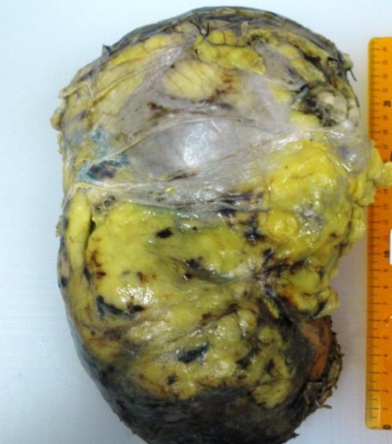
A 59-years-old woman with history of diabetes, obesity, hypertension and urolithiasis consults to the emergency department for acute onset of severe pain in the right lumbar region, irresponsible to analgesics, associated with hematuria and countless emetic episodes. It is initially focused as a renal colic. Physical examination shows a firm, circumscribed and painless palpable mass in the right upper quadrant. Kidney ultrasound shows a lobed-edge defined mass measuring 14x11x13 cm with hypoechoic areas with blood flow inside in right the flank. The CT urography scan revealed a markedly increased right kidney accompanied with hyperdense image of 14 mm of diameter and evidence of ipsilateral ureteral dilatation, indicating urolithiasis, accompanied by a great hydronephrotic bag with isodense and hypodense components suggesting hidropionefrosis. The patient underwent a right renal nephrectomy. Macroscopic features indicate stum or lesion with a varied surface showing whitish, fleshy, and friable, bleeding, with necrotic areas, compromising the entire specimen. Histological examination revealed a Malignancy with biphasic morphology consisting of proliferation of spindle cells with varying from mild to severe atypia.
There are other areas of fibrosis and necrosis. The tumor showed imnunorreactivity for BCL2 and vimentin with diffuse pattern and, P-63, CD-10 and CD-99 with focal pattern (Image 1-2) and negative for S100, miogenin, CD34, and epithelial membrane antigen (EMA). With those findings, pathology service diagnosed synovial sarcoma. Three months after surgery, the patient revisits the hospital with abdominal pain and emesis; due to her background, a CT scan is performed. Hetero geneous tumor mass with recurrent tumor appearance sized 8.2 x 6.7cm was found in right renal fossa compromising and in filtrating hepatic flexure and diaphragm without associated lymphadenopathy. The clinical course is torpid and the patient died during hospitalization.
This neoplasm was first described by Faria, et al in [5, 6]. These tumors tend to occur in men more frequently than women with a male-to-female ratio of 1.7:1. The mean age at diagnosis is 37 years (range, 13‑67 years) but it was also described in a child [7], almost 1% of cases are located in the kidney. The mean tumor diameter is 11 cm (range, 3‑21 cm), and the rate of metastasis upon admission is likely to be low [3]. According to published data, our patient met the average. The most common symptomatology are flank or back pain, emesis and hematuria as in this case. Fei-X, et al [8] showed in their study group of two women and three men the same symptomatic presentation that include lower back pain (n=3), hematuria (n = 2), nausea and vomiting (n =1) [8]. There is no clinical or imaging characteristic that can indicate the diagnosis [4, 6], but the physicians could be able to recognize some imaging patterns for the suspicion of this neoplasm. A study protocol shows some generally features of the tumor through CT scan imaging. They performed at least once in all five patients and twice in one patient. Unenhanced CT showed completely or partly well-defined masses, with heterogeneous (n=5) or homogeneous (n =1) patchy low density. On multiphase contrast-enhanced CT, in five of the six CT examinations, the tumors appeared as solid-cystic masses with cyst walls or pseudo-capsules, and demonstrated moderately heterogeneous (n =5) and/or septate enhancement (n= 2). No lymphadenopathy where reported [8]. Other findings at ultra sonography image are huge cystic circumscribed masses with or without multi loculated shape and heterogeneous content. That means the physicians should be aware of the possibility of malignancy when a young adult presents with a cystic renal tumor [2, 9, 10, 11, 12, 13]. Histologically, synovial sarcoma is divided into monophasic synovial sarcoma (MSS) and biphasic synovial sarcoma (BSS)types on the basis of the presence of a well-defined glandular element and the epithelial and spindle cell components [some authors also call them unidirectional and bidirectional synovial Sarcoma [2]. By the other hand, poorly differentiated synovial sarcomas (PDSS) represent a form of tumor progression seen in both MSS and BSS [1]. PDSS is formed from sheets of un differentiated round cells with hyperchromatic nuclei and frequent mitoses, and is associated with the poorest outcome [3, 12]. Some inmuno hystochemical aspects of Synovial sarcoma are immunoreaction for epithelial markers such as cytokeratin, vimentin, proteins CD-99, CD-56, Bcl-2, TLE1 and EMA, generally with focal distribution [4, 10], as in this case but do not take up stain for action, desmin, S-100 and CD-34 [1, 2]. Macroscopically, the differential diagnosis have to be done from Cistic lessons [12], renomegaly [14] and other neoplasms like adult Wilms' tumors, transitional cell carcinoma, renal cell carcinoma, hemangiopericytoma and primitive neuroectodermal tumors (PNET) [3, 4, 11]. Congenital mesoblasticnephroma, sarcomatoid renal cell carcinoma (SRCC) and undifferentiated carcinoma must be taken into account [2]. The differential diagnosis is most challenging in the monophasic and poorly differentiated types that mimics SRCC, solitary fibrous tumor, and PNET. Like synovial sarcoma, SRCC can show partial immunoreactivity with antibodies to epithelial markers, but in contrast to synovial sarcoma, SRCC does not express TLE1 [1]. Leiomyosarcoma and fibro sarcoma are also rare in the kidney, and are solid, have no small cysts with a spike shaped cell lining, and few hemangiopericytoma shaped structures, often express SMA, but do not express BCL2, CD99, or CD56. Adult Wilm’s tumor will also show primary spindle cell predominance as renal synovial sarcomas and should be taken in account at differential diagnosis in young people. However, the SYT-SSX gene fusion will be absent [2]. Establishing a correct diagnosis is difficult and requires immune his to chemical and molecular pathological methods. This type of tumor is related with some gene alterations as SYT-SSX gene fusion demonstrated by PCR. The translocation t(X; 18)(p11.2;q11.2) which is specific for synovial sarcoma regardless of location or type and grade of differentiation and can be demonstrated in about 90% of all synovial sarcomas [11, 12] Hence, definitive diagnosis can be achieved by identifying the trans location through reverse transcriptase polymerase chain reaction with fluorescence in situ hybridization (FISH) [2, 4, 6]. In this particular case, diagnosis was only established based on conventional morphologic microscopic analysis helped by immuno histochemistry. No definite standard treatment guidelines are available at present. Treatment is based on the occurrence of scattered cases published in literature. Generally, the treatment includes chemotherapy and surgical interview. It has been published that ifosfamide-based chemotherapy plus doxorubicin, mostly adjuvant to radical nephrectomy, allowed the remission and overall survival of patients even in patients with PRSS, developing metastases in the lung [13, 14]. Patients are recommended to undergo adjuvant chemotherapy after radical surgery. However, regardless of the treatment, the prognosis remains unclear due to the limited number of reported cases [2].
| Case Report | ||||
|---|---|---|---|---|
| Volume 1 Issue 2 | ||||
| Received Date: May 24, 2016 | ||||
| Published Date: August 31, 2016 | ||||
| 1Department of Diagnostic, University of Cartagena, Colombia | ||||
| 2School of Medicine, University of Cartagena, Colombia | ||||
| 3Department of Urology, University of Cartagena, Colombia | ||||
| 4Department of Pathology, University of Cartagena, Colombia |
Conclusion
Synovial sarcoma (SS) is a rare entity, with special predilection for certain anatomic sites. Renal location is so unusual, there are very few cases reported in the literature. It behaves very aggressively, with little response to treatment, causing high morbidity and mortality.
References
-
Schoolmeester JK, Cheville JC, Folpe AL (2014) Synovial sarcoma of the kidney: a clinicopathologic, immunohistochemical, and molecular genetic study of 16 cases. Am J Surg Pathol 38(1): 60-65.
-
Ozkan EE, Mertsoylu H, Ozardali HI (2011) A case of renal synovial sarcoma treated with adjuvant ifosfamide and doxorubicin. Intern Med 50(15): 1575-1580.
-
Zijian Wang, Zhaohui Zhong, Liang Zhu, Wei Xiong, Cizhong Pan, et al. (2015) Primary synovial sarcoma of the kidney: A case report. Oncol Lett 10(6): 3542-3544.
-
Schaal CH, Navarro FC, Moraes Neto FA (2004) Primary renal sarcoma with morphologic and immunohistochemical aspects compatible with synovial sarcoma. Int Braz J Urol 30(3): 210-213.
-
Faria P, Argani P, Epstein J, Reuter V, Beckwith B, et al. (1999) Primary synovial sarcoma of the kidney: a molecular reappraisal of a subset of so-called embryonal renal sarcoma. Lab Invest 79(94A).
-
Argani P, Faria PA, Epstein JI, Reuter VE, Perlman EJ, et al. (2000) Primary renal synovial sarcoma: molecular and morphologic delineation of an entity previously included among embryonal sarcomas of the kidney. Am J Surg Pathol 24(8): 1087-1096.
-
Radhakrishnan V, Dhanushkodi M, Narayanswamy K, Raja A, Sundersingh S, et al. (2016) Synovial sarcoma of kidney in a child: A rare presentation. J Indian Assoc Pediatr Surg 21(2): 75-77.
-
Lv XF, Qiu YW, Han LJ, Cao J, Zhang C, et al. (2015) Primary renal synovial sarcoma: computed tomography imaging findings. Acta Radiol 56(4): 493- 499.
-
Mishra S, Awasthi N, Hazra SP, Bera MK (2015) Primary synovial sarcoma of the kidney. Saudi J Kidney Dis Transpl 26(5): 996-999.
-
Hirose M, Mizuno K, Kamisawa H, Nishio H, Moritoki Y, et al. (2015) Clear cell sarcoma of the kidney distinguished from synovial sarcoma using genetic analysis: a case report. BMC Res Notes 8(8): 129.
-
Kohle O, Abt D, Rothermundt C, Ohlschlegel C, Brugnolaro C, et al. (2015) Soft tissue sarcomas of the kidney. Rare tumors 7(1): 5635.
-
Nishida T, Inamoto T, Uehara H, Ibuki N, Koyama K, et al. (2011) Monophasic primary renal synovial sarcoma accompanied with a hemorrhagic cyst. Urol J 8(3): 244-247.
-
Park SJ, Kim HK, Kim CK, Park SK, Go ES, et al. (2004) A case of renal synovial sarcoma: complete remission was induced by chemotherapy with doxorubicin and ifosfamide. Korean J Intern Med 19(1): 62-65.
-
Modi G, Madabhavi I, Panchal H, Anand A, Patel A, et al. (2014) Primary synovial sarcoma of kidney: a rare differential diagnosis of renomegaly. Case rep pathol 2014: 657497.
- Results of 6-Month Follow-Up of Patients After B-Turp and Thulep
- The Effect of Drinking Water with a High Content of Antimony and Arsenic on the Dynamics of their Distribution in the Kidneys and the Renal Excretory Function in Rats
- Effectiveness and Safety of Tansurethral Thulium Laser Enucleation of the Prostate in the Treatment of BPH: Review
- A Systematic Review on Molecular Pathophysiology Involved in Chronic Kidney Disease and the Role of Animal Models in Drug Discovery to Manage in Chronic Kidney Disease - An Update
- Functional Development of Kidneys in Human Ontogenesis
- Testicular Metastasis: Uncommon Prostate Cancer Case Report