It’s not always The Airway! – Learning Points in Airway Management
Task fixation has been identified in aviation and medical emergency management as an obstruction to successful resolution of a situation1, 2. The 4th National Audit Project of the Royal College of Anaesthetists and the Difficult Airway Society- Major complications of airway management in the United Kingdom (NAP4) suggest that the most frequent human factors contributing to death were: poor communication, poor teamwork, poor leadership and task fixation3. We consider three cases where focus on the airway distracted from non-upper airway causes of hypoxia.
L Fraser1, E Sproson1, A Baldock2, G Jones2, A Burgess1 and H
Ismail-Koch1*
Letter to Editor
Task fixation has been identified in aviation and medical emergency management as an obstruction to successful resolution of a situation1, 2. The 4th National Audit Project of the Royal College of Anaesthetists and the Difficult Airway Society- Major complications of airway management in the United Kingdom (NAP4) suggest that the most frequent human factors contributing to death were: poor communication, poor teamwork, poor leadership and task fixation3. We consider three cases where focus on the airway distracted from non-upper airway causes of hypoxia.
Case Reports
Case 1
A 2 week-old female, born at 38 weeks gestation presented with respiratory distress from birth. A prenatal diagnosis of cardiac issues was noted. Co-existing anomalies found at birth following investigations included choanal atresia, renal cysts and clinically CHARGE syndrome. During anaesthesia for the choanal atresia repair the anaesthetist was unable to intubate due to a difficult view. Otorhinolaryngological emergency endotracheal intubation was performed using a laryngoscope with an endotracheal tube threaded over a Hopkins rod under direct vision. The child was ventilated, however loss of cardiac output ensued leading to cardiorespiratory arrest. The left side of the chest was noted not to be moving. Consequently the endotracheal tube was pulled back as it was thought to be in too far.
Letter to Editor
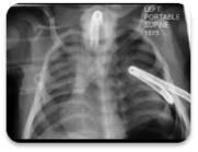
This resulted in accidental extubation. The child was re- intubated once again by the otorhinolaryngologist under direct vision confirming placement in the trachea. Due to lack of left chest excursion aspiration of the left chest with a 23-gauge needle was undertaken to exclude a pneumothorax and produced no air. Thus the endotracheal tube was thought to be blocked. Crash tracheostomy was undertaken with no change in output. Oxygenation was continued throughout. Thoracostomy and left chest drain insertion was performed as there was no movement of the left chest and a left pneumothorax was suspected, restoration of output followed. The diagnosis was confirmed as a left pneumothorax. The child is currently doing well, decannulated with no neurological compromise (Figure 1).
Case 2
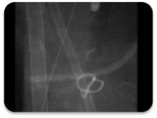
A 1 day-old male born at 36 weeks gestation with respiratory distress was transferred to the Paediatric Intensive Care Unit. Co-existing anomalies found at birth were retrognathia, cleft palate, anal atresia, a single kidney and a double outlet right ventricle. Genetics subsequently revealed a 15q11 deletion. It was not possible to bag and mask ventilate the child and a grade 4 Cormack-Lehane anaesthetic view was found. Otorhinolaryngological intubation with an endotracheal tube threaded over a Hopkin’s Rod under direct vision was undertaken. Good chest movement was noted but a poor Carbon Dioxide (CO2) trace was observed. Endotracheal tube placement was reconfirmed. Following cardiac review and bronchoscopy a diagnosis of near pulmonary atresia and long segment tracheal stenosis was found. The child underwent cardiac surgery and a slide tracheoplasty, but sadly died of cardiac complications (Figure 2).
Case 3
A 52 year-old lady was admitted via the accident and emergency department following a road traffic accident. Computed Tomography imaging revealed a C1 burst fracture and T1-3 stable anterior superior vertebral border fractures. Development of a chest infection and type 2 respiratory failure necessitated Intubation. Following failure of extubation tracheostomy was planned on day 8. Jet ventilation was performed during the tracheostomy procedure. However 40 minutes of hypoxaemia ensued thought to be related to the tracheostomy procedure and tube issues. However chest X-ray intraoperatively revealed a diagnosis of a right pneumothorax thought to be secondary to high ventilation pressures. Thoracostomy and chest drain insertion was performed and the lung re-expanded. Ventilation was weaned at day 17 and she was discharged from hospital (Figure 3).
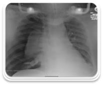
Figure 3: Chest X-ray showing a right pneumothorax. These cases illustrate how task fixation in upper airway management can result in failure to diagnose and manage other synchronous pathologies. We have reflected on these cases in our local practice, to consider learning points and strategies for avoidance of fixation on one particular pathology, and move on to other differentials once the airway is secured. It is important for both the surgeon and wider multidisciplinary team to be aware of non-upper airway causes of hypoxia; their presentation and management. Airway management should be a team- based approach, similar to aviation practice. This allows all members of the team to contribute equally, and consider alternative diagnoses.
References
-
Harmer M The case of Elaine Bromiley. Anonymised report. The clinical human factors group.
-
(2010) 9 Determination of Sheriff Linda Margaret Ruxton in fatal accident inquiry into the death of Gordon Ewing.
-
(2011) 4th National Audit Project of the Royal College of Anaesthetists & the Difficult Airway Society. Major complications of airway management in the UK.
- 4th Branchial Cleft Sinus Anomaly Presenting as Recurrent Thyroid Abscess in A Child: A Case Report
- Parotid Duct Injury Repaired Using an Angiocatheter Stent: A Case Report
- Organization and Functionality of the Referral and Counter-Referral System for ENT Disorders in District Hospitals of N’Djamena, Chad: A Cross-Sectional Analytical Study
- Facial Metastases from a Gastrointestinal Stromal Tumor: A Case Report
- Panorama of Ent Cancers and Literature Review: Epidemiological Profile and Therapeutic Management
- Could Antimicrobial Resistance Prove to Be Both a Threat and an Opportunity for us?