Autism
The term "autism," comes from the ancient Greek word "autos" which means "self" and "ismus", which means "condition". So, the term describes conditions in which the individual becomes withdrawn within self, an isolated self.
Background
The term "autism," comes from the ancient Greek word "autos" which means "self" and "ismus", which means "condition". So, the term describes conditions in which the individual becomes withdrawn within self, an isolated self. Autism is an umbrella term that describes a range of conditions characterized, by a difficulty in forming relationships, communicating and managing repetitive behaviours. Neurobiological Background: The human brain is divided into the cerebrum (telencephalon) and the cerebellum. The cerebrum is composed of 5 pairs of lobes; occipital, parietal, temporal, frontal and limbic.
The occipital lobes of the brain control the sense of sight. The parietal lobes are concerned with the sensations like temperature, pain and pressure, furthermore, visuospatial processing. The temporal lobes are associated with the control of hearing, smell, language and social perception. The frontal lobes are related to the thinking, planning and memory. The part of the frontal lobe immediately in front of the premotor cortex concerned mainly with the associative cognitive functions like attention, motivation and most importantly the behaviour. Scientists believe that this part is the one involved in the behavioural changes in autism [1].
![Figure 2: A diagram illustrating the location of the Prefrontal cortex At the cellular level, after differentiation into neurons, they migrate to their functional positions and grow into axons and dendrites. Inter neuronal connection via synapses leads to the functional neuronal communications that moderate motor and sensory handling, and control the behaviour, learning and memory [2]. From neurobiology point of view, neurons send electro-chemical signals after being stimulated by some kind of stimulus down the length of the cell, what is known as electric potential [3]. This is the electrical part of the electro-chemical signal. Once the electric current reaches the axon, it activates the release of some chemical mediators (neurotransmitters), and this is the chemical part of the signal. These neurotransmitters allow one neuron to connect to one another. The gap between the two neurons is called the synapse which is an important part in any neuronal connection. Neurotransmitters can increase (excitatory) or decrease (inhibitory) the trans- synaptic ion flow and then convey the neuronal massage. An example of excitatory neurotransmitters is glutamate and of inhibitory neurotransmitters is Gamma- aminobutyric acid (GABA). Some neurotransmitters have both actions like acetylcholine. After childhood, the brain shows striking changes in its morphology. Functional MRI (fMRI) has displayed reduction in the grey matter of the cerebral cortex in certain areas of the temporal lobes as well as in the prefrontal lobe [4]. Animal showed extensive synaptic pruning during this age group [5]. Other neuroimagings have revealed expansion in the white matter mass during adolescence in cortical and sub cortical regions [6] coming from augmented myelination or increased axon diameter in white matter [7].](/fulltextimages/127/fig_2.jpeg)
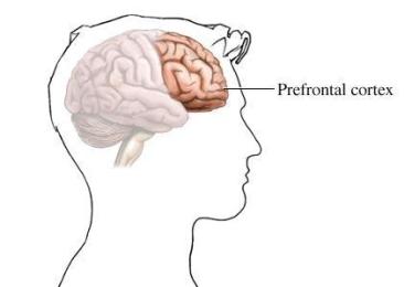
Figure 2: A diagram illustrating the location of the Prefrontal cortex At the cellular level, after differentiation into neurons, they migrate to their functional positions and grow into axons and dendrites. Inter neuronal connection via synapses leads to the functional neuronal communications that moderate motor and sensory handling, and control the behaviour, learning and memory [2]. From neurobiology point of view, neurons send electro-chemical signals after being stimulated by some kind of stimulus down the length of the cell, what is known as electric potential [3]. This is the electrical part of the electro-chemical signal. Once the electric current reaches the axon, it activates the release of some chemical mediators (neurotransmitters), and this is the chemical part of the signal. These neurotransmitters allow one neuron to connect to one another. The gap between the two neurons is called the synapse which is an important part in any neuronal connection. Neurotransmitters can increase (excitatory) or decrease (inhibitory) the trans- synaptic ion flow and then convey the neuronal massage. An example of excitatory neurotransmitters is glutamate and of inhibitory neurotransmitters is Gamma- aminobutyric acid (GABA). Some neurotransmitters have both actions like acetylcholine. After childhood, the brain shows striking changes in its morphology. Functional MRI (fMRI) has displayed reduction in the grey matter of the cerebral cortex in certain areas of the temporal lobes as well as in the prefrontal lobe [4]. Animal showed extensive synaptic pruning during this age group [5]. Other neuroimagings have revealed expansion in the white matter mass during adolescence in cortical and sub cortical regions [6] coming from augmented myelination or increased axon diameter in white matter [7].
![Figure 3: Functional MRI in normal and autistic individuals A study has showed many differences at neurotransmitters level during adolescence [8]. For instance, there are changes in the secretion and flow of Dopamine and there are peaks of Dopamine neural activity in the midbrain that slow down later in life. Studies have revealed huge difference in the balance between excitatory and inhibitory neurotransmission in adolescence compared to adults. The levels of GABA which is the chief inhibitory neurotransmitter have risen smoothly in the adolescent forebrains. The previously mentioned differences if impaired will result in many behavioural implications. At the functional level, fMRI images have shown massive differences in the functional activity in many areas of adolescent forebrains especially in these areas where emotional and sensory data are encrypting [9]. Behavioural development may be affected by maturity within different brain regions and between them and improvement in inter-neuronal coordination. Studies have shown direct relation between the increase in frontal white matter and the inhibition in the control performance [10]. Also, the development of white matter is found to be in direct relation to the improvement in the Factors like white matter enhancement, accelerated pruning of excitatory synapses and improvement in interneuron activity; together may promote better functional cortical coordination through adolescence. Some age-related electrophysiological studies have revealed clue of improvement in interneuron responses and better local coordination. Another study has shown the development of positive peak of about 300 ms when responding to a certain stimulus at the age of 13 [3]. These findings give another proof for the ongoing progression in the prefrontal cortical signal processing through adolescence. In vivo electrophysiological recording of the wave taken when electrodes placed in active behaving animals gives another evidence of neural activity in adolescence. Again, using techniques like fMRI in human samples and electrophysiological studies in animals, it became clearer that aspects of stimulated behaviours are different in various age groups. Regarding the total brain volume, it has been found that it is atypical in autistics in the form of high prevalence of macrocephaly (unusually large sized head; AJM, 2006) coexisted with larger brain volume and / or accelerated growth of brain compared to static total brain volume till the age of 13 years that found to decrease during early years of adulthood (Neuroimage, 2007). This atypical brain growth in childhood is assumed due to the expansion of the total white matter compared to grey matter (Brain, 2003). White matter continues to increase in terms of volume in adolescence while grey matter peaked before adolescence reached [11]. Some studies suggest that over all brain size in adulthood is unchanged in autistic individuals without intellectual impairment [12].](/fulltextimages/127/fig_3.jpeg)
Figure 3: Functional MRI in normal and autistic individuals A study has showed many differences at neurotransmitters level during adolescence [8]. For instance, there are changes in the secretion and flow of Dopamine and there are peaks of Dopamine neural activity in the midbrain that slow down later in life. Studies have revealed huge difference in the balance between excitatory and inhibitory neurotransmission in adolescence compared to adults. The levels of GABA which is the chief inhibitory neurotransmitter have risen smoothly in the adolescent forebrains. The previously mentioned differences if impaired will result in many behavioural implications. At the functional level, fMRI images have shown massive differences in the functional activity in many areas of adolescent forebrains especially in these areas where emotional and sensory data are encrypting [9]. Behavioural development may be affected by maturity within different brain regions and between them and improvement in inter-neuronal coordination. Studies have shown direct relation between the increase in frontal white matter and the inhibition in the control performance [10]. Also, the development of white matter is found to be in direct relation to the improvement in the Factors like white matter enhancement, accelerated pruning of excitatory synapses and improvement in interneuron activity; together may promote better functional cortical coordination through adolescence. Some age-related electrophysiological studies have revealed clue of improvement in interneuron responses and better local coordination. Another study has shown the development of positive peak of about 300 ms when responding to a certain stimulus at the age of 13 [3]. These findings give another proof for the ongoing progression in the prefrontal cortical signal processing through adolescence. In vivo electrophysiological recording of the wave taken when electrodes placed in active behaving animals gives another evidence of neural activity in adolescence. Again, using techniques like fMRI in human samples and electrophysiological studies in animals, it became clearer that aspects of stimulated behaviours are different in various age groups. Regarding the total brain volume, it has been found that it is atypical in autistics in the form of high prevalence of macrocephaly (unusually large sized head; AJM, 2006) coexisted with larger brain volume and / or accelerated growth of brain compared to static total brain volume till the age of 13 years that found to decrease during early years of adulthood (Neuroimage, 2007). This atypical brain growth in childhood is assumed due to the expansion of the total white matter compared to grey matter (Brain, 2003). White matter continues to increase in terms of volume in adolescence while grey matter peaked before adolescence reached [11]. Some studies suggest that over all brain size in adulthood is unchanged in autistic individuals without intellectual impairment [12].
![Figure 4: Macrocephaly in an autistic child. Neuro developmental differences were investigated with Voxel-based morphometry (VBM) which is a neuro imaging technique that allows the study of focal differences in brain anatomy; it showed in autistic individuals that there are considerable anatomical differences in many brain areas. One of these areas that showed increase in grey matter size was the anterior part of temporal lobe. Temporal lobe in the left hemisphere was noticed to be larger to the rest of the brain regions and that was associated with more social, but not repetitive, symptoms. The temporal lobe was drawing much attention in further studies because it was found to be linked in high level cognitive development like in mentalizing or in the semantic processing (deep memory processing) either in childhood, adolescence or early adulthood [13]. These findings were further studied using fMRI and the results suggest the role of the anterior temporal lobe in moderating social impairments in autistic individuals, a study that was conducted by Nature Reviews Neuroscience and was published in 2007. Other researches revealed that the grey matter increases in size in the prefrontal brain cortex especially in the pre-central and post-central gyri. This neuro developmental difference could be the cause in atypical motor responses, fine motor progression and visuo-motor adaptation observed in autistic people. Christine found that there are differences in the basal ganglia and thalamus volumes in autistic individuals compared to neurotypicals. Also, in autistics there are differences in frontostriatal systems starting in childhood and continuing into adulthood and the severity of the repetitive behaviours noticed in autistics are associated with anatomical differences in frontal brain regions.](/fulltextimages/127/fig_4.jpeg)
Figure 4: Macrocephaly in an autistic child. Neuro developmental differences were investigated with Voxel-based morphometry (VBM) which is a neuro imaging technique that allows the study of focal differences in brain anatomy; it showed in autistic individuals that there are considerable anatomical differences in many brain areas. One of these areas that showed increase in grey matter size was the anterior part of temporal lobe. Temporal lobe in the left hemisphere was noticed to be larger to the rest of the brain regions and that was associated with more social, but not repetitive, symptoms. The temporal lobe was drawing much attention in further studies because it was found to be linked in high level cognitive development like in mentalizing or in the semantic processing (deep memory processing) either in childhood, adolescence or early adulthood [13]. These findings were further studied using fMRI and the results suggest the role of the anterior temporal lobe in moderating social impairments in autistic individuals, a study that was conducted by Nature Reviews Neuroscience and was published in 2007. Other researches revealed that the grey matter increases in size in the prefrontal brain cortex especially in the pre-central and post-central gyri. This neuro developmental difference could be the cause in atypical motor responses, fine motor progression and visuo-motor adaptation observed in autistic people. Christine found that there are differences in the basal ganglia and thalamus volumes in autistic individuals compared to neurotypicals. Also, in autistics there are differences in frontostriatal systems starting in childhood and continuing into adulthood and the severity of the repetitive behaviours noticed in autistics are associated with anatomical differences in frontal brain regions.
The reduction in white matter volume in autism has often been explained as one of neurobiological theories in abnormal neuroconnectivity in autism [14]. It has been claimed that in autism, during neurodevelopment there is abnormal connections in the frontal lobes that could be over connectivity or under connectivity that ultimately will cause what is known as Developmental Disconnection Syndrome. Another region that could be affected in autistic individuals is the medial part of the temporal lobe that showed aberrant functional connectivity during panicky face processing especially in Asperger's syndrome [15]. Also, there is a clue that under connectivity between the frontal and the parietal lobes may influence executive functioning.
Historical perspective
Jean-Marc-Gaspard Itard, was a French physician with particular impute to Victor, the so-called “The Wild Boy of Avalon.” who showed several signs of behavioural disturbances and was thought to have lived his entire childhood alone in the French woods. Itard treated him with a special behavioural program started in 1797 and showed some improvement then died in 1828. Then Paul Eugen Bleuler, who was a Swiss psychiatrist. He was the first one to use term; Autism. He started using it around 1908 to refer to a group of patients with schizophrenic symptoms. In 1938, Leo Kanner, an American child psychiatrist, studied 11 children with features of difficulties in social interactions, who were sensitive to auditory stimuli with good intellectual potential and echolalia and put the label early infantile autism. His paper Autistic Disturbances of Affective Contact in 1943, together with the work of Hans Asperger, forms the basis of the modern concept of autism. Hans Asperger, was an Austrian paediatrician, in 1944 he studied a group of children who resembled Kanner’s descriptions. The children he studied did not have echolalia as a linguistic problem but spoke like normals. The term Asperger's syndrome was given after him. Asperger called children with Asperger's syndrome “little professors,” because of their ability to talk about their favourite subject in great details. He was convinced that many would use their special talents in adulthood. He followed one of them, Fritz V into adulthood who became a professor of astronomy. Bruno Bettelheim, an Austrian child psychologist, studied the effect of therapeutic sessions on children who he called autistic. He attributed this condition to parenting style and he was the one who described the refrigerator mother theory of autism in 1950 in which lack of maternal warmth may leads to autism in susceptible individuals. After that Bernard Rimland; an American research psychologist and parent of an autistic child. He disagreed with Bettelheim that the cause of his son’s autism was due to either his or his wife’s parenting style. In 1967, he founded the Autism research institute and Autism society of America. Lorna Wing with Christopher Gillberg at Children's Neuro-Psychiatric Clinic in Sweden in the 1980’s found the Wing’s triad of disturbed social contact, disturbed social communication and limited imagination. In the 1990’s they added another factor making it a square; limited planning ability. Ole Ivar Lovaas, a Norwegian clinical psychologist, considered the father of applied behavioral analysis (ABA) and provided evidence that teaching autistic children can modify their behavior_._ Also, Frith, a German researcher, had clinical training at King's medical college, in 1991 translated the work of Asperger and contributed much in research of autism.
Diagnosis of Autism
According to The International Classification of Diseases - version 10 (ICD-10) definitions, Autism is a complex neurobehavioral condition that is usually manifested in early childhood (before 30 months age) with impaired social interactions, communication and repetitive behaviours. Health care professionals think of autism as a spectrum "Autism Spectrum Disorder" with Asperger syndrome is the milder form on the spectrum. Spectrum is category of behaviour with certain key features, but with important differences within it, ranging from mild to severe. In the case of autism, the term spectrum describes the range of people with symptoms affecting their communication, social interaction and social imagination, and recognizes that these differ significantly in how people experience them. One out of 10 of autism spectrum disorder will have the severe form of the disorder. The Autism lasts throughout a person's life. There is no definite cure for autism, but treatment can help.
- 1. Cognitive Development: this is the skill to learn and solve problems. For example, a two-month-old infant learning to explore the surrounding environment with hands.
- 2. Social and Emotional Development: this is the ability to communicate with others. Like a three months old baby smiling.
- 3. Speech and Language Development: this is the capability to both understand and use language. For instance, a
- 12-month-old baby saying his first words; mama or dada.
- 4. Gross Motor Skill Development: this is the skills of using big muscles. Like, a six-month-old baby learns how to sit up with some support and a five-year-old learns to skip.
- 5. Fine Motor Skill Development: this is the child's ability to use small muscles, specifically of hands and fingers, to pick up small objects, hold a spoon, turn pages in a book, or use a crayon to draw.
Table 1: The five areas of human development.
Normal child development is the process in which child goes through alterations in skill development during certain time frames, these alterations called developmental milestones. Abnormal development refers to developmental delay which happens when the child has not reached these milestones by the proposed time period. For instance, if the normal time frame for learning how to walk is between 9 and 15 months, and a 2 years old child has still not begun walking, this would be considered as a developmental delay. The child attains skills in five main areas of development: Some developmental milestones and the normal time frame for each one:
- Pediatrics & Neonatal Biology Open Access
- Speech:
- Coo -------------------------------------------1-3m
- Babble ---------------------------------------6-9m
- Mama/Dada without meaning ---------10-11m
- Mama/Dada with meaning --------------12m
- Jargon -------------------------------------13-15m
- 2 – 6 words with meaning ----------------15m
- 7 - 20 words with meaning ----------------18m
- > 50 words/2 word sentences ----------------2y
- 200 words/4 word sentences -----------------3y
- Use pronouns ----------------------------------2y
- Distinguish past & present/right & left -------4y
- Distinguish morning/afternoon -------------5y
- Count to 20 ------------------------------------4y
- 50% of speech understood by strangers --20m
- 100% of speech understood by strangers –3y
- Colors :
- Name 1 color -------- 2.5y
- Name 2 colors ------- 3y
- Name 3 colors ------- 4y
- Name 4 colors ------- 5y
- Motion :
- Run ---------------------------------------2y
- Jump (off step both feet together) ----3y
- Hop (15 feet) ----------------------------5y
- Skip with rope ( 3 rounds) ------------7y
- Skip with rope ( 12 rounds) ----------8y
Table 2: The time frame of some developmental milestones.
There are various general indicators of possible delay. These include:
| Behavioural signs | Not paying attention to any activity as other children of the same age do |
|---|---|
| Focuses on uncommon objects for long periods of time | |
| Refrains eye contact with others | |
| Gets easily upset when trying to do simple tasks that most children of the same age can do | |
| Shows aggressive behaviours appears to be very obstinate compared with other children | |
| Stares into nowhere, sways body, or talks to self more often than other children of the same age | |
| Does not have emotions to the caregiver or parent | |
| Gross motor signs | Has rigid extremities |
| Has a flaccid body posture compared to other children of the same age | |
| Uses one side of body more than the other | |
| Have difficulty finding or picking up small objects dropped on the floor | |
| Visual signs | Not following objects or people with eyes |
| Rubs eyes frequently | |
| Turns, tilts or holds head in an unusual position when trying to look at an object | |
| Has difficulty making eye contact | |
| Focusing with one eye when trying to look at far objects | |
| Putting objects too close to eyes to see | |
| One or both eyes appear abnormal in size or position | |
| Hearing signs | Talks in a very loud or very quite manner |
| Turns the body so that the same ear is always turned toward sound |
Table 3: Indicators of possible developmental delay. The delay in development of basic skills observed during early childhood in
Doesn't startle to loud noises or gets frustrated with noisy sounds Ears appear small in size or abnormal in shape Fails to develop sounds or words that would be appropriate at their age Table 3: Indicators of possible developmental delay. The delay in development of basic skills observed during early childhood in particular the ability to socialize with others, to communicate with others and to use imagination; this constitutes a group of disorders known as Pervasive Developmental Disorders (PDD). These disorders divided into five categories as appears in the following diagram.
![Figure 5: A diagram illustrating the Pervasive Developmental Disorders. AD: Autistic Disorder, also known as Kanner's syndrome; AS: Asperger's Syndrome; PDD-NOS: Pervasive Developmental Disorder-Not Otherwise Specified, also called Atypical Autism; CDD: Childhood Disintegrative Disorder, also known as Heller's syndrome; RS: Rett's Syndrome. The first three conditions are referred to Autism Spectrum Disorders (ASD) and the last two are much more uncommon [16,17]. It is obvious that the number of cases with autism spectrum disorders is growing. In one century, the prevalence increased from 1 in every 15,000 to 1 in every 100 in the UK, which means more than 700,000 persons in the UK living with autism with varying degree of severity, boys affected 4 times than girls. The general prevalence is on the rise. Many factors are may put the person at risk of having autism, like:](/fulltextimages/127/fig_5.png)
Figure 5: A diagram illustrating the Pervasive Developmental Disorders. AD: Autistic Disorder, also known as Kanner's syndrome; AS: Asperger's Syndrome; PDD-NOS: Pervasive Developmental Disorder-Not Otherwise Specified, also called Atypical Autism; CDD: Childhood Disintegrative Disorder, also known as Heller's syndrome; RS: Rett's Syndrome. The first three conditions are referred to Autism Spectrum Disorders (ASD) and the last two are much more uncommon [16, 17]. It is obvious that the number of cases with autism spectrum disorders is growing. In one century, the prevalence increased from 1 in every 15,000 to 1 in every 100 in the UK, which means more than 700,000 persons in the UK living with autism with varying degree of severity, boys affected 4 times than girls. The general prevalence is on the rise. Many factors are may put the person at risk of having autism, like:
A sibling with autism. A monozygotic twin with autism. Chromosomal disorders like Trisomy 21(Down's syndrome). Genetic disorders like Fragile - X syndrome. Neurocutaneous diseases like Neurofibromatosis and Tuberous sclerosis. Maternal use of Sodium Valproate during pregnancy.
Gestational age less than 35 weeks at delivery. Central nervous system malformations including Cerebral Palsy. Antenatal exposure to some drugs like Mercury or Metallothionine. Parental schizophrenia-like psychosis or affective disorder. Neonatal encephalopathy or epileptic encephalopathy, including infantile spasms.
Intellectual disability.
Muscular dystrophy. Table 4: The autism risk factors. Mass screening is not recommended as false positive or false negative results may hinder the appropriate diagnosis but surveillance throughout the preschool years is recommended. Referral to specialist assessment should be done if warning signs appear on the child. In the first two years of life, there are usually no warning signs but parental concerns should be evoked if any of these sings appear on the child:
By 6 months: No social smiles or other warm expressions. By 9 months: No back-and-forth sharing of sounds or smiles. By 12 months: Lack of response to name. : No babbling (baby talk) : No back-and-forth gestures, such as pointing or waving. By 18 months: No spoken words. By 24 months: No meaningful two-word sentences. Table 5: The warning signs in the first 2 years of life. In pre-school children (between 2 and 3 years), some of the developmental warnings should alter referral, like [18]:
| Social impairments | .Lack of imitation of actions like hand clapping |
|---|---|
| .Lack of playing with toys | |
| .Lack of interest in other children | |
| .Lack of pretend play | |
| .Minimal responsiveness to other people's feelings like happiness |
Table 4: The warning signs in the first 2 years of life. In pre-school children (between 2 and 3 years), some of the developmenta
.Odd relationship with older people( too friendly or ignores) .Impairment in language development .Poor response to name Communication .Lack of pointing .Absence of social smile .Loss of any developed social skills impairments .Over-sensitivity to sound or touch .Biting or hitting .Aggressive behaviour to peers Behaviour impairments .Inability to cope with change .Repetitive play with toys In school age children, features differ; like:
| Social impairments | .Inability to join in or inappropriate joining with play of other children |
|---|---|
| .Lack of awareness of classroom norms | |
| .Easily overwhelmed by social stimulation | |
| .Failure to relate to adults normally | |
| Communication impairments | .Abnormal language development |
| .Limited use of language for communication | |
| .Persistent echolalia | |
| .Reference to self as you, she or he | |
| .Unusual vocabulary for age | |
| Behaviour impairments | .Lack of flexible cooperative imaginative play |
| .Difficulty in organizing self in relation to unstructured space | |
| .Inability to cope with change or unstructured situations, even ones that other children enjoy like school trips |
professional at the 18 month routine developmental check-up. It consists of two sections: section A; nine items in the form of questions asked to the parents, and section B; five items in the form of observations made by the physician or the health care professional. If a child passes the CHAT from the first session, no further action to be taken. However, passing the CHAT does not mean that the child will not develop a social-communication problem of some form, at that time the parents should seek specialist referral. If the child fails the CHAT, he should be re- screened after a month. If the child fails the CHAT for the second time, he should be referred to a specialist clinic for diagnosis since the CHAT is not a diagnostic tool. The CHAT report put the suspected child in one of three risk levels, low, medium or high and gives recommendation as follows; low risk group to be re-tested in one month, medium risk group if low suspicion to be re-tested after a month and if highly suspicious to be referred to developmental clinic as well as Educational Services Department (ESD). In high risk group, referral to developmental clinic and ESD is recommended. The M-CHAT-R is a 2 stages screening tool to be done by one of the parents. The first stage is 20 questions about how the child usually behaves. The second stage is another 20 questions follow-up screening sheet. The American Academy of Pediatrics (AAP) recommends that all children at 18 and 24 months of age should receive autism screening, and the M-CHAT-R is one of the AAP’s recommended tools, however, the advantages of the CHAT over the M-CHAT-R are that it's an easy, quick and cheap tool. Also, autism is difficult to be detected before the age of three years. The earlier a diagnosis can be made, the earlier intervention can be carried out. The most two diagnostic systems concerning with autistic disorders are: the Diagnostic and Statistical Manual of Mental Disorders - the fifth edition (DSM-V) developed by the American Psychiatric Association (APA) and the International Classification of Diseases - version 10 (ICD-10) designed by the World Health Organization (WHO). The pathway put by the National Institute for Health and Care Excellence (NICE) for recognition, referral and diagnostic assessment of children with possible autism is as follows: A local autism multi-agency strategy group (autism team) should be set up, with managerial, commissioner and clinical representation from child health and mental health services, education, social care, parent and carer service users, and the voluntary sector. This group should appoint a lead professional to be responsible for the local autism pathway for recognition, referral and diagnosis of children and young people with possible autism. The goal of this group is: A. Improve the early recognition of autism by raising awareness of the signs and symptoms of autism through multi-agency training. B. Make sure the relevant professionals (healthcare, social care, education and voluntary sector) are aware of the local autism pathway and how to access diagnostic services. C. Support the smooth transition of young people to the adult services. D. Ensure data collection and audit of the pathway takes place. E. The autism team core members are [19], F. Paediatrician and/or child and adolescent psychiatrist. G. Speech and language therapist. H. Clinical and/or educational psychologist. Also, the team should include or has a direct access to: i. Paediatric neurologist. ii. Child and adolescent psychiatrist. iii. Educational psychologist.
iv. Clinical psychologist.
v. Occupational therapist. vi. Teacher.
In addition, the autism team should include other relevant professionals who may be able to contribute to the autism diagnostic assessment. Like a specialist health visitor or nurse, specialist teacher or social worker. The role of the autism team is to: a. Provide advice to professionals about whether to refer children and young people for autism diagnostic assessments. b. Decide on the assessment needs of those referred or when referral to another service will be needed. c. Carry out the autism diagnostic assessment. d. Share the outcome of the autism diagnostic assessment with parents and carers, and with children and young people if appropriate. e. With parent or carer consent and, if appropriate, the consent of the child or young person, share information from the autism diagnostic assessment directly with relevant services, for example through a school visit by an autism team member. f. Offer information to children, young people and parents and carers about appropriate services and support [20].
According to NICE (2014), to recognize children with autism, the possibility should be considered if there are concerns about development, language or behavior. These concerns can be taken from parents, carers or from the child himself. The profession who consider the possibility of autism should be competent enough before referring the child to the autism team and may ask advice from a colleague if in doubt. Before referring the child to the autism team, the signs and symptoms of the child should be seen in the context of the child's general development taking into consideration the cultural variation. Also, autism may be missed in cases of intellectual disability and might be under diagnosed in females. These signs and symptoms should not be accounted for if there is parental physical or mental disease or in cases of disturbed family experiences. Autism can be suspected even if there are good eye contact, smiling, normal language development or if there is a previous assessment that reported no autism. When parents or carers are not aware about their child's signs and symptoms, then suggesting the possibility of autism may cause distress to them and subsequently they may not accept this possibility. According to NICE (2014), any child younger than 3 years old should be referred to the autism team if there is regression in his social or language skills. But if the child is older than 3 years or showing regression in his motor skills then to be referred to the paediatrician or paediatric neurologist. On referral, some points to be considered like the duration and severity of the signs and symptoms, the magnitude of these signs and symptoms at different situations (as at home or school), the influence of these signs and symptoms on the child and his family, the level of concern of the parent / carer or the child himself, the possibility of any different diagnosis and the presence of any factor associated with increasing prevalence of autism (mentioned before). Whenever the parent or the clinician has concerns about the child development or behaviour, referral to the autism team or another service should be considered. In the referral letter, any data taken from parents, carers or professionals together with the observations from the referring clinician, should be included. The data and observations should include: antenatal and perinatal history, developmental history, if any factor associated with increased prevalence of autism present in the child, the medical observations and investigations and if there are previous assessments. In case that the concerns are insufficient to refer the child to the autism team or the parents / carers refuse the referral, then to consider a period of vigilant waiting. After referral to the autism team, autism diagnostic assessment should be carried out if the child has regression in development or language. If the information collected are insufficient to decide that the child needs autism diagnostic assessment or not, collect any available information from healthcare workers, from the school and from the parents / carers. After that, if in doubt whether autism diagnostic assessment is needed or not so further consultation is required. Autism diagnostic assessment should start within 3 months from the referral to the autism team. The team coordinator must be identified at the beginning of the assessment. The autism team coordinator should be the only contact of the parents / carers and should update them about the timing and progression of the assessment. Moreover, the coordinator should arrange the plan of support for the child and his parents. The autism team should not rely on one autism specific diagnostic tool in diagnosing autism. In some situations, the diagnosis of autism could be doubtful, like in the following conditions: a. Children younger than 24 months. b. Children with developmental age of less than 18 months. c. Children for whom there is a lack of information about their early life (for example some adopted children). d. Older teenagers. e. Children with complex mental health disorder (for example ADHD, conduct disorder, a possible attachment disorder), sensory impairment (for example severe hearing or visual impairment), or motor disorder such as cerebral palsy. The possible differential diagnoses of autism must be considered and they are:
| Disease Category | Example | ||||
|---|---|---|---|---|---|
| * Neurodevelopmental disorders | . Specific language delay | ||||
| . Intellectual disability or global developmental delay | |||||
| .Developmental coordination disorder (DCD) | |||||
| * Mental and behavioural disorders | . Attention deficit hyperactivity disorder (ADHD) | ||||
| . Mood disorder |
| . Anxiety disorder | |
|---|---|
| . Attachment disorders | |
| . Oppositional defiant disorder (ODD) .Conduct disorder obsessive compulsive disorder (OCD) | |
| .Psychosis. | |
| * Developmental regression conditions | . Rett syndrome |
| . Epileptic encephalopathy [21] | |
| * Other conditions | . Severe hearing impairment |
| .Severe visual impairment | |
| .Maltreatment | |
| .Selective mutism. |
References
-
Courchesne E, Karns CM, Davis HR, Ziccardi R, Carper RA, et al. (2001) unusual brain patterns in early life in patients with autistic disorder: an MRI study. Neurology 57(2): 245-254.
-
Ashburner J, Ziviani J, Rodger S (2008) Sensory processing and classroom emotional, behavioural, and educational outcomes in children with Autism Spectrum Disorder. The American Journal of Occupational Therapy 62(5): 564-573.
-
Segalowitz SJ, Davies PL (2004) Charting the maturation of the frontal lobe: an electrophysiological strategy. Brain and cognition 55(1): 116-133.
-
Gogtay N, Giedd JN, Lusk L, Hayashi KM, Greenstein D et al. (2004) Dynamic mapping of human cortical development during childhood through early adulthood. Proc Natl Ac Sci USA 101(21): 8174-8179.
-
Rakic P, Bourgeois JP, Goldman-Rakic PS (1994) Synaptic development of the cerebral cortex: implications for learning, memory, and mental illness. Prog Brain Res 102: 227-243.
-
Asato MR, Terwilliger R, Woo J, Luna B (2010) White matter development in adolescence: a DTI study. Cereb Cortex 20(9): 2122-2131.
-
Paus T (2010) Growth of white matter in the adolescent brain: myelin or axon. Brain Cogn 72(1): 26-35.
-
Hedner T, Iversen K, Lundborg P (1984) Central GABA mechanisms during postnatal development in the rat: neurochemical characteristics. Journal of neural transmission 59(2): 105-118.
-
Galvan A, Hare TA, Parra CE, Penn J, Voss H, et al. (2006) Earlier development of the accumbens relative to orbitofrontal cortex might underlie risk- taking behavior in adolescents. J Neurosci 26(25): 6885-6892.
-
Liston C, Miller MM, Goldwater DS, Radley JJ, Rocher AB et al. (2006) Stress-induced alterations in prefrontal cortical dendritic morphology predict selective impairments in perceptual attentional set- shifting. J Neurosci 26(30): 7870-7874.
-
Olesen J, Leonardi M, (2003) The burden of brain diseases in Europe. Eur J Neurol 10: 471-477.
-
Piven J, Arndt S, Bailey, et al. (1996) Regional brain enlargement in autism: A magnetic resonance imaging study. J Am Acad Child Adolesc Psychiatry 35: 530-536.
-
Patterson K, Nestor PJ, Rogers TT (2007) Where do you know what you know? The representation of semantic knowledge in the human brain. Nat Rev Neurosci 8(12): 976-987.
-
Geschwind DH, Levitt P (2007) Autism spectrum disorders: developmental disconnection syndromes_._ Curr Opin Neurobiol 17(1): 103-111.
-
Welchew DE, Ashwin C, Berkouk K, Salvador R, Suckling J, et al. (2005) Functional disconnectivity of the medial temporal lobe in Asperger's syndrome. Biol Psychiatry 57(9): 991-998.
-
Fombonne E (2003) Epidemiological surveys of autism and other pervasive developmental disorders: an update. J Autism Dev Disord 33(4): 365-382. [INLINE_TABLE:9:0]
-
Williams PG, Sears LL, Allard A (2004) Sleep problems in children with autism. Journal of Sleep Research 13(13): 265-268.
-
https://www.autismspeaks.org/what- autism/treatment/teach
-
https://www.nice.org.uk/
-
Tuchman RF, Rapin I, Shinnar S (1991) Autistic and dysphasic children, II: epilepsy. Pediatrics 88(6): 1219-1225.
- Understanding Pediatric Multiple Sclerosis: Clinical Presentation, Diagnostic Criteria, Therapeutic Advances, and Supportive Care Approaches
- Hemophilia in Children
- Xia-Gibbs Syndrome- A Case Report
- A Study to Assess Effectiveness of Play Therapy in Reducing Post-Operative Pain among Children Age 2 To 5 Year who have Undergone General Surgeries in Selected Pediatric Hospitals of Vadodara
- Preterm Birth: Scope of the Problem, Cost of Care, Potential Complications and Current Guidelines for Management
- Noradrenaline: Can we Use it to Manage Hemodynamic Instability among Neonatal Septic Shock at the NICU?