A Case of Erythema Multiforme Associated with Acute Acquired Cytomegalovirus Infection
Erythema multiforme (EM) is an acute self-limiting disease that is characterized by target-like lesions and may exhibit cutaneous and mucosal involvement. It is known to be caused most commonly by infections, although several factors have been impeached for etiology. In this manuscript, a case of EM caused by cytomegalovirus (CMV) infection, which is a rare agent in etiology, is reported. In the literature, immunocompetent children with CMV infection are known to not admit commonly with cutaneous symptoms. Pediatric CMV infection usually is asymptomatic, although it may also lead to different clinical manifestations. CMV infection should be considered in determining the etiology of EM.
Introduction
Erythema multiforme (EM) is a mucocutaneous disease that is self-limiting, usually includes infections and medications in its etiology, and is characterized by target-like lesions [1]. Infections account for approximately 90% of the cases and the most commonly identified microorganism is herpes simplex virus (HSV). Other infectious agents involved in the etiology include Mycoplasma pneumoniae, Group A Streptococcus, Mycobacterium tuberculosis, Hepatitis A, B and C viruses, human immunodeficiency virus, Epstein-Barr virus (EBV), Cytomegalovirus (CMV), enteroviruses, fungi, and parasites [2, 3, 4].
Target lesions, the characteristic cutaneous sign, are lesions composed of an erythematosus external layer, with a pale periphery and a dark red-purple center, although their morphology is extremely variable. It may manifest as an erythematous macule, papule, vesicle, bulla or urticarial plaques. It is characterized by acute symmetrical rashes which usually begin from the extensor surfaces of the upper extremities and may spread to other parts of the body, which may be accompanied by burning and itching sensation, and may involve mucosal involvements [5].
In the literature, it has been reported that immunocompetent children rarely present with cutaneous symptoms. In this manuscript, a case diagnosed with EM
caused by CMV infection, which is a rare agent in etiology, is reported to make clinicians have its characteristic appearance in their minds.
Case Report
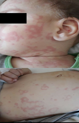
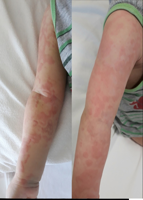
A 1-year-old male patient was admitted with an erythematous pruritic rash which had begun from his face one day before and spread to the arms, legs, and trunk. The patient without history of similar complaints, known systemic disease, recent vaccination or allergy was found to have been hospitalized shortly for febrile seizures and treated with ceftriaxone for three days ten days ago. Physical examination of the patient with insignificant family history revealed that his general condition was fine and the vital signs were stable. The patient had widespread, multiple, erythematosus target-like lesions 2-10 mm in diameter all over the body (Figures 1-3). He had no mucosal lesion. The laboratory work-up results were determined to be as follows: Hemoglobin: 9 g/dl, leukocytes: 12.000/ mm3, and platelets: 684.000/mm3. Liver-kidney function and coagulation tests were normal. C-reactive protein was 5.8 mg/L and procalcitonin 0.09 mcg/L. The patient had normal sedimentation rate, complement-immunoglobulin levels, and ANA profile. Anti-CMV IgM was found to be positive with 1.01 in the patient with negative serological tests for Mycoplasma pneumonia, HSV, Parvovirus B19, EBV, Hepatitis A, B, C viruses, and adenovirus.Our patient was monitored for symptoms for 5 days while on 1 mg/ kg/day of systemic metylprednisolone, 4 mg/kg/day of pheniramine, and 0.5 mg/kg/day of cetirizine. During the follow-up, time-to-time aggravations and alleviations in rashes were observed, with a significant remission of rashes within approximately 10 days. Given these findings, the patient was thought to develop EM caused by the CMV infection he was having.
Discussıon
Erythema multiforme (EM) is an immune-mediated disease characterized by acute, target-like skin lesions [6]. In EM minor, single mucosal ulceration is characterized by typical target lesions, whereas multiple ulcerations in the mucous membranes accompanying cutaneous target lesions in EM major [7].
The target lesions are characteristic lesions, though they may not always be present [8]. In EM, both cutaneous and mucosal lesions are usually present simultaneously. Furthermore, it may present with cutaneous lesions or mucous membrane involvement alone [9]. Cutaneous lesions usually begin symmetrically from extensor surfaces of the extremities and then spread [2]. In atypical cases, it may also involve the face, neck, palm soles, flexor surfaces of the extremities, or the trunk [8]. In our patient, there were cutaneous lesions alone without mucosal involvement. Target-like lesions were widespread all over the body. These lesions had begun on the face and then spread to other parts of the body. The rashes were observed predominantly on the flexor and extensor surfaces of the extremities.
The pathophysiology of EM has not been clearly understood; however, a keratinocyte injury induced by a cell- mediated immune response against an antigenic stimulus is being thought of. The released cytokines may lead to epidermal cell injury. The microscopic findings include the presence of dermo-epidermal lymphocyte infiltration and dyskeratotic keratinocytes [5]. Although the etiology cannot be clearly determined in some cases, infections, medications, and malignancies have been reported as the triggering factors in others [10].
EM is a reactive dermatosis, the etiology of which involves HSV and other infections, rather than drugs [11]. Phenytoin, Phenobarbital, carbamazepine, lamotrigin and valproic acid, among anticonvulsants, and many drugs, particularly penicillins, sulphonamides, isoniazide, kinolons, cefaclor and among antibiotics, have been found to be associated with EM [12]. Drugs are thought to act as an hapten, adhering to surface of keratinocytes and making them antigenic [13].
Since the patient had no rash after antibiotherapy administered during hospitalization and had rashes later, the rashes were not considered to be associated with the medication used. The work-up ordered revealed no pathology, but CMV infection, which was thought to cause the rashes.
In immunocompetent children and adolescents, CMV infections usually have an asymptomatic course. The disease usually is self-limiting and, therefore, does not require treatment mostly. However, it may present with different clinical manifestations which may lead to severe infections [14]. In the literature, a very limited number of cases of EM caused by CMV infections have been reported in immunocompetent individuals [15].Our patient is known to have no immune system concerns.
Although the laboratory findings in EM are usually normal, elevated levels of erythrocyte sedimentation rate, leukocyte count, acute phase reactants, and liver function tests may be observed in some severe cases [16]. The diagnosis of EM usually is established based on history and clinical findings, although a skin biopsy might be performed, when necessary, to confirm the diagnosis [6].
Our patient did not have a severe course, with the laboratory findings being normal in consistence with the literature. The diagnosis of our case was made based on history, physical examination and laboratory findings, without obtaining a skin biopsy.
Erythema multiforme usually is self-limiting, with healing within 2 weeks without any sequelae and supportive care is provided [5]. Symptomatic treatment is recommended for patients with skin-limited lesions or simple mucosal involvement. Topical corticosteroids and oral antihistamines are effective in eliminating the symptoms. Oral intake may be diminished due to pain in patients with severe oral mucosal involvement. These patients may require oral prednisone treatment and, when necessary, hospitalization [6].
Treatment with systemic corticosteroids is still controversial. However, steroid use has been shown to reduce the symptoms and have favorable effects on the prevention spreading of the lesions [17, 18, 19, 20]. We administered systemic methylprednisolone and antihistamine treatment for our patient, and followed-up for symptoms. It was observed that, during the follow-up, a favorable response was noted, and that the rashes completely vanished within 2 weeks. During this period, no sequelae were observed in our patient.
In conclusion, EM usually is a self-limiting disease, although it may also lead to severe clinical manifestations. In EM, the lesions usually heal within two weeks without any sequelae, and the treatment is supportive care. Antihistamines may provide symptomatic relief but do not have an impact on the disease course. Except in Mycoplasma pneumoniae and secondary bacterial infections, antibiotherapy is unnecessary. Steroid treatment in EM is controversial. Since its etiology has not yet been elucidated, it should be remembered that, although rare, CMV infections may be found in patients with suspected EM.
Conflict of Interest
The authors declared no conflicts of interest with respect to the authorship and/or publication of the article.
Financial Disclosure
The authors received no financial support for the research and/or publication of this article
References
-
Odom RB, James WD, Berger TG (2000) Diseases of the skin. In: Sounders WB(Eds.), 9th(Edn.), Philadelphia, pp: 146-171.
-
Weston WL, Brice SL, Jester JD, Lane AT, Stockert S, et al. (1992) Herpes simplex virus in childhood erythema multiforme. Pediatrics 89(1): 32-34.
-
Leaute Labreze C, Lamireau T, Chawki D, Maleville J, Taieb A (2000) Diagnosis, classification and management of erythema multiforme and Stevens Johnson syndrome. Arch Dis Child 83(4): 347-352.
-
Maquet J, Strull N, Quatresooz P, Bricteux G (2008) Erythema multiforme caused by a Mycoplasma infection: a case report. Rev Med Liege 63(1): 6-10.
-
Morelli JG (2007) Erythema multiforme. In: Kliegman RM, et al. (Eds.), Nelson Textbook of Pediatrics. 18th(Edn.), Philadelphia, USA, pp: 2685-2687.
-
Wetter DA (2021) Erythema multiforme: In: Jeffrey Callen Pathogenesis, clinical features, and diagnosis.
-
Erguven M , Yilmaz O, Yilmaz N (2007) Evaluation of clinical presentation and laboratory findings in patients with systemic Lupus Erythematosus. Journal of Child 7(4): 240-246.
-
Huff JC (1985) Erythema multiforme. Dermatologic Clinics 3(1): 141-152.
-
Bean SF, Quezada RK (1983) Recurrent oral erythema multiforme. Clinical experience with 11 patients. JAMA 249(20): 2810-2812.
-
Geraminejad P, Walling HW, Voigt MD, Seabury Stone M (2006) Severe erythema multiforme responding to interferon alfa. J Am Acad Dermatol 54(2): 18-21.
-
Warnock JK, Morris DW (2002) Adverse cutaneous reactions to antidepressants. Am J Clin Dermatol 3(5): 329-339.
-
Forman R, Koren G, Shear NH (2002) Erythema multiforme, Steven-Johnson Syndrome and toxic epidermal necrolysis in children: a review of 10 year’s experience. Drug Saf 25(13): 965-972.
-
Serdaroglu S, Uysal S (2002) Erythema multiforme. Dermatose 1: 9-15.
-
Demmler Harrison GJ (2019) Overview of cytomegalovirus infections in children. UpToDate Inc.
-
Vitiello M, Echeverria B, Elgart G, Kerdel F (2011) Erythema multiforme major associated with CMV infection in an immunocompetent patient. J Cutan Med Surg 15(2): 115-117.
-
Odom RB, James WD, Berger TG (2000) Eryhtema and Urticaria. In: Saunder WB, (Ed.), Andrew’s Disease of The Skin. 9th(Edn.), pp: 146-171.
-
Leaute Labreze C, Lamireau T, Chawki D, Taieb A (2000) Diagnosis, classification and management of erythema multiforme and Stevens Johnson syndrome. Arch Dis Child 83(4): 347-352.
-
Zoghaib S, Kechichian E, Souaid K, Soutou B, Helou J, et al. (2019) Triggers, clinical manifestations, and management of pediatric erythema multiforme: a systematic review. J Am Acad Dermatol 81(3): 813-822.
-
Petrosino MI, Attanasi M, Marcovecchio L, Scaparrotta A, Sabrina DP, et al. (2016) Erythema multiforme syndrome associated with acute acquired cytomegalovirus infection. Arch Med Sci 12(3): 684-686.
-
Melissa C, Goldman RD (2013) Erythema multiforme in children: the steroid debate. Can Fam Physician 59(6): 635-636.
- Understanding Pediatric Multiple Sclerosis: Clinical Presentation, Diagnostic Criteria, Therapeutic Advances, and Supportive Care Approaches
- Hemophilia in Children
- Xia-Gibbs Syndrome- A Case Report
- A Study to Assess Effectiveness of Play Therapy in Reducing Post-Operative Pain among Children Age 2 To 5 Year who have Undergone General Surgeries in Selected Pediatric Hospitals of Vadodara
- Preterm Birth: Scope of the Problem, Cost of Care, Potential Complications and Current Guidelines for Management
- Noradrenaline: Can we Use it to Manage Hemodynamic Instability among Neonatal Septic Shock at the NICU?