Abnormal Cytopathological Induced on Datura Metel L. Tissues Reveals Severe Damage after Potato Virus Y (PVY) Infection
Potato virus Y (PVY) is considered as one of the most important plant viruses affecting potato production and many other crops. The present study was conducted to investigate damage caused by PVY infection in Datura metel L., and demonstrated that changes induced by PVY within the infected leaf cells of Datura metel L. can affect the function of the cell. In this study ultra-transverse and a longitudinal thin section of electron microscopy were used and revealed that PVY infection caused significant damage in infected cell of Datura. This damage was pronounced in form of swollen and vacuolation of the mitochondria. Virus’s particles were found in the chloroplasts, mitochondria and in the cytoplasm of the aforesaid plant cells. Moreover, viral particles were forming a cluster in the cytoplasm and inclusion bodies were observed in the infected cell. However, nuclear and plasma membrane are disrupted. Digestion of nucleus chromatin was observed. This study showed that the mitochondria in the infected leaf of Datura metel L. with PVY was severely damaged due to viral infection and led to a rupture in the mitochondria membrane. Longitudinal ultra-thin sections clearly demonstrated that cell organelles in infected leaf of Datura metel L. were deformed after PVY infection, and this damage appeared in form of disintegration of the chloroplast, and decrease in numbers of mitochondria. This study illustrated the damage in infected cell with PVY and suggests this damage cause the external symptoms of PVY infection and lead to decrease in the growth and yield of the infected plants.
Introduction
Potato virus Y (PVY) is one of the most serious viruses infecting potato and other economic crops [1, 2, 3]. It has very wide host range include different solanaceous and non solanaceous crops [3, 4]. PVY causes serious diseases in many economic crops, especially potato as PVY affecting both yield and tuber quality and causes severe reduction in potato yield up to 80% [5, 6, 7, 8]. PVY is transmitted by many types of aphids in non-persistent manner [9].
PVY can induce severe symptoms in infected hosts, these symptoms include external symptoms like mottling, mosaic, leaf deformation, vein clearing and stunting, also can induce internal symptoms either cytopathological and histopathological changes including producing inclusion bodies, and inducing may changes in cell organelles especially mitochondria and nucleus [10, 11]. These changes induced in the cell affects the cell structure and cell function. This study was designed in order to study the cytological changes induced in Datura due to infection with severe PVY isolate. This PVY was originally isolated from Potato plant in Upper Egypt, and biological and molecular characterization proved this strain is necrotic stain PVY-NTN causing Potato tuber necrotic ring spot disease PTNRD in Upper Egypt [11, 12]. Results from this study will help to accurately assess the damage caused in plants infected with this severe PVY-NTN isolate.
Materials and Methods
Source of Viruses
Potato virus Y (PVY) was isolated and identified using serological and molecular tested described and molecular characterization of coat protein gene showed this strain is PVY-NTN [11, 12].
Electron Microscopy Examination
Crude sap from infected leaves was used. Drops from infectious sap were placed on electron microscope grids, which previously covered with collodion membrane. Grids were placed on the top of infectious sap drops for one min. and left to dry. After that, grids were placed on the drop of specific antiserum of the potato group viruses for 2 min. and then left to dry.
Negative Staining Technique
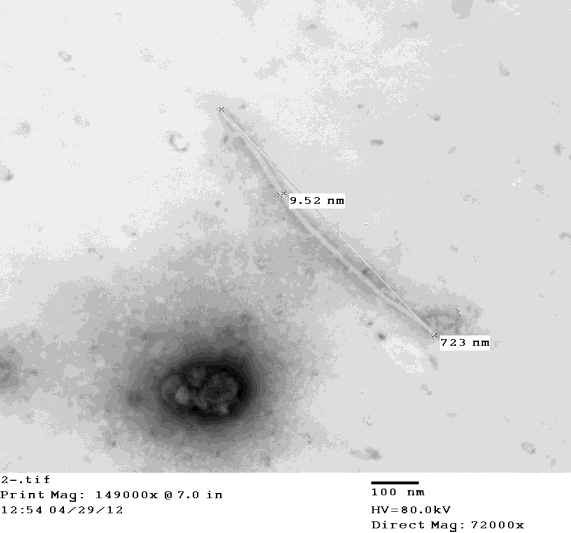
The grids were immersed in 2% phosphotungestic acid pH 7.0 for negative staining. Later the stained grids were raised with a fine tip forceps to the electron microscope for examination and description of the shape of the virus particles and to determine their dimensions. Electron microscope type (JEM 100CX11-EM) at The Electron Microscope Unit, University of Assiut, Assiut, Egypt was used in this study.
Ultra-Thin Sectioning Technique
A pieces of infected Datura leaves showing mosaic and mottling symptoms, which caused by y potato virus Y were cut (1mmX 1mm), fixed in 2.5 glutaraldehyde and in Epon812. Ultra-thin sections of the aforesaid materials thick (55-80 nm) were transversely cut with a glass knife and picked up on 200-mesh grid. Then, the section- mounted grids were positively stained with saturated aqueous urinal acetate 2% for 25 min, and followed by lead citrate for 5 min. After that the sections were observed and photographed under the electron microscope Type (JEM 100CX11-EM) at The Electron Microscope Unit, University of Assiut.
Results
In this experiment Datura metel L. had been used as indicator plant in order to elucidate the effect of the PVY virus on the infected plant cells. Datura metel L. plants 45 days in age were inoculated with PVY obtained from infected "Burna" potato cultivar, by dusting its leaves with 450 mesh carborunum and then infectious sap with cotton pad leaves were inoculated and sprayed with sterilized distilled water. These plants were kept in a greenhouse until the symptoms appeared on the new leaves.
Negative Staining of Potato Virus Y (PVY)
Some of infected leaves were collected for electron microscopy negative staining and ultra-thin sections.
Ultra-thin Sectioning Technique
Cytopathological structures induced in potato Burna variety or Datura metel L. tissues mechanically infected with Potato virus Y (PVY) revealed that severe ultra structural changes induced due Potato virus Y infection.
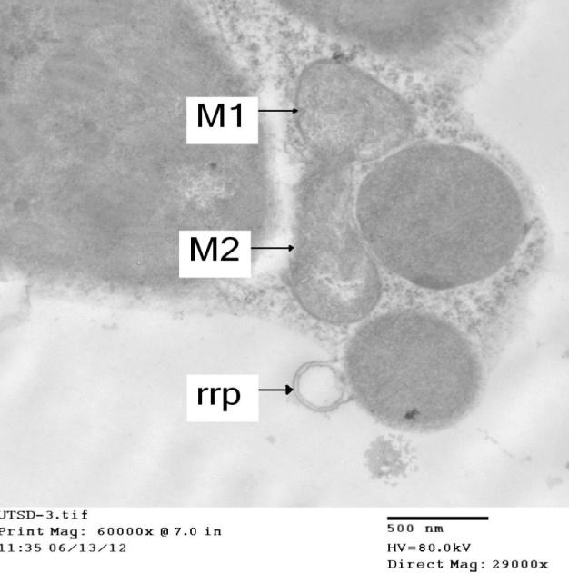
Figure 2: Ultra-thin cross section of electron micrograph: showing the effect of PVY on the cell organelles in infected leaf of Datura metel L. indicated that the mitochondria (M1) was damaged by viral infection . Note that the appearance of ruptures in the mitochondria membrane (M2) clearly demonstrates the lyses of its membrane in one side. However, (rrp) means appearance of ring-like profile was observed due to PVY infection.
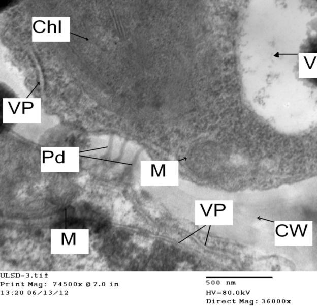
Figure 3: Electron micrograph of longitudinal ultra-thin section: showing the effect of PVY on the cell organelle in infected leaf of Datura metel L. Note that the disintegration of the chloroplast (Chl), little numbers of mitochondria (M) which its membrane greatly damaged due to viral infection. The appearance of filamentous virus particles (VP) of (PVY) near the chloroplast and the cell wall.
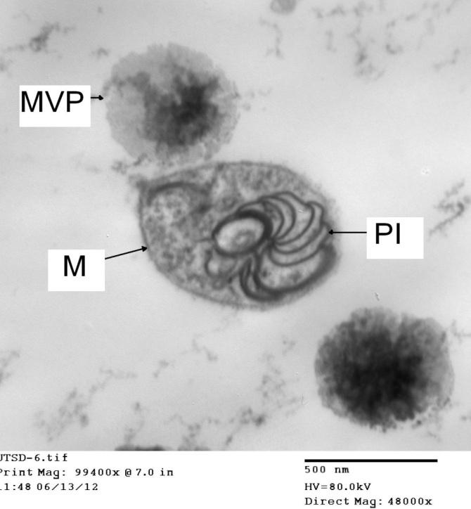
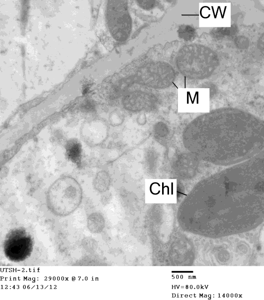
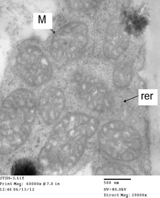
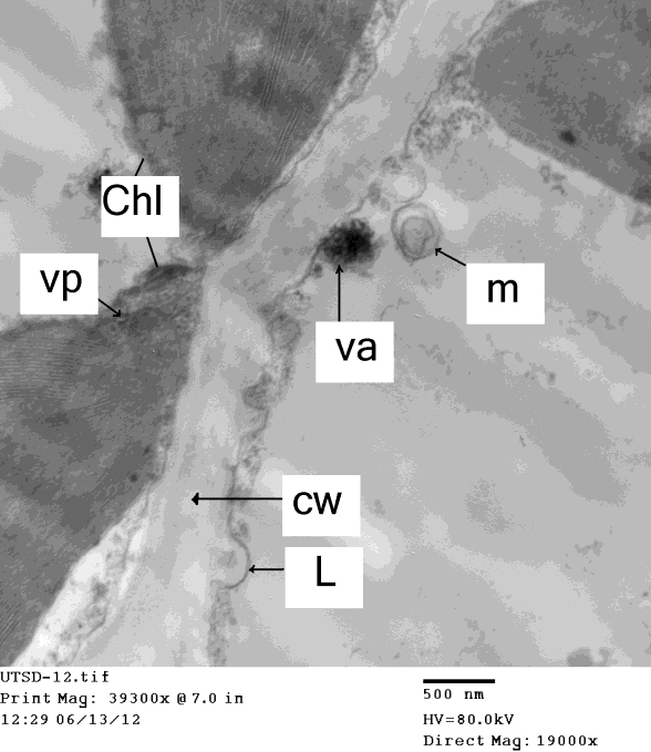
Figure 7: Electron micrograph of ultra-thin cross section showing the chloroplast (Chl) in leaf cell of Datura metel L. infected with PVY. Note that the presence of virus aggregate (va), vacuole due to the viral infection and appearance of micro bodies (m) and virus particles (vp) aggregate beside the cell wall (cw). Note also that the chloroplast beside the grana thylakoid (gt), and the appearance of babble (L) from cell wall due to viral infection, this phenomenon have been not shown in healthy tissues.
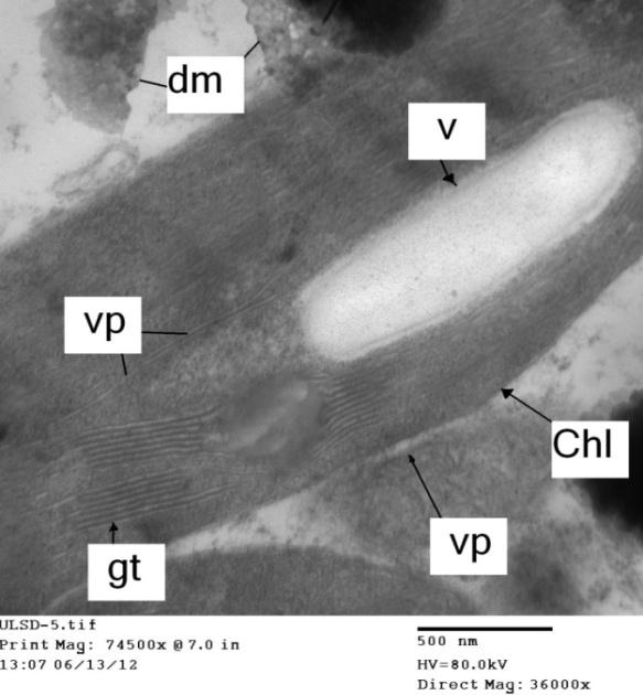
Figure 8: Electron micrograph of longitudinal ultra-thin section showing the effect of (PVY) on the cell organelles in infected leaf of Datura metel L. Note that the swelling of the chloroplast (Chl) and release of its matrix and forming visible vacuole-like structures (V) due to viral infection, .Note also that the appearance of filamentous virus particles (VP) of (PVY) in the iner matrix beside the grana and grana thylakoid (gt). However, viral particles had been shown also near the chloroplast in the cytoplasm. Note also that the viral infection caused mitochondria degeneration (dm) and also had damaged envelopes and lost cristae.
Discussion
The present study demonstrated that great ultra- structural changes induced by PVY within the infected leaf cells of Datura metel L. the sensitive host plant to PVY. Data had been shown that the swollen and vacuolation of the mitochondria was pronounced and this led to alternation within infected potato cells and also Datura metel L. plants. Meanwhile, virus's particles were found in the chloroplasts, mitochondria and in the cytoplasm of the aforesaid plant cells. Moreover, viral particles were forming a cluster in the cytoplasm. However, nuclear and plasma membrane are disrupted and also digestion of nucleus chromatin was happened. Data also showed the effect of PVY on the cell organelles in infected leaf of Datura metel L. with PVY which demonstrated that the mitochondria was damaged by viral infection as well as the appearance of ruptures in the mitochondria membrane. Data also clearly demonstrated that the lyses of its membrane in one side and appearance of ring-like profile was observed due to PVY infection. These results are in agreement with those obtained who indicated the damage and irregularly shaped cytoplasmic inclusions induced by (PVY). Datura metel L. which had been used as sensitive indicator plant to elucidate the cytopathological effects of PVY on the infected plant cells [13, 14, 15, 16]. These cytopathological changes when be detected by electron microscopy examination could be used as a rapid detection method for PVY viruses. The results of ultra-thin sections of infected Datura metel L. leaves with (PVY) are in the line of the results that described by [17]. Data also demonstrated that the viral infection caused mitochondria degeneration, these results were in agreement with the findings reported by [18]. In addition, the presence of papillae like-structure in the cell wall of infected cell plant with ( PVY) may be elucidate the another way for virus entering the cell wall to neighbor cell instead of plasmodesmata, such results in agreement with who indicated that plant microtubules, are being essential for cell-to-cell movement of plant viruses [19]. Furthermore, electron micrograph of ultra-thin cross sections showed that the appearance of pinwheel inclusions in infected leaf cell of Datura metel L. with PVY due to viral infection. These results were in accordance with the findings reported before who reported cytopathological effects due to virus infection [20, 21]. These ultra-structural changes induced by PVY showed a great damage in plant cells resulting a physiological disruptions (symptoms) correspond to changes in chlorophyllous accumulation (mosaics, yellowing and/or reddening), cell or tissue death (necrotic lesions) or tissue distortion (dwarfing, leaf roll, and swelling). And such results also endorse that this plant indicator is very useful for showing the whole damage incited by PVY in infected cell.
References
-
Valkonen JPT (2007) Viruses: economical losses and biotechnological potential. In: Vreugdenhil D, ed. Potato Biology and Biotechnology. New York, NY, USA. Elsevier 619-641.
-
Quenouille J, Vassilakos N, Moury B (2013) Potato virus Y: major crop pathogen that has provided major insights into the evolution of viral pathogenicity. Molecular Plant Pathology 14(5): 439-452.
-
Dupuis B, Cadby J, Goy G, Tallant M, Derron J, et al. (2017) Control of Potato virus Y (PVY) in seed potatoes by oil spraying, straw mulching and intercropping. Plant Pathology 66(6): 960-969.
-
Jeffries CJ (1998) FAO/IPGRI technical guidelines for the safe movement of germplasm Potato. 19: 62-63.
-
Lorenzen JH, Meacham T, Berger PH, Shiel PJ, Crosslin JM, et al. (2006) Whole genome characterization of Potato virus Y isolates collected in western USA and their comparison to isolates from Europe and Canada. Archive of Virology 151(6): 1055-1074.
-
Nolte P, Whitworth JL, Thornotn MK, McIntosh (2004) Effect of Seedborne Potato virus Y on Performance of Russet Burbank, Russet NorKotah, and Shepody potato. Plant Disease 88: 248-252.
-
Kerlan C, Nikolaeva OV, Hu X, Meacham T, Gray SM, et al. (2011) Identification of the molecular make-up of the Potato virus Y strain PVY(Z): genetic typing of PVY(Z)-NTN. Phytopathology 101(9):1052-1060.
-
Cuevas JM, Delaunay A, Visser JC, Bellstedt DU, Jacquot E, et al. (2012) Phylogeography and Molecular Evolution of Potato virus Y. PLOS one 7: 1- 10.
-
Woodford JAT (1992) Virus Transmission by aphids in potato crops. Netherlands Journal of Plant Pathology 98(2): 47-54.
-
Moury B, Morel C, Johansen E, Jacquemon M (2002) Evidence for diversifying selection in Potato virus Y and in the coat protein of other Potyviruses. Journal of general Virology 83(Pt 10): 2563-2573.
-
Abdalla OA, Eraky AI, Mohamed SA, Fahmy FG (2016) Phylogenetic analysis of Potato virus Y (PVY) isolate from Upper Egypt proves the widespread of PVY-NTN strain causing PTNRD disease in Egypt. Annals of Virology and Research 2(3): 1020.
-
Abdalla OA, Eraky AI, Mohamed SA, Fahmy FG (2016) Molecular identification of viruses responsible for severe symptoms on potato (Solanum sp.) growing in Assiut Governorate (Upper Egypt). International Journal of Virology Studies and Research 4(3): 29-33.
-
M'murungl JM (1982) Potato virus S in potatoes (Solanum tuberosum L.) in Kenya. Master of Science Thesis in Plant Pathology. Plant Science & Crop Protection, University of Nairobi.
-
Baker KK, Ramsdell DC, Gillett JM (1985) Electron microscopy: current applications to plant virology. Plant Disease 69: 85-90.
-
Milne RG (1993) Electron microscopy of in vitro preparations. in Diagnosis of plant virus diseases, edited by R.E.F. Matthews. CRC press, Boca Raton, Florida, USA. 215-251.
-
Urbanaviciene L, Zitikite I (2010) Molecular identification of potato X virus in Lithuanian varieties of Solanum tuberosum L. and Lycopersicon esculentum Mill crops. Biologija 56(1-4): 20-23.
-
Kahan RP, Monroe RL (1970) Datura metel L. as a Virus-Indicator Plant. Phytopathology 60: 1183-1185.
-
Edwardson JR, Christie RG, Purcifull DE, Petersen MA (1993) Inclusions in diagnosing plant virus diseases. In Diagnosis of plant virus diseases, edited by R.E.F. Matthews. CRC Press, Boca Raton, Florida, USA. 101- 128.
-
Takemoto D, Hardham AR (2004) The Cytoskeleton as a Regulator and Target of Biotic Interactions in Plants. Physiology 136(4): 3864-3876.
-
Zechmann B, Müller M, Zellnig G (2003) Cytological modification in Zucchini yellow mosaic virus (ZYMV)- infected pumpkin plants. Archives of Virology 148(6): 1119-1133.
-
Navarro B, Russo M, Pantaleo V, Rubino L (2006) Cytological analysis of Saccharomyces cerevisiae cells supporting cymbidium ring spot virus defective interfering RNA replication. Journal of general Virology 87(Pt 3): 705-714.
- hMPV: Is It Another Covid-19 Like Situation?
- Streptomyces: Sources of Novel Discoveries in Antibiotic Research to Combat Antimicrobial Resistance
- A Review of Mosquitoes (Diptera: Culicidae) and Their Biodiversity, Medical and Veterinary Importance
- Past and Current Immunotherapy in Cancer
- Hematological Cancer and Viral Infection
- The Growing Threat of Antimicrobial Resistance in India: Challenges and Solutions