Histoid Leprosy –A Case Report
Histoid leprosy is a rare variant of lepromatous leprosy with unique clinical, immunological, bacteriological &histopathological features. It can occur as relapse following dapsone monotherapy, incomplete therapy, resistance to antileprosy drugs, following MDT & may in rare occassion de-novo. Here, we are presenting a case of histoid leprosy with candidal onychomycosis in middle aged army personnel who had taken incomplete therapy earlier.
Introduction
Histoid leprosy is an uncommon variant of lepromatous leprosy [1] with unique clinical, immunological, bacteriological & histopathological features. Clinically, it is characterized by multiple discrete shiny, smooth, firm, skin colored to yellow brown, cutaneous and or subcutaneous papules, nodules and or plaques on apparently normal skin. Immunologically, it is characterised by increased cell mediated and humoral immunity against M. leprae with focal loss of immunity. Bacteriologically, bacilli are longer than the normal, are uniform in length, more often solid with tapering ends and are arranged in parallel bundles along the long axis of the histiocytes (histoid habitus) without globus formation. Histopathologically, fusiform histiocytes are seen arranged in a whorled, criss-cross, or storiform pattern [2, 3]. It can occur as relapse following dapsone monotherapy, incomplete therapy, resistance to antileprosy drugs, following multidrug therapy (MDT) and may in rare occasion arise de novo [4] but the exact etiopathogenesis is not clarified yet [5]. We are reporting here a case of histoid leprosy with candidal onychomycosis involving all twenty nails in middle aged army personnel who presented with itchy papulonodular eruptions all over body.
A 44-yr-old army man from Punjab, staying at Manipur since five years presented with diffuse itchy nodular lesions all over body associated with dystrophic changes of all twenty nails for 5 months. He had received multibacillary (MB) MDT therapy for 3 months only, 5 yrs back. There was no history of paraesthesia, weakness, epistaxis, constitutional symptoms and lymphadenopathy. None of the family members or colleagues had similar condition. He didn’t have any systemic disorders neither was he on any medication. He was a vegetarian, smoker and occasional alcoholic. On examination, he had bilateral upper and lower limb edema with no other significant systemic findings. Skin was infiltrated over face and forehead with numerous shiny to skin colored, nontender, firm, papules, nodules and few plaques of varying sizes present all over the body (Figures 1a & 1b).
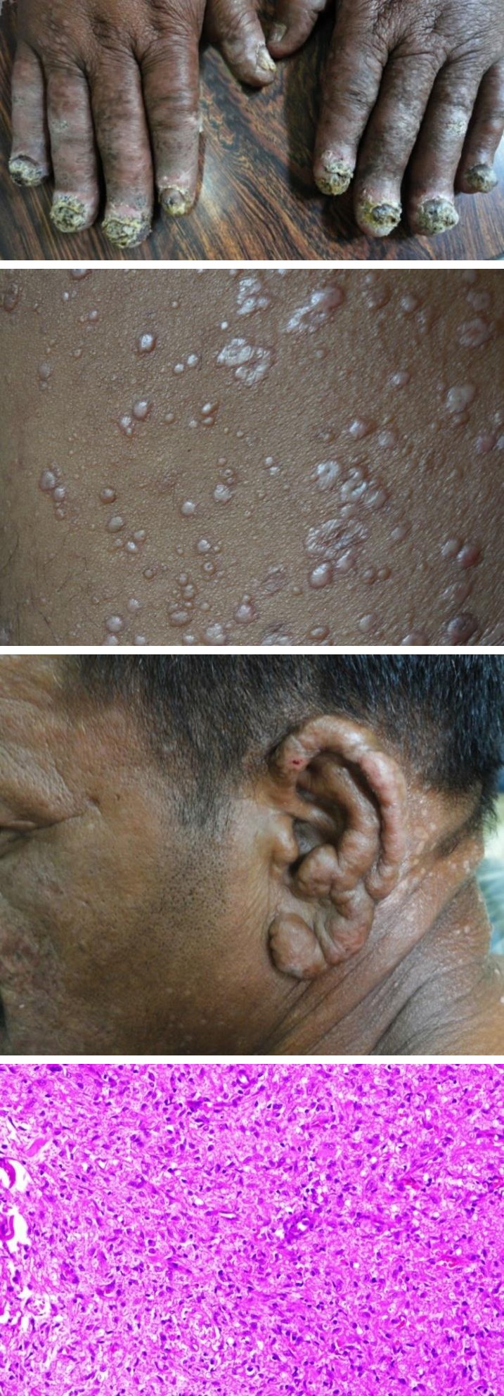
Figure 1a: Showing shiny papulonodular lesions and plaques over trunk.
Figure 1b: Showing infiltrated ear lobules.
Figure 1c: Showing nail dystrophy. Some lesions had central indentation and some were excoriated. Xerosis was present over bilateral lower limbs. Pain, touch and temperature sensations were intact. He had leonine facies, bilateral thickened earlobes and madarosis. Palms, soles and mucosa were spared. On neurological examination, there were no palpable or thickened nerves, neither muscle weakness nor any paraesthesia. There was no orchitis, no lymphedenopathy and no ocular features. Nails showed onycholysis, hyperkeratosis, subungual debris and dystrophy. All the nail folds (proximal and lateral) were distorted, lunula and cuticle were lost. All the twenty nails were affected. Finger nails were affected more than toe nails (Figure 1c). Routine laboratory investigations were normal except raised ESR (70mm/1st hour), Mantoux test and ELISA test for HIV were negative. Slit Skin smear (SSS) examination from nodule and normal skin showed numerous acid fast bacilli present singly and in clumps with bacteriological index (BI) of +4. Histopathological examination (HPE) of biopsy from nodule showed atrophied flattened epidermis and dermis with numerous spindle shaped histiocytes in storiform pattern (Figure 2a). Fite-Faraco stain revealed AFB within the histiocytes (Figure 2b).
Figure 2a: H.P.E. (H&E, 10x) showing diffuse sheets of histiocytes in storiform pattern.
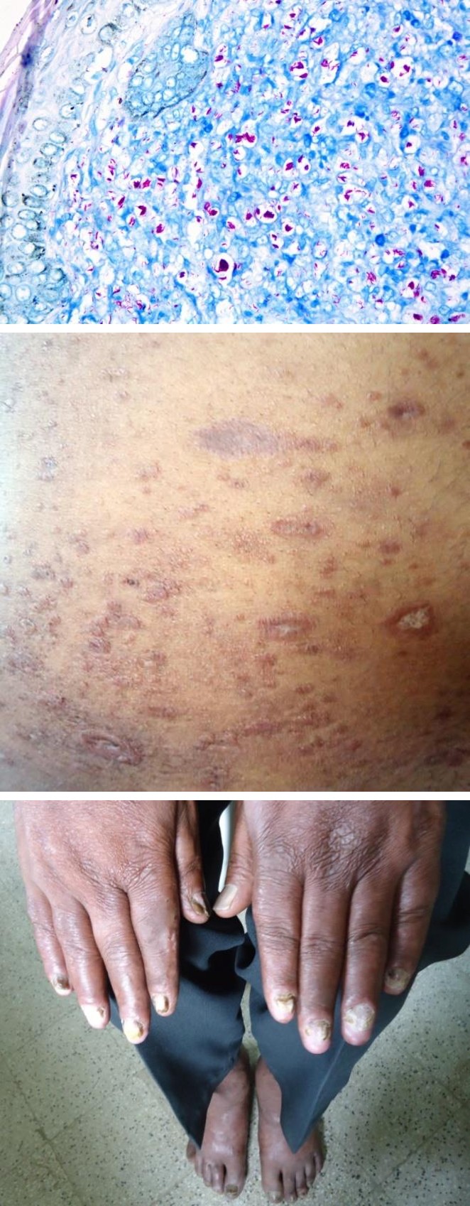
Figure 2b: Wade Fite Faraco stain showing lepra bacilli.
Figure 3a: At 2 months of treatment.
Figure 3b: At 3 months of treatment.
Potassium hydroxide mount (KOH) of nails showed numerous pseudohyphae with spores. Fungal nail culture showed growth of candida albicans. Patient was diagnosed as a case of histoid leprosy with candidal onychomycosis. He was started on MB-MDT with oral fluconazole 300mg weekly to be given for 3months and amorolfine nail lacquer with clinical improvement at three weeks. He was on regular follow-up for few months with significant improvement (Figures 3a & 3b) until he got transferred to another state but was advised to complete MB-MDT for a year and was refered to the nearest leprosy centre for further check-up.
Discussion
Histoid leprosy is a rare variant of lepromatous leprosy, covering 2.79-3.60% of all leprosy. It was originaly described by Wade for the first time in 1960. It commonly occurs at age group of 21-40 yrs with a wide range of 10-80 years with male preponderance [1]. Sites commonly affected are posterolateral aspect of arms, dorsum of hands, thighs, buttocks, lower back and bony prominences. In severe cases mucosa and genitalia may also be involved. The sites usually spared are the palms and soles [2]. An excoriated lesion found in our case was due to itching which could be due to increased proliferation of mast cells and their degranulation in the histoid nodule [6]. The bacilli were believed to be a mutant variant of leprae bacilli. They are not found in globi as they do not secrete glial substance [1, 3] though in our case we found globi in SSS examination though HPE showed spindle shaped histiocytes in storiform pattern. The lesion of histoid leprosy is due to altered growth pattern of bacilli probably due to loss of immunity in local area [7] or there can be interplay of genetic factors, immune response and treatment received in a given patient that seems to influence the manifestations of the disease. Immunohistochemical study revealed increased cell mediated immunity (CMI), with CD4 cells, other activated lymphocytes and macrophages in the lesions. The high number of macrophages, claimed to be due to lack of the capacity to kill bacilli that exist in lesions can be due to reduced bacteriolytic property or production of 'suppressor' cytokines, such as interleukin-10 under the influence of bacillary antigens as IL-10 adversely inhibit T cell-mediated responses to M. leprae [8].
Histoid leprosy clinically simulates molluscum contagiosum, dermatofibromas, xanthomas, neurofibromas, reticulohistiocytosis and cutaneous metastasis. Differentiation can be made by nerve thickening, acid fast bacilli on SSS and characteristic histopathology [1]. HPE may show sub epidermal Grenz
zone with dermis having sheets of round to spindle shaped histiocytes in storiform pattern with vacuolated cytoplasm. Histiocytes here are produced by local multiplication in response to stimulation by rapidly proliferating AFB rather than from circulating monocytes [9]. Histopathologically, histoid leprosy resembles neurofibromatosis, dermatofibromas and fibrohistocytomas [1]. Nail changes in leprosy have similar incidence in both multibacillary (86%) and paucibacillary patients (86%) and could be related to one or more of the following: neuropathy, endarteritis, trauma, drugs or superimposed infections. Patients with leprosy may have a higher prevalence of onychomycosis. This could in part be related to cellular immune dysfunction. Communal housing may contribute to the development of onychomycosis. Adults over 40 years are more likely to have onychomycosis, possible reasons are slower rates of outgrowth and larger surface area of the nail plate in adults compared to children. Adults also have more cumulative trauma and micro trauma to the nail and greater likelihood of peripheral vascular disease [10]. Our patient was diagnosed by history, clinical feature, positive slit skin smear, KOH mount and characteristic histopathological picture.
Conclusion
The case is being reported for its rarity and as it can be a source of infection to the society especially in this post leprosy elimination era since the bacillary load is very high in histoid leprosy. Reason for incomplete treatment could be non-compliance (improper counseling), non-availability of drugs which should be taken care of in leprosy clinic. Apart from pharmacological treatment, it is important to perform contact tracing, rehabilitation and education of the patients to prevent deformity and long-term disability.
References
-
Manoharan R, Madhu R, Srinivasan MS (2008) Histoid Hansen – A Case report. e-Journal of the Indian Society of Teledermatology 2(2): 12.
-
Dimri D, Sethi B, Kumar Y (2012) De Novo Histoid Leprosy in an Elderly: A Case Report and Review of the Literature. Case Reports in Pathology 219421.
-
Murthy SV, Rao SM, Thejaswini, Mannan K (2011) Denovo Histoid leprosy. J Lab Physicians 3(2): 110- 112.
-
SR annigeri, SC Metgud, JR Patil (2007) Lepromatous leprosy of histoid type. Indian J Med Microbiol 25(1): 70-71.
-
Kontochristopoulos GJ, Aroni K, Panteleos DN, Tosca AD (1995) Immunohisto-chemistry in histoid leprosy. Int J Dermatol 34(11): 777-781.
-
Kumar R (1987) Mast cells in histoid lepromatous lesions. Indian J Lepr 59(4): 390-392.
-
Jena S, Swain JP, Mishra S (2005) Fnac study of histoid nodule: an early tool for diagnosis. Indian J Lepr 77(2): 135-139.
-
Kaur I, Dogra S, De D, Saikia UN (2009) Histoid leprosy: a retrospective study of 40 cases from India. Br J Dermatol 160(2): 305-310.
-
Job CK, Chacko CJ, Taylor PM (1977) Electromicroscopic study of histoid leprosy with special reference to its histogenesis. Lepr India 49(4): 467-471.
-
Gupta AK, Konnikov N, Lynde CW, Summerbell RC, Albreski D, et al. (1999) Onychomycosis: predisposed populations and some predictors of suboptimal response to oral antifungal agents. Eur J Dermatol 9(8): 633-638.
- Epithelioid Granuloma; 3cases with Different Clinical Features
- Advancing Representation in Dermatology Clinical Trials: Ethical, Scientific, and Regulatory Imperatives for Inclusion Across all Fitzpatrick Skin Types
- A Case of Atopic Dermatitis with Concurrent Psoriasis Vulgaris: Successful Treatment with Upadacitinib
- Innovation Lifting Eyeshadow: A Synthesis of Makeup and Optical Illusion
- Distinguishing Superficial Actinic Porokeratosis from Actinic Keratosis with UVF Dermoscopy: A Case Report
- High Mobility Group Box 1 (HMGB1) in Cutaneous Inflammation: An Immune Modulator Bridging Cellular Stress, Ferroptosis and Danger Signaling