Case Report: Cutaneous Leishmaniasis
Cutaneous leishmaniasis is a parasitic disease caused by over more than 20 Leishmania species, in both tropical and subtropical regions. There are three main forms of the disease: visceral leishmaniasis (VL), Cutaneous Leishmaniasis (CL) and Mucocutaneous Leishmaniasis (MCL), Leishmania braziliensis predominates in Guatemala. We present a case study of a 15-year-old female patient, referred from the Jutiapa hospital for presenting a lesion in the right ear of 3 months of evolution. When interviewing the patient, she mentions that several months ago she presented a "wheal" on her ear after being bitten by a mosquito, which turned red, grew and then she saw it open and form an ulcer with scabs on the ear. surface, which is not painful. The Giemsa-stained rub revealed the presence of Leishman bodies and the biopsy revealed amastigotes within the histiocytes, thus confirming the initial diagnosis of cutaneous Leishmaniasis. Due to the above, treatment with glucantime was started at a dose of 20mg/kg for 20 days, which improved the condition and resolved it. The patient's sister had the same condition, which was confirmed using the same techniques and is treated with what also resolves it. It is important to take cutaneous leishmania into account as a differential diagnosis in patients who present painless ulcers despite not being in a precisely endemic area.
Introduction
Cutaneous leishmaniasis is a parasitic disease which is endemic in tropical and subtropical countries. This pathology is transmitted by a managed sandfly mosquito. Leishmania braziliensis predominates in Guatemala; however, the patient presented in this case does not come from any endemic department, and she was not the only member of the family affected, since her sister also presented this same lesion.
Clinical Case
A 15-year-old female patient referred for presenting a lesion in the right ear of 3 months of evolution. When interviewing the patient, she reports that several months ago she presented a “wheal” in this place after being bitten by a mosquito, she reports that since its appearance it has been asymptomatic, which turned red, grows and forms an ulcer with crusts on the surface, which asymptomatic. She has applied various triple creams to this injury, however, it does not improve and continues to grow, so she consults the Hospital of Jutiapa, Guatemala, who refer him to the San Juan de Dios General Hospital.
On physical examination, she presented a dermatosis located on the head that affected the right ear, characterized by a 2x3cm ulcer, defined edges, covered by multiple scabs, located on normal skin, chronic evolution, asymptomatic. All records were denied. The clinical impression is of cutaneous leishmaniasis due to the history and findings of the physical examination. Bearing the above in mind, a rub and incisional punch biopsy is requested (Figure 1).
The Giemsa-stained rub revealed the presence of Leishman bodies and the biopsy revealed amastigotes within the histiocytes, thus confirming the initial diagnosis
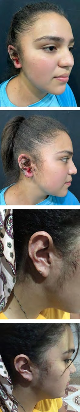
$$ \mathrm {E} = \frac {1}{2} \mathrm {A} ^ {2} + \frac {1}{2} \mathrm {B} ^ {2} + \frac {1}{2} \mathrm {C} ^ {2} $$ Figure 1: First consult.
Figure 2 One year after.
Discussion
Cutaneous leishmaniasis is a parasitic disease which is endemic in tropical countries. This pathology is transmitted by an infected sandfly mosquito. In Guatemala, Leishmania braziliensis predominates; however, the patient presented in this case does not come from any endemic department, and she was not the only member of the affected family, since her sister also had the same lesion [1]. In Guatemala, leishmaniasis continues to be a public health problem, being endemic in the departments of Petén, Alta Verapaz, Izabal, Quiché, Huehuetenango, and El Progreso [2].
of cutaneous Leishmaniasis. Due to the above, treatment with Meglumine Antimoniate was started at a dose of 20mg/kg for 20 days, which improved the condition and resolved it. It is worth mentioning that the patient’s sister had the same condition, which was confirmed using the same techniques and is treated with what also resolves it (Figure 2).
$$ \mathrm {E} = \frac {1}{2} \mathrm {A} ^ {2} + \mathrm {B} ^ {2} $$
$$ \mathrm {E} = \frac {1}{2} \mathrm {A} ^ {2} + \mathrm {B} ^ {2} $$
It is transmitted by the bite of infected female phlebotomine sandflies (infective stage: promastigotes), promastigotes phagocytized by macrophages and other types of mononuclear phagocytic cells and then they transform in these cells into the tissue stage of the parasite (i.e., amastigotes). Parasite, host and other factors affect whether the infection becomes symptomatic and whether it results in cutaneous or visceral leishmaniasis. Sandflies become infected by ingesting infected cells during blood meals. In sandflies, amastigotes transform into promastigotes, develop in the gut, then migrate to the proboscis. Though, no confirmed cases have been found in the literature in the region where the patient presented in this case was affected. It is important to take cutaneous leishmaniasis into account as a differential diagnosis in patients who present painless ulcers despite not being in a precisely endemic area [2]. Likewise, In Guatemala, this pathology has occurred at different ages, so it is important to keep it in mind as a differential diagnosis.
There are multiple treatments that can be used like organic salts of pentavalent antimony, antimoniate of N methylglucamine and sodium stibogluconate, Amphotericin B and lysosomal preparation, Miltefosine. In this case report, we used Meglumine Antimoniate and the patient had a great outcome.
References
-
Arenas R (2015) Dermatología Atlas, diagnóstico y tratamiento. 6th (Edn.), México D.F./Dermatología en medicina general, Fitzpatrick. Editorial médica panamericana.
-
Chang P, Marroquín R, Barillas S, Ovalle J, Samayoa T (2021) Leishmaniasis cutánea en extremidad superior. Rev méd (Col Méd Cir Guatem) 160(2): 191-193.
- Epithelioid Granuloma; 3cases with Different Clinical Features
- Advancing Representation in Dermatology Clinical Trials: Ethical, Scientific, and Regulatory Imperatives for Inclusion Across all Fitzpatrick Skin Types
- A Case of Atopic Dermatitis with Concurrent Psoriasis Vulgaris: Successful Treatment with Upadacitinib
- Innovation Lifting Eyeshadow: A Synthesis of Makeup and Optical Illusion
- Distinguishing Superficial Actinic Porokeratosis from Actinic Keratosis with UVF Dermoscopy: A Case Report
- High Mobility Group Box 1 (HMGB1) in Cutaneous Inflammation: An Immune Modulator Bridging Cellular Stress, Ferroptosis and Danger Signaling