Multiple Eruptive Dermatofibromas in a Healthy Female: A Case Report
Dermatofibromas (DF) are benign fibrohistiocytic tumours usually manifesting as solitary brown nodules on lower limbs of young-middle aged females. Multiple DFs are rare and associated with autoimmune conditions and immunosuppression. We report a case of multiple DFs in a healthy 45 year old female. A 45 year old female presented with asymptomatic brown hyperpigmented nodular lesions all over her body. The lesions first appeared on her back 12 years ago and have been progressively increasing in number to involve the rest of her body. Dimple sign was positive. Histopathology revealed acanthosis, large grenz zone with dense collection of fibro-histiocytes in dermis. Family history was negative and routine investigations, HIV, autoimmune and malignancy workup revealed no underlying abnormalities. Patient was counselled regarding benign nature of the tumor and is under regular follow up. Dermatofibromas are benign dermal and subcutaneous proliferations of histiocyte-like and fibroblast-like cells. Multiple DFs is defined as presence of >15 lesions and occur in patients of autoimmune diseases, immunosuppressed states and hematologic malignancies. It is hypothesized that DF arise due to an immune reactive process in which dermal antigen presenting cells activate immune system in response to these conditions. Our case of multiple DFs occurring in healthy female shows that they are not necessarily reactionnal tumors. An extensive workup to rule out any underlying condition should be done in all cases but may not necessarily reveal an abnormality.
Abbreviations
DF: Dermatofibromas; HIV: Human immunodeficiency viruses; MDF: Multiple Dermatofibromas; MEDF: Multiple Eruptive Dermatofibroma; MCDF: Multiple Cluster Dermatofibroma; GCDF: Giant Combined Dermatofibromas.
Introduction
Dermatofibromas are benign tumors clinically seen as hyperpigmented nodules on lower limbs of young- middle aged females. Multiple dermatofibromas are rare and associated with autoimmune conditions and immunosuppression. We report a case of multiple eruptive dermatofibromas in a healthy 45-year-old female.
A 45-year-old female presented with brown lesions all over her body (Figures 1A & 1B). The lesions first appeared on her back 12 years ago and have been progressively increasing in number to involve the rest of her body. They are asymptomatic. On examination, well defined brown hyperpigmented nodules were noted on her bilateral upper limb, lower limb and trunk. Dimple sign was positive. Family history was negative and there were no systemic complaints. Histopathological examination from one of the lesions revealed acanthosis, large grenz zone with dense collection of fibro-histiocytes in dermis. Routine investigations, HIV, autoimmune workup and malignancy screen revealed no underlying abnormalities. Patient is under observation as none of the lesions was causing any symptoms.
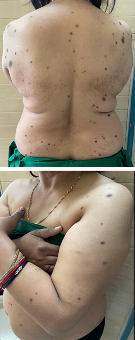
Figure 1A: Hyperpigmented nodules on back of a 45 year old female.
Figure 1B: Hyperpigmented nodules on upper limb of a 45 year old female.
Dermatofibromas are benign dermal and subcutaneous proliferations of histiocyte-like and fibroblast-like cells commonly seen in middle aged females. They are usually solitary, however if there are >15 lesions it is termed as multiple dermatofibroma.1 Multiple dermatofibroma is rare and has four subtypes types: multiple dermatofibromas (MDF), multiple eruptive dermatofibroma (MEDF), multiple cluster dermatofibroma (MCDF), and giant combined dermatofibromas (GCDF) [1]. Multiple Eruptive Dermatofibromas is defined as >5 lesions appear in a span of 4 months [2]. Multiple dermatofibromas usually occur in patients of autoimmune diseases (systemic lupus erythematosus, dermatomyositis, Sjögren syndrome), immunosuppressed states (drugs, HIV infection) and hematologic malignancies.2 Rare reports of association with pulmonary hypertension, atopic dermatitis, Down’s syndrome, hypertriglyceridemia and pregnancy have been mentioned in literature. Some authors believe dermatofibromas to be result of an immune reactive process in which dermal antigen presenting cells activate immune response to a stimulus [3].
Clinically, well defined, firm reddish-brown nodules are seen commonly on the extremities although any part of body may be involved. They may be asymptomatic or associated with pain or pruritis. Upon applying pressure, there is central dimpling over the lesion known as “Dimple sign”.
Histopathologically, localized proliferation of fibrohistiocytic cells are seen within the dermis which may extend to subcutaneous tissue. Epidermis may show hyperkeratosis, acanthosis and increased basal layer pigmentation with an underlying grenz zone. At the periphery of the lesion, entrapped collagen bundles are a characteristic finding. A central white scar like patch with delicate pigment network at the periphery is seen on dermoscopy, although many other patterns have been reported [4].
There is no specific treatment and usually only observation is recommended for multiple dermatofibromas. Excision may be done for lesions causing symptoms or cosmetically disfiguring.
Conclusion
Our case of multiple dermatofibromas occurring in a healthy female shows that multiple eruptive dermatofibromas are not necessarily reactionnal tumors linked to immune dysregulation and some other etiopathogenetic mechanisms may be involved in their development.
Conflict of Interest
None
Patient consent
Written informed consent was obtained from the parents for publication of this case report and accompanying images.
Ethics approval
Not required
References
-
Yao MX, Wang YT, Zhou NH (2024) Multiple Eruptive Dermatofibroma: A Case Report. Clin Cosmet Investig Dermatol 17: 457-461.
-
Haber JS, Meehan S, Orlow SJ (2022) Multiple eruptive dermatofibromas in an adolescent with a history of pityriasis lichenoides et varioliformis acuta. JAAD Case Rep 21: 26-28.
-
Samlaska C, Bennion S (2002) Eruptive dermatofibromas in a kindred. Cutis 69(3): 187-190.
-
Camara MF, Pinheiro PM, Jales RD, Trindade Neto PB, Costa JB, et al. (2013) Multiple dermatofibromas: dermoscopic patterns. Indian J Dermatol 58(3): 243.
- Epithelioid Granuloma; 3cases with Different Clinical Features
- Advancing Representation in Dermatology Clinical Trials: Ethical, Scientific, and Regulatory Imperatives for Inclusion Across all Fitzpatrick Skin Types
- A Case of Atopic Dermatitis with Concurrent Psoriasis Vulgaris: Successful Treatment with Upadacitinib
- Innovation Lifting Eyeshadow: A Synthesis of Makeup and Optical Illusion
- Distinguishing Superficial Actinic Porokeratosis from Actinic Keratosis with UVF Dermoscopy: A Case Report
- High Mobility Group Box 1 (HMGB1) in Cutaneous Inflammation: An Immune Modulator Bridging Cellular Stress, Ferroptosis and Danger Signaling