Clinical Anatomy of the Face
<p>Replacement and redistribution of face volumes is an essential part of facial aesthetic treatment, however, updated concepts of the aging process and the anatomy on which treatment is based are still confused and conflicting in the literature. The authors summarize the updated concepts of the aging process and anatomy, based on recent literature, clinical experience and the author’s fresh cadaver dissection study.</p>
Introduction
The most recent advances in facial aesthetics treatment were the better understanding of facial natural aging process, and better comprehension of facial anatomy. With this knowledge, aesthetic and reparative facial treatments advanced, both in plastic surgery and in dermatology [1, 2, 3]. The present paper summarizes the latest concepts of the aging process and anatomy, and this comprehension may enable increased therapeutic possibilities. It is based on recent literature, clinical experience and the author’s fresh cadaver dissection study.
Unaesthetic Characteristics
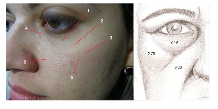
There is a lot of confusion between rejuvenating and beautifying, and many unaesthetic facial characteristics are not due to aging [1]. The face is formed by the reunion of many organs, the eyes, the nose, the mouth and the ears. Beauty is related not only to each organ feature, but also to their relationship and the harmony of the elements as a whole. Even though these organs are contiguous, they are not continuous, and each organ is an anatomically and functionally independent structure. Each facial region has different skin and subcutaneous tissues from the adjacent areas, with or without underlying muscle, which may also vary in strength and vector. Furrows, folds and wrinkles usually appear at the transitional limits of these anatomical units [2]. For example, the malar region is one aesthetic unit, with rounded and smooth surface [1]. However, anatomically, it comprises three distinct regions: the thin-skinned orbital region over the orbicularis oculi muscle, with sparse subcutaneous tissue; the cheek region with thick skin and subcutaneous adipose compartments on top of the superior lip elevator muscles; and the zygomatic region, of intermediate thickness skin, on top of the subcutaneous adipose compartment, orbicular oculi muscles and origin of the greater zygomatic muscle on the zygomatic bone prominence [1, 2, 4]. The boundary between the orbital, the nasal, and the buccal regions is the nasolacrimal sulcus. The zygomatic region is separated from the oral region by the buccal zygomatic groove and from the orbital region by the palpebromalar groove [5] (Figures 1 and 2). These characteristics are already present in the youth, butonlybecome more evident with excess skin and with hypertrophy and atrophy of the fat compartments, and with skin aging itself1. The aesthetic treatment consists in making these three anatomical structures in a single homogeneous aesthetic unit, which is of extreme difficulty [1] (Figure 3).
![Figure 2: Aesthetic treatment of the malar area: uniting the three anatomical structures into one homogeneous aesthetic unit. Preoperative of autologous lipoaspirate graft (left); 2 years postoperative (right). Gravity was thought to cause a lower positioning of anatomical structures, and the "gravitational" theory was created to explain aging. Today, it is known that aging is genetic, not gravitational [6-9]. The skin becomes thinner and atrophic, with less cellular elements, less vascularity, decreased collagen and elastic fibers, flattening of the dermoepidermal papillae, and increased radial extension. Together with variations of the underlying volume, these changes result in grooves, folds and wrinkles [10].](/fulltextimages/419/fig_2.jpeg)
Figure 2: Aesthetic treatment of the malar area: uniting the three anatomical structures into one homogeneous aesthetic unit. Preoperative of autologous lipoaspirate graft (left); 2 years postoperative (right). Gravity was thought to cause a lower positioning of anatomical structures, and the "gravitational" theory was created to explain aging. Today, it is known that aging is genetic, not gravitational [6, 7, 8, 9]. The skin becomes thinner and atrophic, with less cellular elements, less vascularity, decreased collagen and elastic fibers, flattening of the dermoepidermal papillae, and increased radial extension. Together with variations of the underlying volume, these changes result in grooves, folds and wrinkles [10].
Adipose compartments, which have independent metabolism, become atrophic or hypertrophic, and along with the continuous remodeling of facial bones, alter the shape of the face [8, 9]. The change in skin quality and its consequences constitute the major difference between youth and old age, along with the redistribution of volumes caused by altered adipose metabolism [1, 3]. There is little muscle alteration in the aging process [11, 12]. There is no treatment for genetic, or intrinsic aging [13]. Prevention is the best therapy for extrinsic aging, such as avoiding sun exposure, which is the major external cause of skin aging, smoking, etc. Cell therapy, perhaps the only non-ablative method, such as autologous lipoaspirate grafting, is currently the focus of extrinsic aging treatment research. In fact, researches are still initiating with only slight advances in the process of cutaneous regeneration [14, 15, 16]. Ablative methods may, in the long term, cause atrophy, and should be applied with prudence [17]. The most significant results in facial aesthetics are the anatomical ones. The major unaesthetic anatomical changes of the face are illustrated and analyzed (Figure 3).
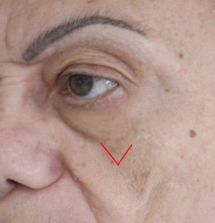
Nasojugal Groove, Nasolacrimal or Lacrimal Gut (Tear Trough)
There is much controversy regarding anatomy, and tear trough is inaccurately thought to be caused by aging. Tear trough is a personal anatomical characteristic, present from childhood [1] (Figure 4).
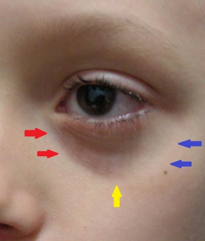
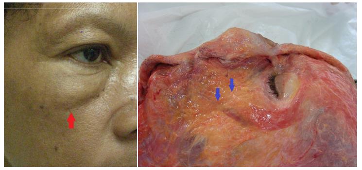
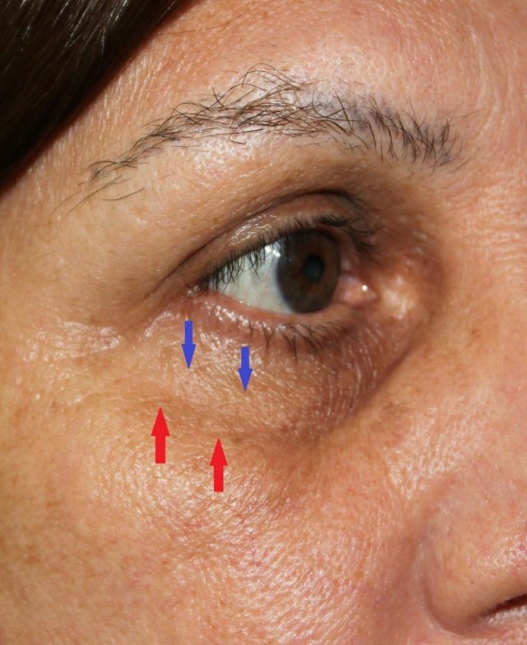
![Figure 4: 6-year-old child with nasolacrimal sulcus (red arrows), palpebromalar groove (blue arrows) and V- shaped deformity (yellow arrow). It is located below the medial canthus of the eye and is directed inferiorly and laterally to a little more than the lateral half of the orbit, a few millimeters below the orbital rim. It is mostly caused by the transition from the thin palpebral skin, devoid of subcutaneous adipose tissue, to the thicker malar skin with underlying subcutaneous tissue [1,18]. A ligament has been described as its cause; however, other authors argue that the ligament is the bone origin of the pre-orbital part of the orbicularis oculi muscle (Figure 5) [19,20]. It may also be accentuated by a bone depression at the muscle origin, anterior to the anterior lacrimal crest [1,2]. In addition to depth variations, the thin skin is further darkened by abundant blood vessels and increased skin pigment (Figure 6) [1].](/fulltextimages/419/fig_4.jpeg)
Figure 4: 6-year-old child with nasolacrimal sulcus (red arrows), palpebromalar groove (blue arrows) and V- shaped deformity (yellow arrow). It is located below the medial canthus of the eye and is directed inferiorly and laterally to a little more than the lateral half of the orbit, a few millimeters below the orbital rim. It is mostly caused by the transition from the thin palpebral skin, devoid of subcutaneous adipose tissue, to the thicker malar skin with underlying subcutaneous tissue [1, 18]. A ligament has been described as its cause; however, other authors argue that the ligament is the bone origin of the pre-orbital part of the orbicularis oculi muscle (Figure 5) [19, 20]. It may also be accentuated by a bone depression at the muscle origin, anterior to the anterior lacrimal crest [1, 2]. In addition to depth variations, the thin skin is further darkened by abundant blood vessels and increased skin pigment (Figure 6) [1].
Palpebromalar Groove
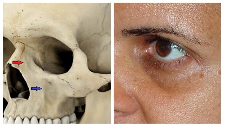
It can be continuous with the tear trough, or continuity can be interrupted by the V-shaped deformity. It is directed lateral and superiorly, along with, but below the orbital border. It is mainly caused by the difference in the malar adipose compartment, the transition from the palpebral skin to the malar skin, and by the zygomatic bone prominence (Figure 7) [1, 2, 18].
Buccal Zygomatic Groove
It begins at the V-shaped deformity, or at the junction of the tear trough with the palpebromalar groove, moving in an inferior and laterally, obliquely dividing the cheekbone (Figure 8) [1, 2]. It have several peculiarities: the groove accompanies the convexity of the malar bone in the transition to zygomatic prominence; There is a clear difference in the cutaneous characteristics of the malar region and the region of the nasolabial compartment; And, internally, it is the anterior limit of the oral cavity, which means it is the limit between the mobile part of the cheek and the fixed malar area (Figure 9) [1, 2]. The described zygomatic ligament as cause of the sulcus, however, is uncertain [21].
V-Shaped Deformity
It is a triangular depression in the middle region of the inferior orbital border, where the tear trough, the palpebral-malar and zygomatic-buccal grooves meet. It is caused by a lack of adipose tissue between the nasolabial and malar compartments associated to the zygomatic bony prominence (Figure 10) [1, 2, 5].
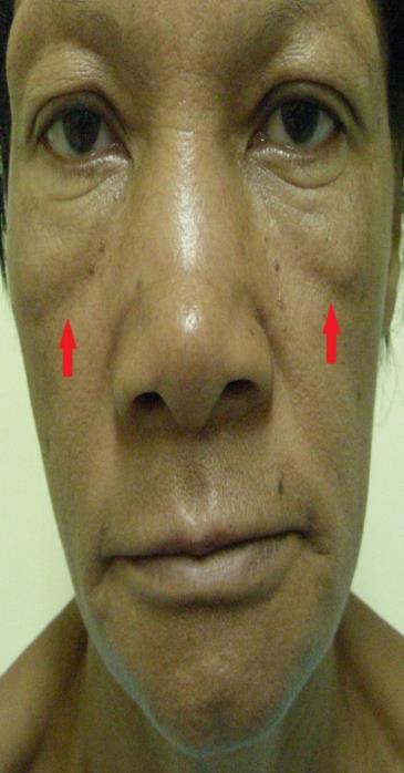
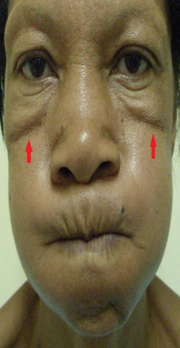
Nasolabial groove
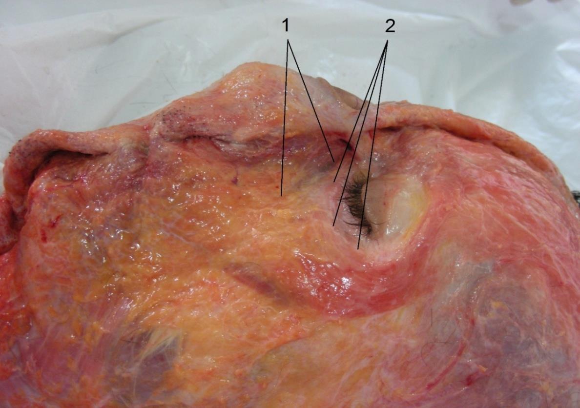
It starts laterally to the nasal wing and is inferior and laterally directed, ending laterally to the modiolus. Sometimes it may join with the lip-mandibular groove (Figure 11). It is caused by the transition of the (redundant) skin of the cheek with the nasolabial adipose compartment, to the labial skin devoid of subcutaneous adiposity and well adhered to the orbicularis oris muscle. The groove demarcates the limit of the orbicularis oris muscle and the insertions of the upper lip elevator muscles and the zygomaticus minor (Figure 12) [1, 2].
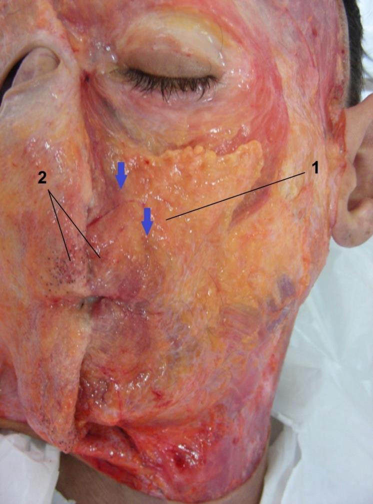
![Figure 14: Labiomental groove demonstrated in a fresh cadaver dissection. It is formed by the transition of the cheek skin with the subcutaneous adipose compartment of the jowl to the labial skin, firmly adhered to the orbicularis oris muscle and with scarce subcutaneous. The insertion of the depressor anguli oris muscle also contributes to its formation. Based on current concepts of anatomy and of the aging process, facial aesthetic treatment consists on adipose volume redistribution and contour restoration by rebalancing the existing volume to the amount of skin through redundant skin excision. Improving skin texture and its quality, with less or no ablative methods. For a complete and long-lasting result, it is necessary to combine modern surgical techniques, that cover the upper, middle and lower thirds of the face, with more vertical vectors, and scars extension as required, with cell therapy in volumetric replacement and tissue "regeneration" (Figure 15) [22].](/fulltextimages/419/fig_14.jpeg)
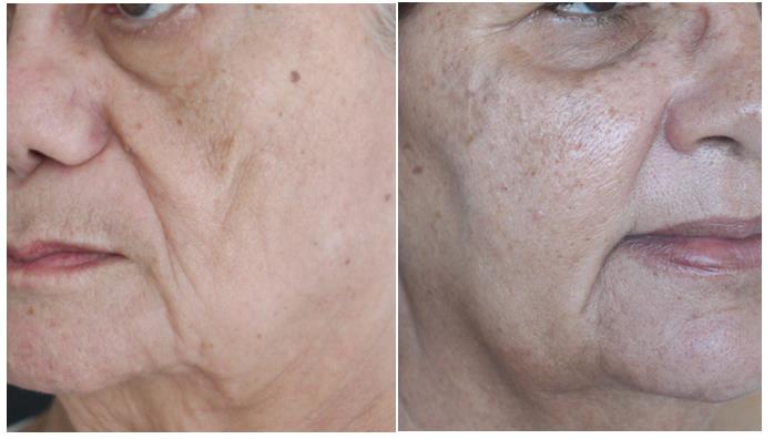
Figure 13: Labiomental groove formed by the transition between the excess skin along with the subcutaneous adipose compartment of the jowl, laterally, and the lower labial skin with scarce subcutaneous tissue. The crease may start in the corner of the mouth (left - red arrows), or can be continuous with the nasolabial groove, lateral to the modiolus (right - yellow arrows).
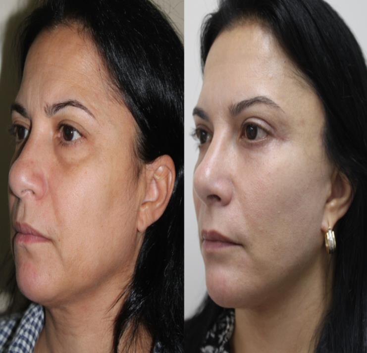
Figure 14: Labiomental groove demonstrated in a fresh cadaver dissection. It is formed by the transition of the cheek skin with the subcutaneous adipose compartment of the jowl to the labial skin, firmly adhered to the orbicularis oris muscle and with scarce subcutaneous. The insertion of the depressor anguli oris muscle also contributes to its formation. Based on current concepts of anatomy and of the aging process, facial aesthetic treatment consists on adipose volume redistribution and contour restoration by rebalancing the existing volume to the amount of skin through redundant skin excision. Improving skin texture and its quality, with less or no ablative methods. For a complete and long-lasting result, it is necessary to combine modern surgical techniques, that cover the upper, middle and lower thirds of the face, with more vertical vectors, and scars extension as required, with cell therapy in volumetric replacement and tissue "regeneration" (Figure 15) [22].
References
-
Chang YC (2016) Tear trough – Anatomy and treatment by autologous fat grafting. J Surg Dermatol 1(2): 116-122.
-
Chang YC, Ramos HNS (2016) Anatomia. In: Chang YC (ed), Rejuvenescimento facial, conceitos e técnicas. Dilivros, Rio de Janeiro, pp. 2-3.
-
Chang YC, Sanchez FH (2016) Processo de envelhecimento facial. In Chang YC (ed), Rejuvenescimento facial, conceitos e técnicas. Dilivros, Rio de Janeiro, pp. 44-56.
-
Ha RY, Nojima K, Adams WP, Brown SA (2005) Analysis of facial skin thickness: Defining the relative thickness index. PlastReconstr Surg 115(6): 1769- 1773.
-
Goldberg RA (2005) The three periorbital hollows: A paradigm for periorbital rejuvenation. Plast Reconstr Surg 116(6): 1796-1804.
-
Donofrio LM (2000) Fat distribution: A morphologic study of the aging face. Dermatol Surg 26(12): 1107- 1112.
-
Lambros V (2007) Observations on periorbital and midface aging. Plast Reconstr Surg 120(5): 1367- 1376.
-
Rohrich RJ, Pessa JE (2007) The fat compartments of the face: Anatomy and clinical implications for cosmetic surgery. Plast Reconstr Surg 119(7): 2219- 2227.
-
Pessa JE, Desvigne LD, Lambros VS, Nimerick J, Sug- unan B, et al. (1999) Changes in ocular globe-to- orbital rim position with age: Implications for aesthetic blepharoplasty of the lower eyelids. Aesth Plast Surg 23(5): 337-342.
-
Naylor EC, Watson REB, Sherratt MJ (2011) Molecular aspects of the skin ageing. Maturitas 69(3): 249-256.
-
Pottier F, El-Shazly NZ, El-Shazly AE (2008) Aging of orbicularis oculi anatomophysiologic consideration in upper blepharoplasty. Arch Facial Plast Surg 10(5): 346-349.
-
Yun S, Son D, Yeo H, Kim S, Kim J, et al. (2014) Changes of eyebrow muscle activity with aging: functional analysis revealed by electromyography. Plast Reconstr Surg 113(4): 455e-463e.
-
Kim WS, Park BS, Park SH, Kim HK, Sung JH (2009) Antiwrinkle effect of adipose´derived stem cell: Activation of dermal fibroblast by secretory factors. J Dermatol Sci 53(2): 96-102.
-
Tonnard P, Verpaele A, Peeters G, Hamdi M, Cornelissen M, et al. (2013) Nanofat grafting: basic research and clinical applications. Plast Reconstr Surg 132(4): 1017-1026.
-
Majollal A, Lequeux C, Shipkov C, Breton P, Foyatier JL, et al. (2009) Improvement of skin quality after fat grafting: Clinical observation and an animal study. Plast Reconstr Surg 124(3): 765-774.
-
Covarrubias P, Cárdenas-Camarena L, Guerrerosanto J, Valenzuela L, Espejo I, et al. (2013) Evaluation of the histologic changes in the fat-grafted facial skin: Clinical trial. Aesth Plast Surg; 37(4): 778-783.
-
Prado A, Andrades P, Danilla S, Benitez S, Reyes S, et al. (2008) Full-face carbon dioxide laser resurfacing: a 10-year follow-up descriptive study. Plast Reconstr Surg 121(3): 983-993.
-
Haddock NT, Saadeh PB, Boutros S, Thorne CH (2009) The tear trough and lid/cheek junction: Anatomy and implications for surgical correction. Plast Reconstr Surg 123(4): 1332-1340.
-
Wong CH, Hsieh MKH, Mendelson B (2012) The tear trough ligament: Anatomical basis for the tear trough deformity. Plast Reconstr Surg 129(6): 1392–1402.
-
Pessa JE (2012) Discussion. The tear trough ligament: Anatomical basis for the tear trough deformity. Plast Reconstr Surg 129(6): 1403-1404.
-
Mendelson BC, Muzzaffar AR, Adams WP Jr (2002) Surgical anatomy of midcheek and malar mounds. Plast Reconstr Surg 110(3): 885-911.
-
Chang YC (2016) Face complete. In: Chang YC (ed), Rejuvenescimento facial, conceitos e técnicas. Dilivros, Rio de Janeiro, pp. 309-315.
- Epithelioid Granuloma; 3cases with Different Clinical Features
- Advancing Representation in Dermatology Clinical Trials: Ethical, Scientific, and Regulatory Imperatives for Inclusion Across all Fitzpatrick Skin Types
- A Case of Atopic Dermatitis with Concurrent Psoriasis Vulgaris: Successful Treatment with Upadacitinib
- Innovation Lifting Eyeshadow: A Synthesis of Makeup and Optical Illusion
- Distinguishing Superficial Actinic Porokeratosis from Actinic Keratosis with UVF Dermoscopy: A Case Report
- High Mobility Group Box 1 (HMGB1) in Cutaneous Inflammation: An Immune Modulator Bridging Cellular Stress, Ferroptosis and Danger Signaling