Congenital Primary Cutaneous Rhabdomyosarcoma of the Perineum with Mixed Histopathological Features and Unusual Expression of Smooth Muscle Actin: Report of a Case
<p>Rhabdomyosarcoma (RMS) affecting the soft tissue of the perianal and perineal regions in children is uncommon and account for 2% of all rhabdomyosarcomas. Primary cutaneous presentation in this anatomical location is extremely rare. We report a 9-month-old female infant with a congenital primary cutaneous RMS presenting as a polypoidal exophytic mass in the perineum mimicking giant condyloma acuminatum (Buschke and Löwenstein tumor). Histopathological examination revealed a predominantly alveolar RMS with areas showing embryonal and pleomorphic features. In addition to expressing desmin and myogenin, the tumor cells also expressed smooth muscle actin.</p>
Introduction
Rhabdomyosarcoma (RMS) is a malignant mesenchymal neoplasm that arises from mesenchymal precursors of striated (skeletal) muscles [1]. It typically arises in deep soft tissue or in visceral organs and thus seldom presents to dermatologists. It represents 53% of soft tissue tumors in the pediatric population [2]. Histopathologically, RMS includes three main types: embryonal (including botryoid, spindle and anaplastic variants), alveolar and pleomorphic with the first two types being the most commonly encountered types [3, 4, 5]. While the embryonal type tends to involve the soft tissues or viscera of the head and neck or the genitourinary tract, the alveolar type tends to affect the deep soft tissue of extremities [6]. Rare histopathological types of RMS include a distinctive spindle cell variant [7] and an epithelioid variant [8].
Compared to conventional RMS, tumors presenting in the skin are extremely rare and were found to represent 0.7% of all RMS cases collected at two large specialized institutes [9]. On the other hand, among all cases of cutaneous malignant solid tumors (primary or metastatic) studied in a Spanish pediatric dermatology department over duration of 14 years, cutaneous RMS represented 32% of the cases [10]. The skin can be involved by RMS either as a primary or a metastatic event. Primary cutaneous cases are very rare with less than 50 cases reported so far in the literature [11]. Cases are classified as primary cutaneous only after clinical and radiological exclusion of metastasis from RMS elsewhere or extension to the skin from an underlying deep soft tissue lesion. Primary cutaneous rhabdomyosarcomas (PC-RMS) tend to affect both children and adults with a bimodal age distribution [12]. While conventional RMS has a slight male predominance, PC-RMS tends to be more common in females [11]. Among pediatrics, the face tends to be the most frequently affected site with PC-RMS [1, 2, 9, 12, 13, 14, 15]. Other rarely involved sites include the hand [16], chest [17], feet [12], anus [18, 19], and the perineum [20]. Congenital cases are rare [2, 20, 21]. Among pediatrics, PC- RMS is usually of the alveolar or embryonal types while in adults most primary cutaneous cases are pleomorphic, epithelioid or not otherwise specified (RMS-NOS) [12]. Immunohistochemically, all types of RMS show skeletal muscle differentiation and are at least focally positive for desmin, muscle specific actin (HHF-35), myogenin and myo D1 with the last two being the more specific diagnostic markers [3, 4]. Herein we report a female infant with congenital primary cutaneous rhabdomyosarcoma affecting the perineum and showing mixed histopathological features and unusual strong positive staining with smooth muscle actin.
Case Report
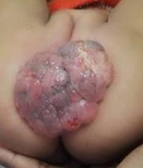
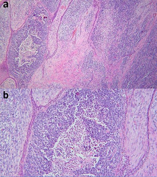
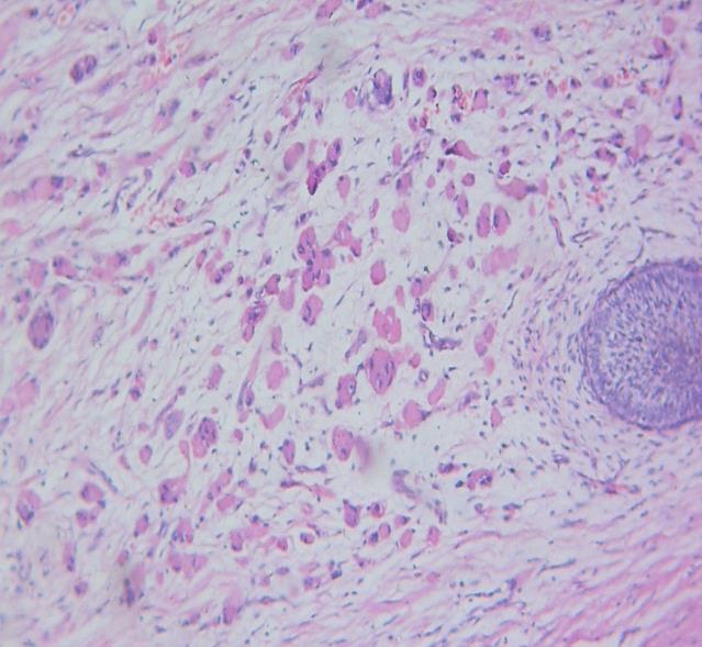
A 9-month-old female infant presented to the outpatient clinic of the Dermatology Department of Al- Zahraa Hospital (Faculty of Medicine for Girls, Al Azhar University) with a perineal mass. The mass was present at birth and underwent rapid progressive increase in size over time with history of spontaneous bleeding. Examination revealed a large (8X12 cm) polypoidal exophytic firm mass with a lobulated pink to dusky erythematous surface showing focal ulceration (Figure 1). Other than the mass, her medical history and clinical examination were unremarkable. A suggested clinical diagnosis of giant condyloma acuminatum (Buschke and Löwenstein tumor) was done and the patient was referred to the Pediatric Surgery Department for excision of the mass. Histopathological examination revealed: hyperplastic focally ulcerated epidermis and a dermal neoplastic infiltration. The tumor extended to the underlying subcutaneous fat and reached the lower margin of the specimen. In the majority of the lesion, the neoplastic growth was composed of discrete nests separated by fibrous septa (Figure 2a). Although most of the nests revealed solid growth pattern, some showed characteristic central cellular dissociation (Figure 2b).
Abdel-Halim MRE, et al. Congenital Primary Cutaneous Rhabdomyosarcoma of the Perineum with Mixed Histopathological Features and Unusual Expression of Smooth Muscle Actin: Report of a Case. Clin Dermatol J 2017, 2(1): 000109.
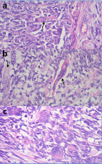
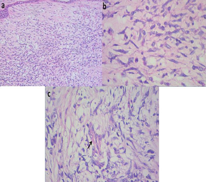
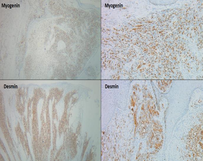
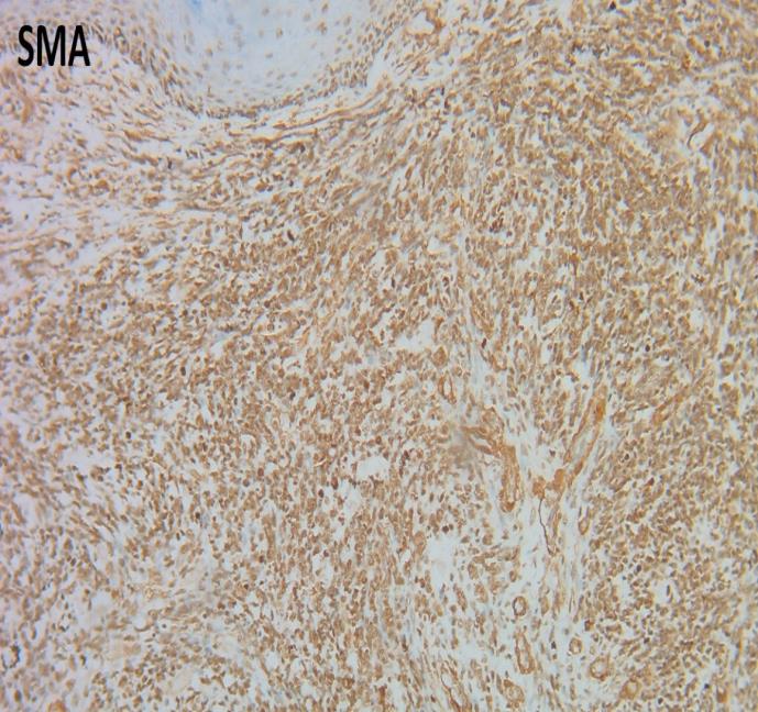
The nests were made of large cells with pleomorphic vesicular nuclei and contained many easily recognizable rhabdomyoblasts with abundant esinophilic cytoplasm and characteristic 'strap" or "tadpole" appearance (Figure 3 a&b). Many multinucleated cells were detected within the nests (Figure 3c), some with wreath like morphology. In some parts of the tumor the growth appeared as small rounded to spindle-shaped undifferentiated cells arranged in loose myxoid stroma with scattered identifiable rhabdomyoblasts (Figure 4). Focally, aggregates of large number of bizarre spindle shaped cells admixed with readily identifiable polygonal and pleomorphic rhabdomyoblasts were identified (Figure 5). The morphological features were typical of RMS with a predominant alveolar morphology admixed with areas of embryonal and pleomorphic features. The diagnosis was confirmed by the diffuse positive staining of tumor cells with myogenin and desmin (Figure 6). The tumors cells showed also strong diffuse positivity with smooth muscle actin (SMA) (Figure 7). All photomicrographs presented are according to their original magnification. Following diagnosis the patient was referred to the Cancer Institute for further assessment, re-excision and further treatment. Radiological workup revealed no lesions elsewhere and no extension in the perineal soft tissue.
Copyright© Abdel-Halim MRE, et al.
Abdel-Halim MRE, et al. Congenital Primary Cutaneous Rhabdomyosarcoma of the Perineum with Mixed Histopathological Features and Unusual Expression of Smooth Muscle Actin: Report of a Case. Clin Dermatol J 2017, 2(1): 000109.
Copyright© Abdel-Halim MRE, et al.
Abdel-Halim MRE, et al. Congenital Primary Cutaneous Rhabdomyosarcoma of the Perineum with Mixed Histopathological Features and Unusual Expression of Smooth Muscle Actin: Report of a Case. Clin Dermatol J 2017, 2(1): 000109.
Discussion
Rhabdomyosarcoma affecting the soft tissue of the perianal and perineal regions in children is uncommon and account for 2% of all rhabdomyosarcomas [22]. On the other hand, primary cutaneous presentation in this anatomical location is extremely rare. Primary cutaneous embryonal RMS presenting with polypoidal masses mimicking condyloma acuminatum have been reported in the anus [18, 19]. Moreover, Gong et al. [20] reported a newborn with abnormal symmetrical perineal overgrowth causing ambiguous genital morphology and histologic examination revealed cutaneous alveolar RMS admixed with areas of embryonal and pleomorphic RMS features. Only a relatively small number of mixed-type RMS have been reported and are usually associated with poor prognosis. They usually affected soft tissue and visceral organs [23, 24, 25]. The presence of many readily identifiable rhabdomyoblasts with characteristic 'strap cell' or 'tadpole' morphology in our case was typical of RMS on morphological level. Further immunohistochemical studies confirmed the diagnosis. However, in undifferentiated cases with undetectable rhabdomyoblasts, the diagnosis RMS can be difficult and requires extensive immunohistochemical workup. In children, the alveolar type can be mistaken with other tumors of the "small round blue cell" category such as Ewing sarcoma/primitive neuroectodermal tumors, metastatic neuroblastoma, hematopoeitic malignancies and extra renal Wilm's tumor, while in adults Merkel cell carcinoma and small cell carcinoma of the lung can also be a differential diagnosis. The embryonal type can be mistaken with leukemia cutis and the pleomorphic variants of RMS should be differentiated from other pleomorphic tumors with rhabdoid features such as: atypical fibroxanthoma, superficial pleomorphic undifferentiated sarcoma, proximal type epithelioid sarcoma, epithelioid angiosarcoma, high grade synovial sarcoma, melanoma, metastatic carcinoma and sarcomatoid squamous cell carcinoma [12, 26, 27, 28, 29, 30]. An unusual finding in our case was the presence of strong diffuse SMA expression by tumor cells. Although this finding has been previously reported in literature in pleomorphic rhabdomyosarcomas [31, 32, 33, 34], the positivity with SMA in our case was diffuse throughout the tumor and not restricted only to the pleomorphic areas. The exact significance of this SMA expression is unknown and whether it indicates a true smooth muscle differentiation Copyright© Abdel-Halim MRE, et al.
or just an anomalous immune reactivity could not be verified as ultrastructural studies were not feasible. Similar to conventional soft tissue RMS, primary cutaneous forms are also aggressive and can lead to distant metastases, usually to the lung. According to the largest available series of PC-RMS, the mortality rate was estimated to be 36% [12]. This necessitates rapid diagnosis and evaluation for metastasis. The treatment of PC-RMS usually includes a multidisciplinary approach with a combination of surgical excision, chemotherapy and radiotherapy [12]. Awareness of this rare tumor and its variable presentations is important to allow for early diagnosis and proper treatment.
References
-
Brecher AR, Reyes-Mugica M, Kamino H, Chang MW (2003) Congenital primary cutaneous rhabdomyosarcoma in a neonate. Pediatr Dermatol 20(4): 335-338.
-
Lima LL, Rodrigues CA, Pereira PM, Schettini AP, Tupinambá WL (2011) Primary cutaneous alveolar rhabdomyosarcoma in a pediatric patient. An Bras Dermatol 86(2): 363-365.
-
Tsokos M (1994) The diagnosis and classification of childhood rhabdomyosarcoma. Semin Diagn Pathol 11(1): 26-38.
-
Cui S, Hano H, Harada T, Takai S, Masui F, et al. (1999) Evaluation of new monoclonal anti-MyoD1 and anti- myogenin antibodies for the diagnosis of rhabdomyosarcoma. Pathol Int 49(1): 62-68.
-
Parham DM (1994) The molecular biology of childhood rhabdomyosarcoma. Semin Diagn Pathol 11(1): 39-46.
-
Fletcher CDM, Uni KK, Mertens F (2007) World Health Organization classification of pathology and genetics of tumors of soft tissue and bone. IARC Press, London.
-
Mentzel T, Kuhnen C (2006) Spindle cell rhabdomyosarcoma in adults: clinicopathological and immunohistochemical analysis of seven new cases. Virchows Arch 449(5): 554-560.
-
Jo VY, Mariño-Enríquez A, Fletcher CD (2011) Epithelioid rhabdomyosarcoma: clinicopathologic Abdel-Halim MRE, et al. Congenital Primary Cutaneous Rhabdomyosarcoma of the Perineum with Mixed Histopathological Features and Unusual Expression of Smooth Muscle Actin: Report of a Case. Clin Dermatol J 2017, 2(1): 000109. analysis of 16 cases of a morphologically distinct variant of rhabdomyosarcoma. Am J Surg Pathol 35(10): 1523-1530.
-
Schmidt D, Fletcher CD, Harms D (1993) Rhabdomyosarcomas with primary presentation in the skin. Pathol Res Pract 189(4): 422-427.
-
Figueroa Tovar MI, Laterza AM, Tamayo L, Ruiz- Maldonado R (1989) Incidence of malignant, primary and metastatic solid skin tumors at a pediatric dermatology service. Med Cutan Ibero Lat Am 17(1): 52-57.
-
GardnerJM, Smolle BR (2015) Primary Cutaneous Rhabdomyosarcoma. In: Rongioletti F et al. (eds.), Rare Malignant Skin Tumors, pp. 189-192.
-
Marburger TB, Gardner JM, Prieto VG, Billings SD (2012) Primary cutaneous rhabdomyosarcoma: a clinicopathologic review of 11 cases. J Cutan Pathol 39(11): 987-995.
-
Bröcker EB, Hamm H, Ritter J, Apple R, Schmidt D (1992) Rhabdomyosarcoma: differential diagnosis of cutaneous tumors in childhood. Hautarzt 43(9): 590- 593.
-
Tari AS, Amoli FA, Rajabi MT, Esfahani MR, Rahimi A (2006) Cutaneous embryonal rhabdomyosarcoma presenting as a nodule on cheek; a case report and review of literature. Orbit 25(3): 235-238.
-
Kim YS, Lee JH, Lee JY, Park YM (2015) Primary cutaneous rhabdomyosarcoma: Case report and review of published work. J Dermatol 42(10): 1014- 1015.
-
Bianchi L, Orlandi A, Iraci S, Spagnoli LG, Nini G (1995) Solid alveolar rhabdomyosarcoma of the hand in adolescence: a clinical, histologic, immunologic, and ultrastructural study. Pediatr Dermatol 12(4): 343-347.
-
Chang Y, Dehner LP, Egbert B (1990) Primary cutaneous rhabdomyosarcoma. Am J Surg Pathol 14(10): 977-982.
-
Gökdemir G, Ekmen S, Gungor S, Singer R (2013) Perianal rhabdomyosarcoma: report of a case in an infant and review of the literature. Pediatr Dermatol 30(1): 97-99. Copyright© Abdel-Halim MRE, et al.
-
Lee MW, Chung WK, Choi JH, Moon KC, Koh JK (2009) A case of botryoid-type embryonal rhabdomyosarcoma. Clin Exp Dermatol 34(8): e737- e739.
-
Gong Y, Chao J, Bauer B, Sun X, Chou PM (2002) Primary cutaneous alveolar rhabdomyosarcoma of the perineum. Arch Pathol Lab Med 126(8): 982-984.
-
Kuroiwa M, Sakamoto J, Shimada A, Suzuki N, Hirato J, et al. (2009) Manifestation of alveolar rhabdomyosarcoma as primary cutaneous lesions in a neonate with Beckwith-Wiedemann syndrome. J Pediatr Surg 44(3): e31-e35.
-
Raney RB Jr, Crist W, Hays D, Newton W, Ruymann F, et al. (1990) Soft tissue sarcoma of the perineal region in childhood. A report from the Intergroup Rhabdomyosarcoma Studies I and II, 1972 through 1984. Cancer 65(12): 2787-2792.
-
Lee HY, Tsai CC, Huang CH, Li WM, Yeh HC, et al. (2011) Mixed-type paratesticular rhabdomyosarcoma--a case report. Kaohsiung J Med Sci 27(6): 239-241.
-
Cohen J, Rybak L (1983) Mixed embryonal and alveolar type rhabdomyosarcoma of the sphenoid sinus. Arch Otolaryngol 109(1): 64-65.
-
Treetipsatit J, Kittikowit W, Zielenska M, Chaipipat M, Thorner PS, et al. (2009) Mixed embryonal/alveolar rhabdomyosarcoma of the prostate: report of a case with molecular genetic studies and literature review. Pediatr Dev Pathol 12(5): 383-389.
-
Parham DM, Dias P, Kelly DR, Rutledge JC, Houghton P (1992) Desmin positivity in primitive neuroectodermal tumors of childhood. Am J Surg Pathol 16(5): 483-492. Abdel-Halim MRE, et al. Congenital Primary Cutaneous Rhabdomyosarcoma of the Perineum with Mixed Histopathological Features and Unusual Expression of Smooth Muscle Actin: Report of a Case. Clin Dermatol J 2017, 2(1): 000109.
-
Folpe AL, Goldblum JR, Rubin BP, Shehata BM, Liu W, et al. (2005) Morphologic and immunophenotypic diversity in Ewing family tumors: a study of 66 genetically confirmed cases. Am J Surg Pathol 29(8): 1025-1033.
-
Adhikari LA, McCalmont TH, Folpe AL (2012) Merkel cell carcinoma with heterologous rhabdomyoblastic differentiation: the role of immunohistochemistry for Merkel cell polyomavirus large T-antigen in confirmation. J Cutan Pathol 39(1): 47-51.
-
Luzar B, Calonje E (2010) Morphological and immunohistochemical characteristics of atypical fibroxanthoma with a special emphasis on potential diagnostic pitfalls: a review. J Cutan Pathol 37(3): 301-309.
-
Banerjee SS, Harris M (2000) Morphological and immunophenotypic variations in malignant melanoma. Histopathology 36(5): 387-402.
-
Schürch W, Bégin LR, Seemayer TA, Lagacé R, Boivin JC, et al. (1996) Pleomorphic soft tissue myogenic sarcomas of adulthood. A reappraisal in the mid- 1990s. Am J Surg Pathol 20(2): 131-147.
-
Gaffney EF, Dervan PA, Fletcher CD (1993) Pleomorphic rhabdomyosarcoma in adulthood. Analysis of 11 cases with definition of diagnostic criteria. Am J Surg Pathol 17(6): 601-609.
-
Furlong MA, Mentzel T, Fanburg-Smith JC (2001) Pleomorphic rhabdomyosarcoma in adults: a clinicopathologic study of 38 cases with emphasis on morphologic variants and recent skeletal muscle- specific markers. Mod Pathol 14(6): 595-603.
-
Eyden B (2010) Pleomorphic rhabdomyosarcoma showing smooth-muscle and fibrohistiocytic differentiation: a single case report. Ultrastruct Pathol 34(1): 42-47. Copyright© Abdel-Halim MRE, et al.
- Epithelioid Granuloma; 3cases with Different Clinical Features
- Advancing Representation in Dermatology Clinical Trials: Ethical, Scientific, and Regulatory Imperatives for Inclusion Across all Fitzpatrick Skin Types
- A Case of Atopic Dermatitis with Concurrent Psoriasis Vulgaris: Successful Treatment with Upadacitinib
- Innovation Lifting Eyeshadow: A Synthesis of Makeup and Optical Illusion
- Distinguishing Superficial Actinic Porokeratosis from Actinic Keratosis with UVF Dermoscopy: A Case Report
- High Mobility Group Box 1 (HMGB1) in Cutaneous Inflammation: An Immune Modulator Bridging Cellular Stress, Ferroptosis and Danger Signaling