A Cadaveric Case Report of the Four-Headed Bicep Brachii Muscle and its Clinical Importance: A Case Study
Objective: The motive of this cadaveric case report is to demonstrate a variation of the Bicep Brachii Muscle identified in Asian origin adult male cadaveric specimen and its eventual clinical consequences. Method: Left Upper extremity of Asian origin male cadaver has been dissected to show four headed Bicep Brachii Muscle. Adjacent neurovascular structures as well as muscles were isolated, and their courses were observed for possible areas of compression also for anatomical variation. Result: Four headed Bicep Brachii Muscle was founded on the left upper extremity of formalin fixed Asian origin an 84-yearold male cadaver. The median nerve and brachial artery maintained their common neurovascular path. The musculocutaneous nerve passed deep to the third head of the anatomical variant before distributing its cutaneous branches as the lateral antebrachial cutaneous nerves were observed. Conclusion: The presence of a four headed Bicep Brachii Muscle in anterior compartment of upper extremity may cause neurovascular compression of the median nerve, musculocutaneous nerve, or brachial artery, resulting in peripheral nerve difficulties, so the practitioners should pay more careful to existence of this type of variation in clinical practice. Hence, these findings are very important not only for anatomical education but can also to serve as useful data for clinical diagnosis, treatment and research purpose.
Introduction
Comprehensive information of the human musculoskeletal system relies on a thorough knowledge and understanding of normal human anatomy. Morphologic variations are frequently encountered during clinical practice that could affect diagnosis and subsequent treatment. The presence of a four headed Bicep Brachii Muscle may compresses the adjacent neurovascular structures, leading to motor and sensory difficulties. Anatomical variations in the upper extremity are more common finding which affect both muscle and neurovascular structures. The Biceps Brachii is one of the most frequently affected muscles regarding the variation. A third, or fourth, head of the Bicep Brachii Muscle has occasionally been reported. This may generate additional power, as well as cause peripheral nerve compression. Variations in the distribution of the Musculocutaneous Nerve have been reported in people with extra heads of the biceps brachii. There have also been cases reported where the Musculocutaneous Nerve passes between the Biceps Brachii and Triceps Muscles, resulting in a potential site of nerve compression. The path of the Brachial Artery and Median Nerve, which course together adjacent to the tendons of the Bicep Brachii Muscle and the Brachialis muscle, may also be at risk for neurovascular compression.
Nerve entrapments of the upper extremity are a common finding, especially in athletes. Compression of peripheral nerves may present with varying degrees of complications, which result in paresthesia, muscle weakness, and even atrophy of the muscle. Peripheral nerve compression may result in sensory difficulties leading to a decrease in proper functioning, joint control, and stabilization, all of which are predisposing factors for injury. The etiology of these compartment syndromes varies, from muscular or fascial adhesions to inflammatory compression of nerve or neurovascular bundles to anatomical variations of muscles or nerves. Variant muscle patterns may be considered when assessing patients with nerve entrapment symptoms, those with unusual clinical presentations, or those who do not respond favorably to care. This study investigates anatomical variations of the Bicep Brachii Muscle and discusses potential neurovascular compromise and its clinical implications [1].
Case Report
The presence of unilateral four headed Bicep Brachii Muscle was observed during a routine dissection of anterior compartment of left upper-extremity of formalin fixed Asian origin an 84-year-old male cadaver which having no any trauma or surgery noted for BAMS Graduate Scholars in the Department of Rachana Sharir, National Institute of Ayurveda Deemed University, Jaipur. The dissection procedure of left upper extremity was begun by removing skin and adipose tissue from the both right and left upper extremities. Neurovascular tissue was identified and preserved in order to understand its anatomical distribution. The proximal and distal attachments of the muscles of the anterior brachial region were identified, and each muscle was separated and observed along with its fascial planes. During this process we came across with four headed Bicep Brachii Muscle. Adjacent neurovascular structures were traced along their anatomical pathways in order to identify if any areas of possible compression due to four headed variation of Bicep Brachii Muscle (Figure 1).
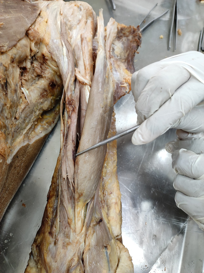
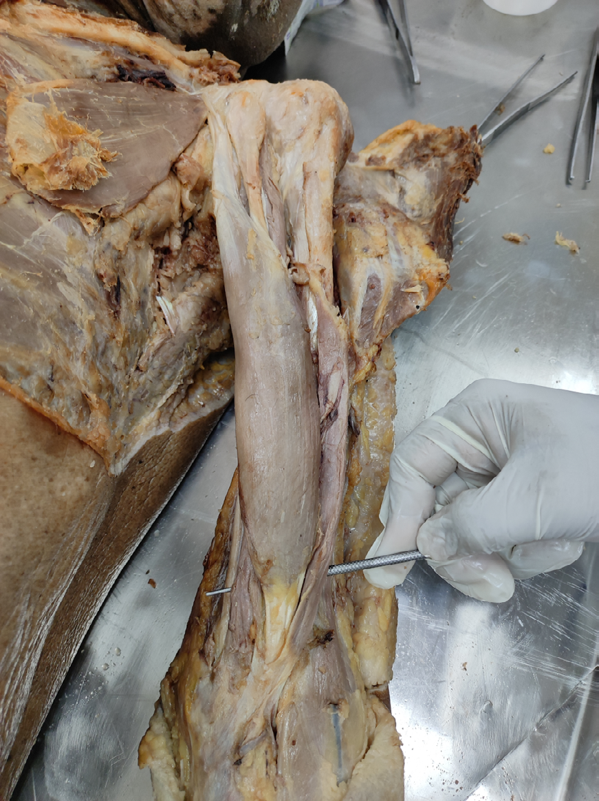
While dissecting the anterior compartment of left sided upper arm four headed Bicep Brachii Muscle and 3 headed Bicep Brachii Muscle on the right side has been found. The common proximal and distal attachments of the Brachialis and Bicep Brachii Muscle were noted. The third head of the Bicep Brachii Muscle was originated from the distal 2/3 of the anteromedial surface of the Humerus and fourth head was originated from the distal 2/3 of the anterolateral surface of Humerus. Both heads merged with the common tendon of the short and long heads of the muscle and inserted into the radial tuberosity of the Radius along with formation of Bicipital apponurosis. The Musculocutaneous Nerve passed deep to the four heads of the Biceps Brachii Muscle for nerve supply, while remaining superficial to the Brachialis Muscle, and terminated as the Lateral Anterior Cutaneous Nerve. Notably, the Musculocutaneous nerve traversed deep to the 4th head of Biceps Muscle at the most distal region of the left Brachium and supply the 4th head (Figure 2). This type of configuration may be a site of compression of Musculocutaneous Nerve, brachial artery or median nerve. In the right upper extremity, the Musculocutaneous Nerve terminated as the Lateral Antebrachial Cutaneous Nerve
at the middle third of the brachium, passing between the muscle bellies of the Brachialis and Biceps Brachii [2].
Discussion
The Biceps Brachii Muscle is a long fusiform muscle of the anterior compartment of the upper extremity which is responsible for the flexion and supination of the forearm as well as flexion of the Shoulder joint. The Bicep Brachii Muscle normally originates as two separate heads that join into a single tendon inserting distally into the Radial tuberosity of the Radius bone. The proximally tendon of the long head attaches to the Supraglenoid tubercle of the Scapula bone, becoming intracapsular as it traverses the shoulder joint and then within the Bicipital Groove. The proximally short head of Bicep Brachii Muscle attaches on the Coracoid process of the Scapula bone and travels as extra capsular along with tendon of Coracobrachialis muscle. Along with the Brachialis and Coracobrachialis Muscle, it receives nerve supply from the Musculocutaneous Nerve and its arterial supply is from the Brachial Artery. The Brachial Artery, which courses together with the Median Nerve between the Biceps Brachii and the Brachialis muscles, may undergo compression in individuals with variations of the Biceps Brachii Muscle. The cadaver in present study did not reveal a definitive or obvious neurovascular entrapment, but the Musculocutaneous Nerve may have been at risk for compression at the distal aspect of the 4th head of Biceps Brachii Muscle. The presence of these two extra bellies of the Bicep Brachii Muscle may cause muscular compression forces to shift laterally over the area where the Musculocutaneous Nerve terminates into the lateral Antebrachial Cutaneous Nerve [1].
Variation of three-headed Biceps Brachii Muscle, high- originated Radial Artery and communication between the Median and Musculocutaneous nerves have been well acknowledged in the available literature. However co- existence of these variations is very rare. They aimed to describe multiple variations in the upper limb and discuss their co-existence from clinical and embryological points of view [3]. A very rare case of a four-headed biceps Brachii Muscle associated with a dual piercing of one of the supernumerary heads by the Musculocutaneous Nerve was seen in the right arm of an 87-year-old female cadaver. Out of them one of the supernumerary heads of the Biceps Brachii originated from the Humerus, in the area between the lesser tubercle and the Coracobrachialis and Brachialis muscles and joined the long head at the level where the latter joined the short head and second supernumerary head originated from the Humerus at the point where the Coracobrachialis Muscle inserted and joined the Biceps Brachii tendon and its Bicipital Aponeurosis at the inferior third of the arm [4].
As per Embryological point, very well co-ordination of muscles, tendons and cartilage from the early stages of development suggests that these tissues could be co- ordinated and interdependent during the induction, differentiation and growth of the Musculoskeletal System [5]. According to Haines, R. Wheeler, development of the muscles of the Arm begins at the early Embryonic stage (about 5–6 weeks), and the growth of these muscles depends on the bone to which they attach [6]. Piagkou, et al. stated that it is possible that these conditions would be controlled by a system of genes, nervous impulses, or hormones, keeping the individual cells of the bones and each part of the muscles and tendons successfully modelled in response to each other in touch with the parts around it. Thus, occurrence of accessory heads of Biceps Brachii Muscle accompanied with multiple neurovascular variations might occur when any one of the conditions changed [7].
In the early embryonic stage, we know that the Biceps Brachii Muscle blends with the Brachialis muscle and the flexor mass at the distal end. The Brachialis muscle is attached closely overlying the Biceps Brachii Muscle, and one can hardly determine the line between these two muscles. According to Yamamoto, et al. Out of these muscles developing from the arm, the pre-muscular sheath of the more proximal ones is more developed than that of the ones more distal. Recently, cases of Brachialis Muscle
having superficial and deep heads were reported [8]. The superficial heads of the Brachialis Muscle may be the origin of the accessory heads of the Biceps Brachii Muscle, when the distal part of the superficial muscular band fuses with the Biceps Brachii Muscle. Thus, the accessory heads of the Biceps Brachii Muscle may be derived from the Brachialis muscle during embryological development [9].
In Present unusual variant of normal anatomy may be clinically significant in cases of neurovascular compromise with unknown etiology. This is a rare finding; this study highlights the need for Clinicians, Academicians and Researchers to recognize potential variations in Anatomy, which may present unilaterally, bilaterally, or in combination with other Anatomic variation. Additional examinations or focused diagnostic testing, including imaging or electro diagnostic testing, may enhance the diagnoses and treatment results.
Functional Effect
The Bicep Brachii Muscle generates actions responsible for Flexion at the Shoulder joint, Supination of the forearm, and Flexion at the elbow. Depending on the size of the third and fourth head, the additional mass may provide a substantial contribution to strength in flexion and Supination of the forearm [10]. Thus increased in muscle size may also create compression of peripheral nerves. Specifically, compression of the median nerve may be associated with an aberrant muscle attachment of a supernumerary head [11]. Additionally, compression of the Musculocutaneous Nerve may cause decreases in strength in the Biceps Brachii, Coracobrachialis, or Brachialis Muscles, or Neuro sensory changes of the lateral side of the Forearm and anterior capsule of the Elbow joint.
Clinical Implications
This variation of the Bicep Brachii Muscle may contribute to changes in strength, sensation, and common peripheral neuropathies of the Upper Extremity. With the addition of the rare muscular variant discussed in this study, altered Bicipital kinematics may be present, increasing Flexion and Supination of the Elbow Joint [12]. The wide variability in presentation of neurovascular structures associated with muscular variants predisposes individuals to compression syndromes. These compartment syndromes may complicate Symptomatology usually associated with median or Musculocutaneous Nerve injuries, creating diagnostic challenges. Anatomical variations of the Biceps Brachii Muscle and other structures within the anterior compartment of Arm are more common. The presence of a four headed Biceps Brachii Muscle may cause neurovascular compression of the Median Nerve, Musculocutaneous Nerve, or Brachial Artery, resulting in Neurosensory and Motor complications [13].
Limitations of the study
Present cadaveric study describes a single anatomical finding of a four headed Bicep Brachii Muscle in an Asian origin of 84 years old male cadaver. This variation in anatomy is very rare in populations; this single case discovered during a schedule dissection cannot justify a discussion of attribution within the general population. Morphological and histological analysis of tissue samples was not included in this study.
Conclusion
Anatomical variations of the Bicep Brachii Muscle and structures within the anterior brachial compartment are though very common but the presences of a four headed Bicep Brachii Muscle is very rare variation and it may cause neurovascular compression of the median nerve, musculocutaneous nerve, or brachial artery, resulting in vascular, neurosensory and motor deformities, so the clinicians should pay more attention to existence of this type of variation in clinical practice. Hence, these findings are not only for anatomical education but can also serve as useful data for clinical diagnosis, treatment and research rationale.
Declaration of Competing Interest
The authors declare that they have no conflicts of interest.
Acknowledgments
Department of Sharir Rachana, National Institute of Ayurveda, deemed university faculties, PG Scholars who assisted during dissection as well as measurements and Professor Sanjeev Sharma, Head of the Department and Vice-Chancellor, National Institute of Ayurveda Deemed University, Jaipur, Rajasthan for his continuous support and expertise guidance.
References
-
Williams PL (1995) Gray’s anatomy: The anatomical basis of medicine and surgery. 14th (Edn.), Churchill Livingstone Robert Stevenson, Edinburgh EH1 3AF, pp: 827.
-
Tank PW (2012) Grant’s Dissector with the Point Access Scratch Code. 15th (Edn.), Lippincott Williams and Wilkins, pp: 288.
-
Catli MM, Ozsoy U, Kaya Y, Hizay A, Yildirim FB, et al. (2012) Four-headed biceps brachii, three-headed coracobrachialis muscles associated with arterial and nervous anomalies in the upper limb. Anat Cell Biol 45(2): 136-139.
-
Vázquez T, Rodríguez-Niedenführ M, Parkin I, Sañudo JR (2003) A rare case of a four-headed biceps brachii muscle with a double piercing by the musculocutaneous nerve. Surgical and Radiologic Anatomy 25(5-6): 462- 464.
-
Huang AH, Riordan TJ, Pryce B, Weibel JL, Watson SS, et al. (2015) Musculoskeletal integration at the wrist underlies the modular development of limb tendons. Development 142(14): 2431-2441.
-
Haines RW (1932) The laws of muscle and tendon growth. Journal of anatomy 66(Pt 4): 578-585.
-
Piagkou M, Totlis T, Anastasopoulos N, Lazaridis N, Natsis K (2019) An atypical biceps brachii and coracobrachialis muscles associated with multiple neurovascular aberrations: a case report with clinical significance. Folia Morphologica 78(2): 444-449.
-
Yamamoto M, Kojyo U, Yanagisawa N, Mitomo K, Takayama T, et al. (2018) Morphology and relationships of the biceps brachii and brachialis with the musculocutaneous nerve. Surgical and Radiologic Anatomy 40(3): 303-311.
-
Rai R, Ranade AV, Prabhu LV, Pai MM, Prakash (2007) Third head of biceps brachii in an Indian population. Singapore medical journal 48(10): 929-931.
-
Nakatani T, Tanaka S, Mizukami S (1998) Bilateral four‐ headed biceps brachii muscles: The median nerve and brachial artery passing through a tunnel formed by a muscle slip from the accessory head. Clinical Anatomy 11(3): 209-212.
-
Mehta V, Yadav Y, Arora J, Kumar H, Suri R, et al. (2009) A new variant in the brachium musculature with reinforced innervation from a median–musculocutaneous nerve communication. Morphologie 93(301): 63-66.
-
Moore KL, Agur AM, Dalley AF (2011). Essential Clinical Anatomy. 4th (Edn.), Baltimore, Lippincott Williams & Wilkins, pp: 363-365.
-
Higashi N, Sone C (1988) A study of the accessory head of the biceps brachii in man. Journal of Anatomy 63(2): 78-88.
- Pattern of Breast Lesions in Ovu Inland, Delta State, South Southern Nigeria
- Morphometric Analysis of the Human Femur: Exploring Platymetric and Robusticity Indices Among the Nigerian Population
- Anatomical Variation of Arteria Lusoria: Clinical Implications for Dysphagia Lusoria and Surgical Risk
- Morphometric Study of the Vertebral Body and Pedicle of Typical Cervical Vertebrae Using Radiological Image
- Epigenetic Mechanisms Driving Human Evolutionary Changes
- Neuroprotective Effects of Ginkgo Biloba Extract on Bilateral Common Carotid Artery Ischaemic Stroke Induced in Wistar Rat