The Internal Aesthetics of the Human Body – A Multidisciplinary Approach
There is not one single article in the available literature dealing with internal aesthetics. Two brains were dissected by us, a radiogram of 1 brain was performed, 2 heads and 1 trunk were serially sectioned, and vascular casts were made of several organs. Finally, 210 histologic slices of various specimens were made. Historically, some animal and human organs, or their models, especially of the heart and liver, were used for a future prediction or as votive items in ancient times. Later on, the sacred heart was often painted in the portraits of Christ. Da Vinci and Vesalius during the Renaissance artistically presented the aesthetics of many organs, including Bourgery and Pernkopf some 3 and 4 centuries later. Modern digital artists presented them in 3D. In conclusion, gifted individuals in the Paleolithic and Neolithic times, in the ancient civilizations, and in the antique period depicted several organs, mainly for religious reasons. From the Renaissance onward, and particularly in modern times, the aesthetics of virtually all organs was shown in anthropology, fine art, and medicine.
Introduction
There are certain reports about the anatomic, artistic, and anthropologic aspect regarding the external beauty of the human body [1, 2, 3, 4, 5, 6, 7], but not one single article related to the internal aesthetics. However, each organ of the body has a certain morphology, structure, and its own aesthetics [8, 9, 10, 11].
People were interested in some animal and human organs since Paleolithic times, and which continued in the ancient civilizations, mostly with a religious connotation [1, 12]. The Renaissance masters, especially Leonardo and Vesalius, presented many organs from both scientific and artistic aspects [1, 13]. The famous 18th century painter Fragonard made aesthetic dissections of all organs. Many anatomists engaged professional artists in preparing their atlases [9, 10]. Later on, including modern times, more and more artists painted or modeled certain organs, due to the popularization of anatomy and medicine [4, 8, 11, 14] importantly, some organs or body parts have a special cultural significance in certain societies [12, 15]. In any case, we shall present the aesthetics of each organ separately.
The Brain
The human brain (Lat. cerebrum; Gr. enkephalos, hence encephalitis for its inflammation) [16] is a specific organ from many aspects [17]. Yet, the ancient Greek philosopher Plato stated almost 2.5 millennia ago that the brain is the seat of thought and feeling [12].
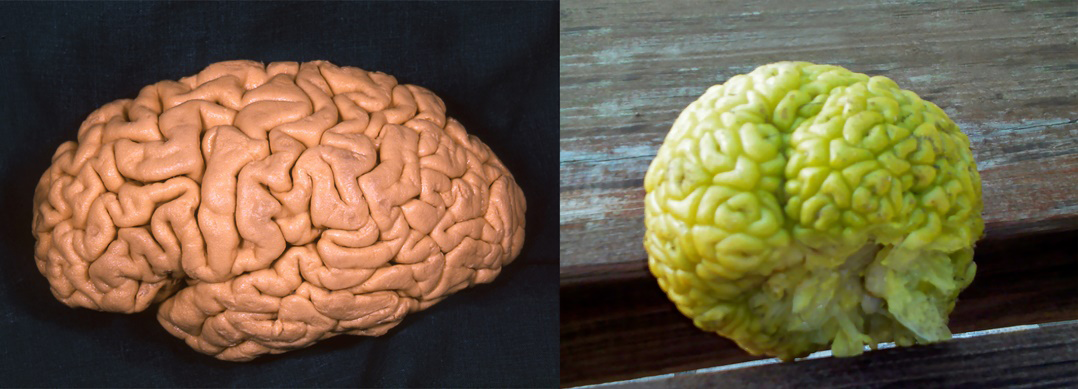
Dozens of the hemispheric gyri (from Gr. gyros = circle) virtually create an original artistic composition (Figure 1, left). The cerebral cortex of Homo sapiens, with 16.3 billion neurons on average, and trillions of synapses [17], was forced to form many convolutions in order to reduce the brain volume during its intense evolutionary development. This is, in fact, a reminiscence of the same biological process used even in some plants, e.g. the brain cactus and rhubarb bud (Figure 1, right), as well as in certain simple animals, such as brain corals [18, 19].
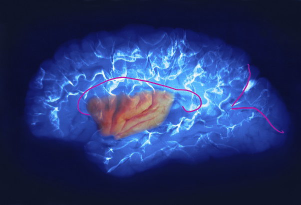
Each part of the brain has its own aesthetic value. In order to illustrate that, we made a photographic composition “An Island in the Brain Ocean” using our specimen of insula and a radiogram of the left hemisphere (Figure 2).
One of the geometrical and mathematical essences of aesthetics is the golden proportion, which is expressed as a value of 1.618 [20]. As regards the brain dimensions, its electrical activiy, and the arterial circle of Willis, there are proportions which correspond to the golden ratio.
Some professional artists made fine illustrations of the brain for certain anatomic atlases [9, 10, 11, 12]. Rembrandt’s 1656 work “Joan Deyman’s Anatomy Lesson” is among the most famous brain paintings [1]. In addition to Vesalius illustrations in the 16th century [12], Gotier D’Agoty’s 1748 painting of the axial section of a girl’s brain also is of a high aesthetic value, as well as Bourgery’s 1850 color drawings, and Pernkopf’s illustrations published almost one century later [9, 10].
Modern painter Ivanjicki presented the brain in some compositions [19]. Her contemporary Friščić created a 3D brain painting, with some vessels and a few gyri forming elongated tortuous structures such as the octopus tentacles. Dalla Benetta modeled several bronze brains with the cerebral gyri [21].
Certain radiologic techniques, such as 3D computed tomography (CT) [22], influenced Stratton to paint a skull with the cerebral arteries. Similarly, radiologic cerebral tractography images [11] inspired Camazine to paint the fiber bundles of the brain. Computer graphics enabled some artists to create fine digital brain images [4].
The Main Senses
The human brain constantly processes information received from the main sense organs. As for the eyes, many thousands of them were painted or modeled in art history, sometimes with inlays of precious stones or glass, as shown in the Roman bust of Brutus [1]. They were depicted in various colors, depending mainly on the quantity of the pigment in the iris (Gr. iris = rainbow; Hebr. = flower) [11]. Loss of vision is a handicap to many artists. For example, the famous painters Monet and O’Keeffe, suffering from cataracts and macular degeneration, respectively, gradually transformed their figural painting to beautiful abstract artworks [1]. Finally, Bruegel created the parable “The Blind Leading the Blind” [1].
The paired ear collects and delivers sound waves to the brain for further processing [17]. John Bacon painted the inner ear which receives sound waves. The spiral cochlea and cochlear duct there clearly correspond to the mathematical Fibonacci sequence [20]. Nevertheless, hearing loss was a big handicap to musicians, such as Beethoven. However, he created music mentally in his temporal and prefrontal cortices and thus managed to compose the brilliant Ninth Symphony [23].
Even the oral cavity with the sense of taste (Lat. gustatio) was the subject of some artists. Thus, Bucci created a long and elegant sculpture of the tongue (Lat. lingua; Gr. glossa), Velazquez depicted “The Lunch,” whilst Chagall painted his sister eating kasha [24].
Finally, the nose (Lat. nasus; Gr. rhis, rhinos, hence rhinitis and rhinoceros) of various morphologies was depicted in thousands of works. Some contemporary artists presented faces with only a nose, or the nose alone, e.g. Baldessari [4]. The olfactory system (Lat. olfacere = to smell) in the nasal cavity was presented digitally by Lu Yang and David Ornitz.
The Internal Organs
The organs (Lat. organum; Gr. organnon) were also in the focus of some ancient peoples and artists. They had a good knowledge of the internal organs, due to the Egyptian embalming their pharaohs, and by Mesopotamians and others as a result of their examination of the sacrificed animals’ organs [12, 25].
The basis of the body and organ aesthetics are the golden ratio, Fibonacci sequence, and similar geometric and mathematical lows [20]. They are also the essence of many living structures and processes, where they enable the stability of the structures and an optimal function at minimum consumption of energy [26].
In addition to the Renaissance artists and later anatomists, modern Damien Hirst made a huge open bronze anatomic sculpture (“Hymn,” i.e. “Him”) [4], 7 m in height, with most of the organs inside, whilst some others painted or modeled isolated organs [1, 6].
Thoracic Organs
Lungs
Among the airways, the nasal cavity, larynx and trachea were sporadically presented by artists, e.g. in the series “Floral Anatomy.” Vocal cords within the larynx producing the voice (Lat. vox, vocis, hence vocal; Gr. phone, hence phonetics and telephone) are used in speech and for singing. Trachea (Gr. tracheia) and bronchi (Lat. bronchus, from Gr. bronchos = windpipe) were occasionally painted and modeled [4].
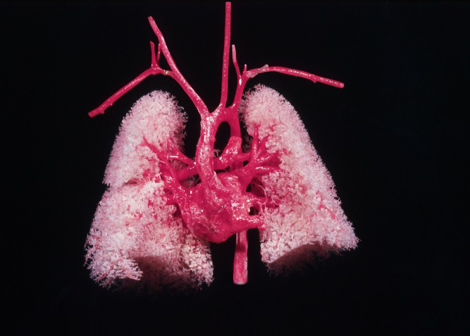
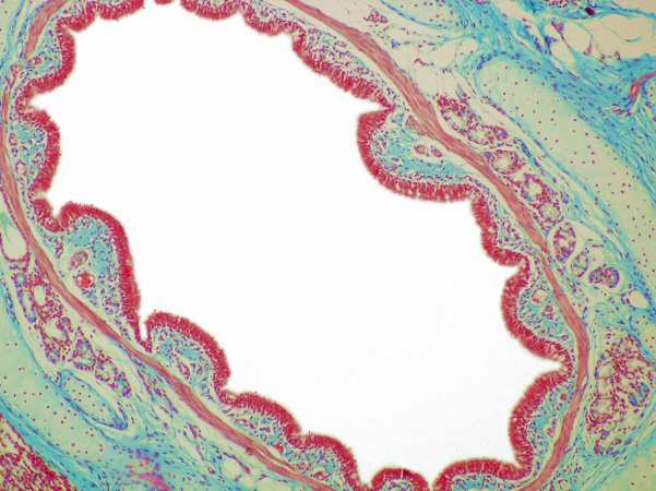
The lungs (Lat. pulmo, pulmonis; Gr. pneumon, from pneuma = air, breath, or spirit – hence pneumonia) (Figure 3) show gradual internal divisions of the tissue and ramifications of deep bronchial branches to the alveolar ducts level [11], This was artistically presented by Elizabeth S. and in the “Floral Anatomy.” Our micrograph of a segmental bronchus (Figure 4) is like an abstract expressionist painting [5]. Nevertheless, the mentioned bronchi divisions and subdivisions correspond to the golden ratio.
Some modern artists created lungs and the bronchial system as sculptures or installations, for example Giusto Pilan in ceramics, and MacDowell in porcelain. The famous artist Hirst designed lungs in the mentioned sculpture “Hymn” [4].
Heart
The heart [Lat. cor, cordis, from Gr. kardia, hence (myo) carditis] is like a rounded pyramid [11]. Several cardiac parts and functions follow the golden rule [27].
The heart was used by Babylonians from the sacrificed animals to predict the future [12]. The Egyptians preserved the pharaohs’ heart during embalming, for their afterlife, and also made thousands of the heart-shaped amulets [25]. Da Vinci and Vesalius presented the heart in a series of anatomic and artistic drawings [12, 13]. In addition, there were many religious paintings of the “Sacred Heart of Jesus,” e.g. by Pompeo Batoni, whilst Zurbaran painted a flaming heart held by a young woman in “Allegory of Charity” [28].
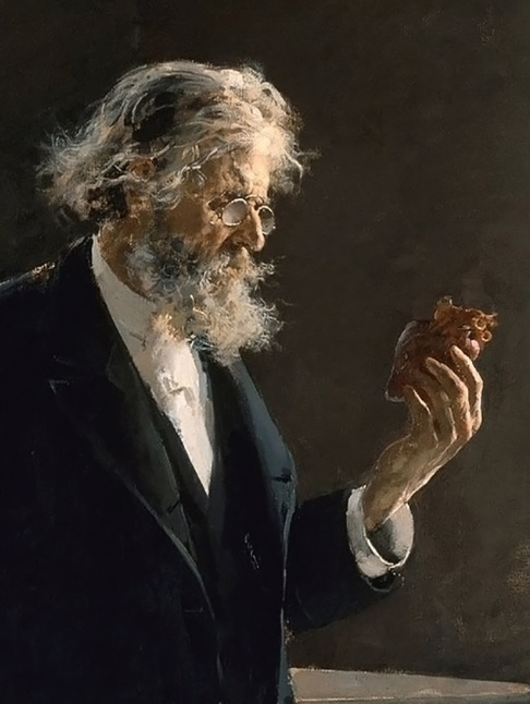
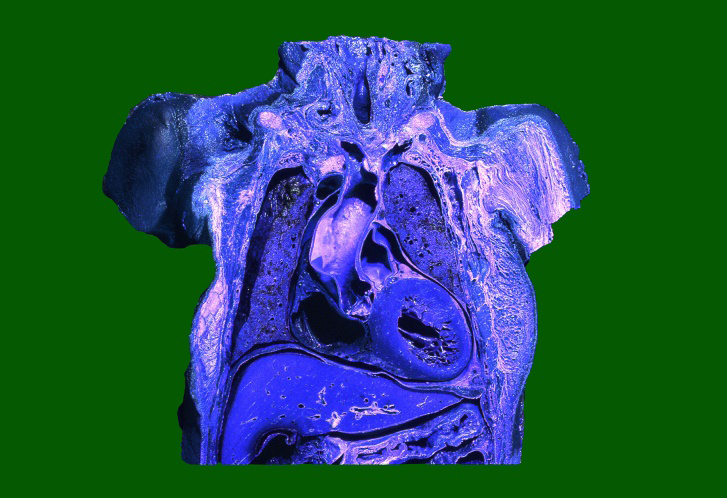
The heart was realistically painted by Spanish artist Lombardo, who presented a professor of pathology (Figure 5) holding a heart during an autopsy of a girl, trying to find the cause of her death. Modern Frida Kahlo depicted a double self-portrait with two interconnected hearts [2]. Marwedel created a heart image by using a special posture of human figures. A wooden heart was modeled by Tsykalov, a glass one by Sarah Band and John Burchetta, and one of metal by Barbora Mastrlova [19]. The surrealist Dali created “The Royal Heart” of gold with rubies, and with a crown at its base [1]. We made a coronal section of the heart, lungs and other organs presented in Figure 6.
The heart was regarded as the site of the soul, love, and bravery for millennia [29]. According to Aristotle, it was even the seat of intelligence [12]. The mentioned flaming heart suggested the outmost Christian religious fervor [28].
Abdominal Organs In general, artists were rarely interested in the digestive system. However, the mentioned Bucci created a tongue sculpture, and Uklański made an installation in the form of an oropharyngeal isthmus [4].
Stomach
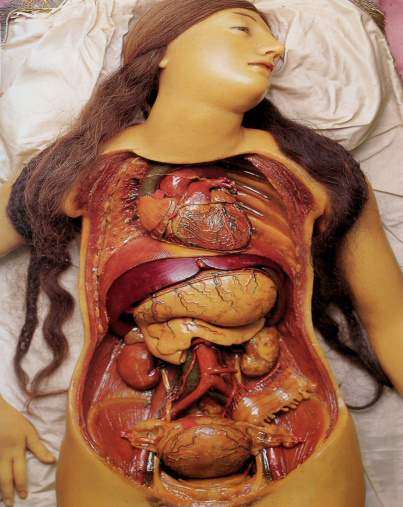
The stomach (Lat. stomachus; Gr. gaster, hence gastritis), the widest part of the alimentary canal, was depicted by Shakti Rinolfi. The stomach was modeled in situ by the mentioned Hirst in his sculpture “Hymn” [4]. It was also presented in some digital works, but also shown in an anatomic wax model (Figure 7).
Intestine
The small intestine (Lat. intestinum; Gr. enteron, hence enteritis) and the large bowel average almost 7 m in length [11]. This is why they form characteristic loops or flexures in the abdominal cavity. Some modern authors presented solely the small bowel loops. Yet others created them either within a skeleton, or inside a transparent digital image of the human body [4]. Paula Braconnot created the “Memento Mori” torso with a part of the intestinal loops.
The large bowel (Lat. colon, from Gr. kolon), with its specific surface morphology, is painted or digitally designed as encircling the small intestine. Nishinaga presented them both with a fine 3D vascular network, as did Paci in his drawings. They were also modeled in the mentioned sculpture by Hirst. Finally, Dongwook Lee created a female figure holding her inflated colorful intestinal loops.
Liver
The liver (Lat. iecur, iecoris; Gr. hepar, hepatis – hence hepatitis), lying below the diaphragm (Figure 6), is like half of an irregular ellipsoid [11]. It gives rise to the extrahepatic biliary system. The ancient Akkadians made some clay models of the liver and gallbladder [12]. Their priests used to examine the liver of the sacrificed animals to predict the future events. The Etruscans had a similar habit, as seen in their bronze model “Piacenza liver,” which was later accepted by the Romans.
According to the Greek myth of Prometheus, an eagle ate his liver every day, as ordered by Zeus [29]. This was painted, for instance, by Giordano, Rubens, and Moreau, and dramatically modeled by the Rococo artist Adam [1]. Some modern authors, e.g. Tsykalov, depicted the liver, whilst Nunzio Paci created it in drawings of a dissected body. Several authors, such as Sebastian Kaulitzki, presented a liver within a transparent digital human body. The liver is also modeled in the Hirst’s figure, and in a wax torso (Figure 7).
As for the biliary system (Lat. bilis = bile; Gr. chole; by adding melas = black, the term melancholia was coined for severe depressive state) [30], several authors created digital images of the gallbladder (Lat. vesica biliaris; Gr. chole kystis, hence cholecystitis). The bile stones were presented in a Ruysch composition a few centuries ago [8].
Spleen
The spleen (Lat. lien; Gr. splen), which predominantly belongs to the immune and blood systems, is like a giant grain of coffee, which is simplistically shown in watercolor and digitally created by various authors. Dr. Fung modified and colored a 3D CT scan of the spleen and liver using a special software. Some authors made the aesthetic scanning electron micrographs (SEM) of the spleen red and white pulp [31].
Pancreas
The elongated pancreas (Lat. pancreas, pancreatis, from Gr. pankreas) contains many microscopic islands (Lat. insulae) with the endocrine cells in its exocrine tissue – hence the name of the insulin hormone [31]. It was painted, along with the adjacent organs, by Elizabeth S. and Norman Trent. Kaulitzki depicted the pancreas digitally. Pancreatic vasculature was presented in a 3D CT image by the mentioned radiologist Dr. Fung.
Kidney
The kidney (Lat. ren, renis, therefore renal; Gr. nephros, hence nephron and nephritis) is a paired been- like retroperitoneal organ [11]. Elizabeth S. painted and Tsykalov D. modeled the kidneys, whilst Paci presented them like an artistic dissection. Dr. Fung created a fine radiologic composition of them. The kidneys were also presented in the mentioned wax model (Figure 7). An original composition was drawn three centuries ago by Rush, which contained the kidney, bile stones and injected blood vessels [8].
Pelvic Organs
Bladder
The bladder (Lat. vesica urinaria; Gr. kystis = sack, bladder – hence cystitis) is ellipsoid in appearance when filled with urine (Lat. urina; Gr. ouro). Flasks with urine were painted many times, e.g. by Teniers and van Ostade, depicting doctors visually examining patients’ urine [1]. Simpson and van Ostade painted some children playing with a blown pig bladder. Elizabet S. and John Bavosi depicted the human bladder. Finally, Nishinaga presented a complex 3D network of the bladder vessels.
Even urination was shown by some famous artists, e.g. by Bosch, Mantegna, Titian, Rembrandt, Boucher, and Gauguin [32], whilst some presented eroticized urination, e.g. Picasso, Dubuffet, and Klee. Duchamp put a pissoir in a horizontal position and entitled it “Fountain” [1]. There is the famous 17th century “Manneken Pis” statue of peeing boy in Brussels [21].
Rectum
The rectum (Lat. rectum = straight; Gr. proktos = anus, hence proctitis and proctoscopy) was presented by Pansaing in the cubist style, and also by several digital artists. Nishinaga showed its 3D vascular network. Even defecation and excrements were depicted by some artists, e.g. by Jan van Meiris, and the famous Catalonian painter Joan Miró [6].
Male Genitalia
They are crucial for gender differences and, along with the female organs, for human reproduction and sexual enjoyment [11, 32]. In any case, sexuality is a question of humans’ survival and pleasure, and thus an important subject for the fine art.
Testis
Testis (Gr. o_rchis_, and therefore orchitis) is a Latin term meaning a witness, and hence the Roman sentence “testis unus, testis nullus“ (one witness, no witness), as well as the English word “testimony” [16]. Testes are de facto the internal organs, situated just below the penis, which produce spermatozoa and male sexual hormones [31, 33].
According to a Greek legend, Kronos (the Roman Saturn) castrated his father, the god Uranus, and threw his genitalia into the sea causing a formation of foam, from which Aphrodite (the Roman Venus, Veneris) was born [29]. The Renaissance artist Botticelli presented this scene beautifully in his painting [1]. Michelangelo was very original when in the “Last Judgement” he painted the naked king Minos wrapped in a serpent which started his castration. This is in fact an ancient illustration of the future Freudian theory of castration anxiety [34].
Goya in “Los Caprichos” showed actual castrated men, something which was done to Spanish victims by French soldiers [1]. A true castrato was, for example, the famous 18th century opera singer Farinelli, who was beautifully presented in the famous 1994 film directed by Gérard Corbiau.
A testis was modeled by Denis Defrancesco, whilst Brancusi created an elegant set of testes made of polished metal [6]. They were also presented in many naked male statues. Genital artworks were depicted in some paintings by Munch, Schiele, Matisse, Giger, and Dali [6, 32]. Fine SEM pictures of the seminiferous tubules and spermatozoa were made by Gschmeissner.
Prostate
This male gland (Lat. prostata, from Gr. prostates), chestnut-like in form [11], was rarely painted, but it was digitally designed by Sciepro and others. The histologist Gschmeissner presented several SEM images of prostate cancer cells, and Hirst painted these cells in his biopsy series.
Female Genitalia
The external genitalia mainly comprise of the mons pubis (mons Veneris), labia majora and minora with the clitoris (from Gr. kleitoris), whilst the internal ones consist of the vagina, uterus, Fallopian tubes, and ovaries [11].
The pudendum (Lat. vulva, hence vulvitis) is located just inferior to the vaginal opening [1]. It was first carved in “The Venus of Willendorf” some 30,000 years ago [1]. Mons Veneris (Germ. Venusberg) was later mentioned by Wagner in his opera “Tannhäuser,” as well as in “The Picture of Dorian Gray” by Oscar Wilde.
The vulva itself was realistically depicted by the Baroque painter Boucher, and later by Klimt and Schiele, naturalistically by Courbet in “The Origin of the World”, cubistically by Picasso, symbolically by O’Keeffe in “Black Iris,” and surrealistically by Dali in “The Vulva” [1, 2] The pudendum was modeled by Victor, Mercier, Wüst, and McCartey” as well as by Notari like a land art [32]. David Hamilton and some others shot many art photographs of the vulva [35].
From the anatomic aspect, labia minora, which surround the vestibule of vagina, are more developed than labia majora in only 10% of women [36]. An artificial elongation of the labia minora is performed in several African tribes [37].
Hymen
The hymen (Gr. hymen = membrane, and also the god of marriage) [16, 29], is a fold of mucous membrane at the level of the vaginal opening. In traditional, conservative, or very religious societies, premarital hymen rupture (Lat. defloration) caused by a sexual activity, is not permitted [15]. If a girl breaks this rule, she is usually punished, and even excluded from the local community. For those reasons, hymenoplasty became very popular in such societies [15].
Nevertheless, the hymen was painted by Lequeau in the “Virgin Girl.” The loss of virginity was symbolically depicted by Gauguin, and described in “The Lover,” a novel by Duras [1]. The asexual goddess Athena was the famous antique virgin (Lat. Virgo intacta = untouched maiden; Gr. parthenos), whose monumental statue in Parthenon, created by Phidias, was decorated with gold and ivory [1, 29].
Vagina
The vagina (Lat. vagina = sheath, i.e. a tubular structure; Gr. kolpos, hence colpitis and colposcopy) is like a flattened fibromuscular tube lined with a mucous membrane forming transverse folds [11]. The vagina was painted by Sabrina PM, and modeled by Kapoor in Versailles close to Paris [32]. Joanna Frueh wrote an essay in 2003 entitled “Vaginal aesthetics” [38].
Uterus
The womb (Lat. uterus) resembles an inverted pear situated above the vagina [11]. It was found that the length and width of the uterus corresponds to the golden proportion (1.618), especially at the age of 21, which is characterized by optimal fertility [39].
Its stylized morphology was used by the Egyptians to create the ankh symbol, whilst Romans produced the womb clay models as votive offerings [12, 25]. The Greek philosopher Aristotle called it hystera and metra [12]. Hence certain medical terms, e.g. hysteria (a somatization disorder) and hysterectomy, as well as endometrium and myometrium within its wall [11, 12, 30].
The uterus was depicted by Sabrina PM, modeled in wax (Figure 7), and designed by some digital artists. Jesse Kanda created a uterus around a naked female torso. The womb may contain an embryo or a fetus in artworks or reality (Figure 8). The Andean Inca modeled the uterus with a fetus inside [40]. Raphael depicted a pregnant woman “La donna gravida” [1], whilst Van Eyck painted Arnolfini and his pregnant wife. Klimt beautifully painted both naked and dressed pregnant women. A fetus in the womb was presented in a fine drawing by da Vinci, and in several paintings by Frida Kahlo and Chagall [1, 13, 24]. Picasso and Quinn modeled pregnant women sculptures. Modern Damien Hirst created a series of sculptures of a sectioned uterus containing a fetus. Schmalz modeled a spiral Virgin Mary’s figure with the fetus of Christ in her womb [4].
![Figure 8: A drawing of a pregnant woman following an opening of the abdominal cavity and a coronal section of the uterus wall. (Modified by S. Marinković after Pernkopf [10]).](/fulltextimages/12226/fig_8.png)
A labor (Lat. partus) or childbirth (Lat. parturitio) was presented in some Neolithic works 6,000 ago. An Aztec figurine of the goddess Tlazolteotl showed a painful face expression due to enormous physical effort while giving birth [40]. Masson, Chagall and Kahlo painted childbirth several times. It was also mentioned in the novel “The Lost Girl” by D. H. Lawrence.
Uterine Tube
The paired Fallopian tube (the oviduct) is a slender trumpet-like duct (Lat. tuba; Gr. salpinx, hence salpingitis) [11]. Bavosi painted a flower-like tube, and Nishinaga made beautiful images of the tube fimbriae, like a rose flower.
Gschmeissner, Nikas and Motta shot fine histologic and SEM images of the oviduct mucosa cells, with a certain aesthetic value. If there is a post-inflammatory obstruction in the oviduct, an ectopic pregnancy (Lat. graviditas extrauterina) following fertilization will occur. Several authors painted this event, and Landau created a corresponding sculpture.
Ovary
The ovary (Lat. ovarium; Gr. oophoron = bearing eggs, hence oophoritis) is a paired female gonad which produces oocytes and female sexual hormones [31]. A mature oocyte leaves the Graafian follicle (Lat. ovulatio, from ovum = egg), which is presented in fine scientific photographs, and in paintings by Rouncefield and Marangio. The ovary itself was depicted by Lingerfelt, whilst Barbara Hepworth modelled a fine ovary-like sculpture [4].
Sexual Intercourse
The intercourse (Lat. intercursus = running between, or coitus) is a very important aspect of human life, which was especially examined and potentiated by Freud [34], and later on by some others [30].
Coitus was also the subject of fine art since the time of rock drawings created 7,000 years ago [32]. It was then modeled in some Egyptian figures, in Akkadian relief, in certain Roman figures, as well as on many Greek items of pottery [1]. It was also presented in the African, Mesoamerican, Chinese, Japanese, and Indian artworks [40, 41]. The latter civilization created the Kama Sutra in Sanskrit language dealing with sexuality, including the intercourse, about 2,000 years ago.
There are also some European artworks, especially a drawing made by Leonardo da Vinci in 1493 [13]. Aretino created a series of the coitus engravings in 1602. In addition to them, some other best world masters also presented the intercourse, e.g. Michelangelo, Rembrandt, Ingres, Rodin, Klimt, Schiele, Masson, and Giger [2]. The American writer Henry Miller described coitus several times in his novel “Tropic of Cancer,” as well as Richard Jonathan in “Mara, Marietta,” and Duvas in “The Lover.” Some artists created erotic (Gr. erotikos) and sexual artworks based on mythology. This was especially true for a Greek story about a woman Leda and Zeus, who transformed himself into a swan to seduce Leda [29]. This myth was beautifully presented by da Vinci, Michelangelo, Cellini, Correggio, Cézanne, and some modern artists, as well as in sculptures by Ammannati, Klinger, and Botero [1].
Correggio painted the original and most tender intercourse in “Jupiter and Io” [1]. In Bernini’s masterpiece regarding St. Teresa, her religious ecstasy in white marble has a pure orgasmic connotation. Finally, Podkowinski painted “Ecstasy” and Scriabin composed a poem with the same title [2].
Neuroscientific Aspect
The male and female genitalia, including the breasts, have a high aesthetic and erotic appreciation. This was confirmed by functional MRI of volunteers imagining sexual excitement [42] or watching sexual videos [42]. The MRI revealed an activation of the bilateral genital sensory area in the paracentral lobule of the cerebral hemisphere, and the secondary somatosensory cortex in the parietal operculum, as well as of the cingulate cortex, hippocampus, hypothalamus, ventral striatum, amygdala, medulla, and cerebellum.
The “erogenous” and orgasmic regions of the brain are also activated, e.g. the medial prefrontal cortex, the left insula, nucleus accumbens, amygdala, ventral tegmental area, and the substantia nigra [43, 44]. They are a part of the brain reward system, i.e. hedonic structures [17, 42, 44]. A coitus performance is regulated by the hypothalamic preoptic area and some other structures [44].
Radiologic Findings
Some radiologists engaged groups of couples as volunteers and performed an MRI during their coitus [45]. It was noted that the orgasmic reflex (Gr. orgasmos) mainly included the anterior vaginal wall, the periurethral tissues, the urinary bladder, and the perineal muscles. In any case, the MRI enabled for the first time a scientific examination of intercourse and sexual excitement.
Cellular Level
Almost each cell contains a nucleus with chromosomes made of a double DNA helix which consists of nucleotides. Each of the latter has one of the four bases: T, A, C, or G [31]. It was revealed that a DNA segment of 144 bases always has 55 T bases, and 89 A, C, or G bases. All these numbers (144, 55, 89) belong to the Fibonacci sequence [20]. Nevertheless, DNA was aesthetically modeled by Soneriu, Fields and some other artists [4].
About 200 different cell types (Lat. cellula; Gr. kytos, i.e. cytos, hence cytology) form the human body [31, 46]. Some of them are of different size and shapes, some others of branching types. Similar cells form certain tissues of various appearance.
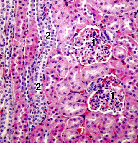
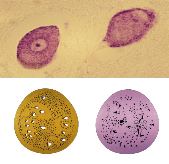
In any case, each type of cells and tissues has their own beauty, as shown in our Figure 9, and by Gschmeissner’s micrographs [31]. Some artists, such as Angela Hopkins, specialized in painting different types of cells and tissues. Similarly, Damien Hirst made a series of biopsy paintings. Certain artists designed sculptures or installations of branching nerve cells [4]. We ourselves chose two ganglion cells of the mesencephalic trigeminal nucleus in our specimen, which are similar to the work of the famous artist Lucio Fontana (Figure 10). This is in fact a combination of natural and artistic aesthetics, that is, a merge of nature and the unlimited creativity of the human brain.
Conclusion
The human need to present the important items from the environment, including the body and some organs, started in the Paleolithic period some 30,000 years ago, when the cognitive abilities of Homo sapiens reached a high evolutionary level. In the first civilizations, the organs had a special significance in rituals related to various divinities, and for the prediction of future events and thus survival. Similar traditions were later expressed among the ancient Greeks and Romans who often modeled several organs as votive offerings to their divinities. Since Christianity had Jesus’ love in the focus, the heart, as the seat of love and spirit, became a very important organ, which is why Christ was often painted with a sacred heart. Renaissance’s spiritual and intellectual deliberation resulted in a fine presentation of the human internal aesthetics. This was continued by means of subsequent artistic styles, with an addition of certain subjectivity, originality, and symbolic or metaphoric meaning of internal organs morphology. Their beauty is now appreciated by most people.
Acknowledgements
We are very grateful to Mrs. Elza Holt for reviewing the English text of our manuscript.
Conflicts of Interest
The author has no competing interest to declare.
Funding
No funding was received in support of this study.
References
-
Davies PJE, Denny WB, Hofrichter FF, Jacobs JF, Roberts AS, et al. (2007) Janson’s history of art: The Western tradition. 7th(Edn.), Upper Saddle River, Pearson Prentice Hall, USA.
-
Gibson M (2006) Symbolism. Taschen, Koln, USA, pp: 1-255.
-
Hogarth W (1753) The analysis of beauty. With a view of fixing the fluctuating ideas of taste. Strahan W, London.
-
Holzwart HW (2009) 100 contemporary artists. Taschen, Köln, Germany.
-
Lucie-Smith E (1977) Art Now From Abstract Expressionism To Superrealism. 1st(Edn.), William Morrow, New York, USA.
-
Ruhrberg K, Schneckenburger S, Fricke C, Honnef K (2005) Art of 20th century: Painting, sculpture, new media, photography. Taschen, Köln, GER.
-
Singh D, Dixson BJ, Jessop TS, Morgan B, Dixson AF (2010) Cross-cultural consensus for waist–hip ratio and women’s attractiveness. Evolution and Human Behavior 31(3): 176-181.
-
Kemp M (2000) Visualizations: The nature book of art and science. Oxford Publishing Press, Oxford, London.
-
le Minor JM, Sick H (2005) A monumental work of the 19th century. In: Bourgery JM, et al. (Eds.), The atlas of anatomy and surgery. Taschen, Köln, Germany.
-
Pernkopf E (1963) Atlas of topographical and applied human anatomy. W.B. Saunders Company, Philadelphia, USA.
-
Standring S (2016) The anatomical basis of clinical practice. In: Standring S (Ed.), Gray’s anatomy. 41th(Edn.), Elsevier Limited, London.
-
Persaud TVN (1984) Early history of human anatomy. From antiquity to the beginning of the modern era. Charles C Thomas Publishers, Springfield, USA.
-
Nathan J (2007) Anatomical drawings. In: Zöllner F (Ed.), Leonardo da Vinci: The complete paintings and drawings. Taschen, Köln, Germany.
-
Mergoupi-Savaidou E, Papanelopoulou F, Carneiro A (2016) Popularization of science, technology, and medicine in the “Periphery”: A step further? Technology and Culture 57(4): 966-977.
-
Ahmadi A (2015) Recreating virginity in Iran: Hymenoplasty as a form of resistance. Medical Anthropology Quarterly 30(2): 222-237.
-
Dornald WAN (2003) Dorland’s illustrated medical dictionary. 30th(Edn.), Saunders, Elsevier, Philadelphia, USA.
-
Kandel ER, Schwarz JH, Jessell TM, Siegelbaum SA, Hudspeth AJ (2013) Principles of neural science. 5th(Edn.), McGraw Hill Medical, New York, USA.
-
Campbell NA, Mitchell LG, Reece JB, Taylor MR (2003) Biology: Concepts and connections. 4th(Edn.), Benjamin Cummings, Philadelphia, USA, pp: 781
-
Lazić D, Marinković S, Tomić I, Mitrović D, Starčević A (2014) Brain and art: illustrations of the cerebral convolutions. A review. Folia Morphologica 73(3): 247- 258.
-
Persaud D, O’Leary JP (2015) Fibonacci series, golden proportions, and the human biology. Austin Journal of Surgery 2(5): 1066.
-
Duby G, Daval JL (2006) Sculptures from the antiquity to the middle ages, and from the Renaissance to the present day. Taschen, Köln, Germany.
-
Kretschmann HJ, Weinrich W (2004) Atlas of MR imaging and computed tomography. In: Kretschmann HJ, et al. (Eds.), Cranial neuroimaging and clinical Neuroanatomy. 3rd(Edn.), Thieme, Stuttgart, Germany.
-
Angulo-Perkins A, Aubé W, Peretz I, Barrios FA, Armony JL, et al. (2014) Music listening engages specific cortical regions within the temporal lobes: Differences between musicians and non-musicians. Cortex 59: 126-137.
-
Baal-Teshuva J (2003) Chagall. Taschen, Köln, Germany, pp: 280.
-
Rose-Marie H, Hagen R (2005) Egypt. People-gods- pharaohs. Taschen, Köln, Germany.
-
Sen SK, Agarwal RP (2008) Golden ratio in science, as random sequence source, its computation and beyond. Computers & Mathematics with Application 56(2): 469- 498.
-
Henein MY, Zhao Y, Nicoll R, Sun L, Khir AW, et al. (2011) The human heart: Application of the golden ratio and angle. International Journal of Cardiology 150(3): 239- 242.
-
Ferguson G (1961) Signs and symbols in Christian art. Oxford University Press, London.
-
Guirand F, Schmidt J (1996) Mythes & Mythologie. Histoire et dictionnaire. Larousse, Paris, France.
-
Sadock BJ, Sadock VA, Kaplan HI (2003) Kaplan & Sadock’s synopsis of psychiatry: Behavioral sciences/ clinical psychiatry. 9th(Edn.), Lippicott Williams & Wilkins, Philadelphia. USA.
-
Gartner LP, Hiatt JL (2001) Color textbook of histology. 2nd(Edn.), W.B. Saunders Company, Philadelphia, USA.
-
Néret G (2005) Erotica universalis. 1st(Edn.), Taschen America Llc, Köln, Germany.
-
Demir F, Sonmez G, Keske M, Mert Ali Karadag MA (2021) Is the penile golden ratio an indicator of normal sperm count and normal hormonal status? A prospective- observational study. Journal of Men’s Health 17(3): 104- 109.
-
Freud S (1997) Sexuality and psychology of love. Touchstone, New York, USA, pp: 224.
-
Misselbeck R (2007) 20th century photography: Museum Ludwig Cologne. Taschen, Köln, Germany, pp: 760.
-
Wildfang LA, Christian DH, Hoa LJU, Rikke G (2017) The size of labia minora and perception of genital appearance: A cross-sectional study. Journal of Lower Genital Tract Disease 21(3): 198-203.
-
Pérez GM, Aznar CT, Bagnol B (2014) Labia minora elongation and its implications on the health of women: A systematic review. International Journal of Sexual Health 26(3): 155-171.
-
Frueh J (2003) Vaginal aesthetics. Hypatia 18(4): 137- 158.
-
Verguts J, Ameye L, Bourne T, Timmerman D (2013) Normative data for uterine size according to age and gravidity and possible role of the classical golden ratio. Ultrasound in Obstetrics & Gynecology 42(6): 713-717.
-
Phillips C (2004) The lost history of Aztec & Maya. Anness Publishing Ltd, London, pp: 256.
-
Fahr-Becker G (2006) The art of East Asia. In: Fahr- Becker G (Ed.), Konemann UK Ltd, Germany, pp: 739.
-
Wise NJ, Frangos E, Komisaruk BR (2016) Activation of sensory cortex by imagined genital stimulation: an fMRI analysis. Socioaffective Neuroscience Psychology 6(1): 1-8.
-
Voon V, Mole TB, Banca P, Porter L, Morris L, et al. (2014) Neural correlates of sexual cue reactivity in individuals with and without compulsive sexual behaviours. Plos One 9(7): e102419.
-
Wiseman S (2023) A very sexy circuit. Nature Neuroscience 26: 1481.
-
Faix AJF, Lapray JF, Callede O, Maubon A, Lanfrey K (2002) Magnetic resonance imaging (MRI) of sexual intercourse: Second experience in missionary position and initial experience in posterior position. Journal of Sex & Marital Therapy 28(sup1): 63-76.
-
Bancroft JD, Gamble M (2002) Techniques in neuropathology_._ In: Bancroft JD (Ed.), Theory and practice of histological techniques. 5th(Edn.), Churchill Livingstone, London.
- Pattern of Breast Lesions in Ovu Inland, Delta State, South Southern Nigeria
- Morphometric Analysis of the Human Femur: Exploring Platymetric and Robusticity Indices Among the Nigerian Population
- Anatomical Variation of Arteria Lusoria: Clinical Implications for Dysphagia Lusoria and Surgical Risk
- Morphometric Study of the Vertebral Body and Pedicle of Typical Cervical Vertebrae Using Radiological Image
- Epigenetic Mechanisms Driving Human Evolutionary Changes
- Neuroprotective Effects of Ginkgo Biloba Extract on Bilateral Common Carotid Artery Ischaemic Stroke Induced in Wistar Rat