Tropical Pyomyositis of Atypical Etiology: A Case Report
Introduction: Tropical pyomyositis is a bacterial infection of the skeletal muscle caused by Staphylococcus aureus in 90% of cases. The most predisposed patients are those who are immunocompromised. Case Report: A 58-year-old male patient with a history of malignant prostate cancer undergoing treatment with hormonal therapy and radiotherapy, went to the emergency room for presenting, 1 week prior to admission, fever of 39°C associated with pain of progressive appearance located in the supracondylar region of the right lower limb, radiating to the inguinal region, of moderate to strong intensity, of an oppressive nature, which limited the movement of the entire right lower limb. After multiple negative tests, an MRI of the abdomen, pelvis, and lower limbs reported myositis of infectious etiology. From there, the relevant antibiotic treatment was indicated, and an ultrasound-guided biopsy of the iliopsoas muscle was performed, which reported Streptococcus agalactiae resistant to ampicillin and penicillin as the responsible pathogen. The evolution of the patient was favorable. Conclusions: Tropical pyomyositis is a disease little known by the medical community, which makes early diagnosis difficult. Immunosuppression plays an important role in its pathogenesis, so a thorough investigation must be carried out to exclude multiple predisposing conditions.
Introduction
Tropical pyomyositis is a bacterial infection of the skeletal muscle that can occur at any time in life, although it is most common in male patients 2 to 5 years of age and 20 to 45 years of age [1]. Its incidence in tropical zones is not clear, but it is an important cause of morbidity that represents 1% of hospitalizations in the Peruvian Amazon. Staphylococcus aureus is implicated in 90% of cases and usually only affects one muscle group, but could cause diffuse involvement in up to 20% of scenarios. The muscles of the lower limbs, especially the quadriceps, are usually the most compromised because they are prone to injury, stress, and constant tension [2]. The most predisposed patients are those immunocompromised with non-infectious pathologies such as neoplasms or diabetes mellitus [1, 3].
Clinically, tropical pyomyositis has 3 phases. The first phase, known as invasive or subacute, lasts from 1 to 3 weeks and is characterized by the insidious appearance of myalgia, mild localized edema, general malaise, and low-grade fever, although the skin remains intact, making diagnosis difficult. Actually, this is usually done in the next phase, called purulent or suppurative, which occurs between 10 and 21 days after the onset of symptoms. This is because it is possible to identify the abscess by imaging and analyze the pathology study, which usually shows suppuration, polymorphonuclear leukocytes, and necrotic cellular debris. In this phase the myalgia is severe, and the fever is high. Finally, the late phase, to which few patients reach, is a severe stage with signs of sepsis, where abscesses with bacteremia occur, culminating in multiple organ failure and death of the patient [2, 3].
The gold standard for the diagnosis is the gram and culture from the aspiration of pus or a biopsy of the affected muscle, but in case of not finding macro abscesses, staining with hematoxylin and eosin of the muscle tissue can be performed. However, imaging tests are more accessible and help us make an early diagnosis, with magnetic resonance being the imaging study of choice to demonstrate deep muscle collections [3, 4].
The cornerstone of treatment is the combination of broad-spectrum antimicrobials for 4 weeks and drainage of the abscess using percutaneous techniques or open surgery [1, 3]. We present the case of a male patient with tropical pyomyositis, of particular evolution and unusual etiology, which required joint management with several specialties for a favorable evolution.
Presentation of the Case
A 58-year-old male patient with a history of malignant prostate cancer undergoing treatment with hormonal therapy and radiotherapy, attended the emergency room for presenting, 1 week prior to admission, evening fever of 39°C associated with pain of progressive onset located in the supracondylar region of the limb. Lower right, radiating to the inguinal region, of moderate to strong intensity, oppressive in nature, which limited the movement of the entire lower right extremity. The physical examination at the time of admission revealed limited mobility of the right lower limb, with pain on right hip flexion (positive lasegue maneuver), no pain on palpation of the quadriceps, no obvious color changes in the hip and right lower limb. The laboratory tests requested on admission showed normochromic normocytic anemia, normal white blood cell count, elevated C-reactive protein table 1 and muscle enzymes without alterations (Table 1). The patient was hospitalized in our internal medicine service, where, in view of being an immunosuppressed patient and presenting fever without an apparent source, 4 blood cultures and rose Bengal serology were performed. Thinking of a presumptive diagnosis of brucellosis, empirical antibiotic therapy was started with doxycycline 100 mg tablets orally every 12 hours and clindamycin 600 mg intravenously. Blood cultures and serology were negative.
MR imaging of the abdomen, pelvis and lower limbs was requested, which reported: myositis of infectious etiology associated with collection at the level of the fibers of the distal segment of the iliopsoas muscle, iliacus, distal end of the psoas major, gluteus minimums and Medius, quadratus femoris, obturator internus and externus, adductors, pectineus, and vastus lateralis (Figure 1); consistent with the diagnosis of pyomyositis. The previously mentioned treatment was suspended and vancomycin 1 g VSV every 12 hours and meropenem 1 g VSV every 8 hours were indicated.
Interconsultation with the interventional radiology service was carried out, who, in view of not observing an evident collection, took an ultrasound-guided biopsy of the iliopsoas muscle, whose culture result at 4 days reported the growth of more than 100,000 CFU of Streptococcus. Agalactiae resistant to ampicillin and penicillin, this is why we continued with the previously initiated antibiotic treatment.
In subsequent controls, a decrease in acute phase reactants and absence of fever were observed, however, there was a slow clinical progression of the patient, evidenced by the inability to mobilize the affected lower limb, given the pain that continued to persist.
Antibiotic treatment was continued, and physical therapy and rehabilitation were performed for a period of 20 days. Control MRI performed on the 24th day after starting antibiotics showed a decrease in the size of the collections in affected muscles. He was discharged with successive check- ups in the office. External where he had a favorable clinical evolution given by active mobility of the hip and both lower limbs with absence of pain but with mild atrophy of the quadriceps muscle to be managed by the physical therapy and rehabilitation service.
| Laboratory Exam | Normal Values | Evolution | ||||||||
|---|---|---|---|---|---|---|---|---|---|---|
| At Admission | Journey 1 | Journey 2 | Journey 3 | Journey 4 | Journey 6 | Journey 7 | Journey 9 | Journey 11 | ||
| Inflammatory Markers | ||||||||||
| PCR | 0 - 0.5 mg/dl | 7,69 | 12,58 | - | 15,3 | 16,07 | 10,85 | 8,07 | 6,13 | 2,85 |
| Blood count | ||||||||||
| Hb | 13.6 - 17.5 g/dl | 10,5 | 9,5 | - | 9,4 | 9,1 | 9,4 | 9,8 | 10,3 | 10,4 |
| HTO | 40 - 52% | 32,2 | 28,7 | - | 28,5 | 27 | 28,8 | 29,8 | 31 | 32,3 |
| Leukocytes | 4400 - 11300/ mm3 | 11320 | 10700 | - | 10000 | 11540 | 7500 | 6390 | 6820 | 6660 |
| Hepatic Profile | ||||||||||
| TGO | 0 - 40 U/L | - | - | 30 | - | - | 79 | - | 63 | - |
| TGP | 0 - 41 U/L | - | - | 77 | - | - | 114 | - | 128 | - |
| FA | 40 - 130 U/L | - | - | 233 | - | - | 548 | - | 638 | 561 |
| GGTP | 8 - 61 U/L | - | - | 285 | - | - | - | - | 488 | 425 |
| Albumin | 3.5 - 5.2 g/dl | - | - | 3,29 | - | - | 3,07 | - | - | - |
| Bilirubins | 0 - 1.2 mg/dl | - | - | 0,93 | - | - | 0,86 | - | 0,56 | - |
| Muscle Enzymes | ||||||||||
| CPK | 26 - 190 U/L | - | - | - | 39 | - | 103 | - | - | - |
Table 1: Laboratory tests
PCR: Protein C Reactive; HB: Hemoglobin; HTO: Hematocrit; TGO: Transaminase Glutamic-Oxaloacetic; TGP: Transaminase Glutamic-Pyruvic; PA: Phosphatase Alkaline; GGTP: Gamma Glutamyl Transpeptidase; CPK: Creatine Phosphokinase Table 1: Laboratory tests
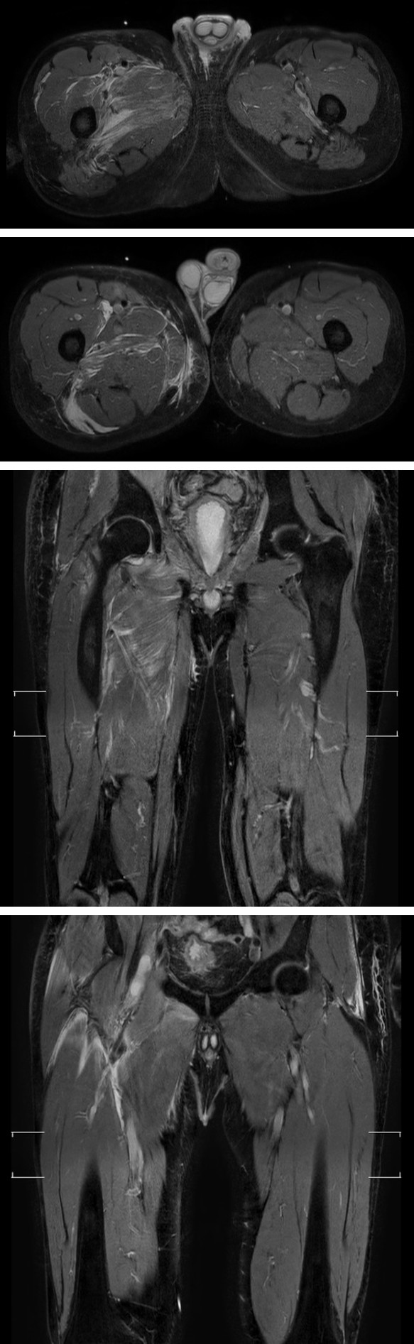
Figure 1: Magnetic resonance imaging of the abdomen, pelvis and lower limbs in transversal and coronal section. Hyperintense images corresponding to an inflammatory process are evidenced in the fibers of the iliopsoas muscle (distal segment), gluteus minimus and medius, quadratus femoris, internal and external obturator, adductors, pectineus and vastus lateralis consistent with pyomyositis of infectious etiology.
Discussion
Tropical pyomyositis continues to be a pathology that represents a challenge for every treating physician. An early diagnosis is essential to avoid complications related to the late phase such as sepsis or muscle necrosis, but often this crucial step is hindered due to different factors. The lack of familiarity with the disease, its variability of presentations, the plurality of differential diagnoses and the absence of specific signs during its evolution are some of them. They could be solved by addressing two main points: the validation of current diagnostic strategies, with respect to bacteriological and imaging techniques, and the definition of strategies that defend empirical treatment.
Going deeper into the diagnostic difficulty, various factors arose in this particular case. Initially, negative blood cultures were a major obstacle, but it turns out that in tropical pyomyositis blood cultures are positive in less than 5% of cases at the time of clinical presentation, with a maximum positivity of 35% [5]. Also, despite being an intrinsic muscle pathology, creatine phosphokinase and aldolase levels are normal or slightly elevated. This means that a muscular pathology like this is not the main suspicion of the treating physicians. Another relevant aspect lies in the form of presentation. The usual is the affection of a single muscle group , but in up to 12-40% of cases an affection of several muscle groups has been seen, either sequentially or simultaneously, which happened in the reported case.
As previously mentioned, Staphylococcus aureus is the predominant microorganism in immunosuppressed and immunocompetent patients. However, new microorganisms such as group A and B Streptococcus are documented more frequently. In recent decades, the incidence of invasive infections by Streptococcus agalactiae in adults has increased, especially in immunosuppressed patients with severe underlying diseases, as in the patient reported [6].
Although tropical pyomyositis is presumed to be caused by bacterial superinfection of a predisposed muscle, its pathogenesis is not well defined. Trauma, immunodeficiency, nutritional alterations, among others, have been postulated as possible etiological factors. In addition, the rise of this pathology in non-temperate climates such as Lima has been fundamentally related to immunodeficiency; particularly with pathologies such as HIV infection, diabetes mellitus, neoplasms, rheumatological conditions or intravenous drug abuse [1, 5].
The proposed treatment for tropical pyomyositis consists of broad-spectrum antibiotics in its initial stage, but when diagnosis is late, drainage of abscesses should be added to antibiotic therapy [7]. In the patient presented, carbapenems were used for 20 days due to the particular resistance of the pathogen involved, which is consistent with the most appropriate empirical therapy that usually lasts from 2 to 9 weeks and involves ampicillin/sulbactam or carbapenems.
Conclusion
Tropical pyomyositis is a rare disease and little known by the medical community, which makes early diagnosis difficult. Antibiotic treatment is essential and surgery should be started early, likewise, when it is considered clinically relevant. Immunosuppression has an important role in the pathogenesis of pyomyositis, and investigation should be performed to exclude multiple predisposing conditions.
Consent
The patient signed a written informed consent for the publication of this case report and the use of accompanying images. A copy of this is available for review by the editor-in- chief of this journal.
Acknowledgment
We want to thank the contribution of all the doctors, nurses and technicians for their help in making the diagnosis and facilitating the management of the patient who could have had a favorable clinical evolution.
References
-
Shittu A, Deinhardt-Emmer S, Vas Nunes J, Niemann S, Grobusch MP, et al. (2020) Tropical pyomyositis: an update. Trop Med Int Health 25(6): 660-665.
-
Narayanappa G, Nandeesh BN (2021) Infective myositis. Brain Pathol _(Zurich, Switzerland), 31_(3), 10-13.
-
Habeych ME, Trinh T, Crum-Cianflone NF (2020) Purulent infectious myositis (formerly tropical pyomyositis). J Neurol Sci _413(3)_, 11-12.
-
Chauhan S, Jain S, Varma S, Chauhan SS (2004) Tropical pyomyositis (myositis tropicans): current perspective. Postgrad Med J _80(9)_: 267-270.
-
Villamil-Cajoto I, Maceiras-Pan F, Villacián-Vicedo MJ (2006) Piomiositis: presentación de 17 casos en niños y adultos. Rev Med Chil 134(1): 31-38.
-
Revuelto-Artigas T, Cabrerizo-García JL, Domene-Moros R, Sanjoaquin-Conde I (2011) Celulitis, piomiositis y artritis esternoclavicular por Streptococcus agalactiae en paciente con cirrosis hepática [Cellulitis, pyomyositis, and sternoclavicular arthritis due to Streptococcus agalactiae in a patient with liver cirrhosis]. Gastroenterol Hepatol 34(1): 53-54.
-
Palacio EP, Rizzi NG, Reinas GS, Júnior MM, Júnior AD, et al. (2010) Open drainage versus percutaneous drainage in the treatment of tropical pyomyositis. prospective and randomized study. Revista Brasileira de Ortopedia (Sao Paulo) 45 (3): 260-268.
- Epidemiological and Clinical Aspects of Intestinal Parasitoses Among Students in the City of Bocaranga, Central African Republic
- Artificial Intelligence Empowers Global Infectious Disease Prevention and Control: Opportunities and Challenges
- Factors that Affect the Incidence of Babesia and Blood Donor Testing in Select States: A Regression Analysis
- Neuro-TB: The Battle between Tuberculosis and the Nervous System
- The Biological and Health Implications of Cat Fleas (Ctenocephalides felis): Assessing Zoonotic Risks and Hygiene Strategies
- Biostatistical Analysis of Medicinal Plants for Treating Schizophrenia