Stem and Root Diseases of Tea (Camellia sinensis (L.) O Kuntze) and their Control Measures with Special Reference to Assam, India: A Review
India is one of the largest contributors of Tea (Camellia sinensis) throughout the globe. This is solely due to the suitable agroclimatic conditions perfect for growth of the plant especially overthe NER (North Eastern Region) of India having appropriate humidity, soil fertility and hilly terrain. The land suitable for tea cultivation is limited and its productivity needs to be increased in order to meet the ever-increasing domestic and international demand for this non-alcoholic beverage. Considering the impact of plant diseases in the overall production of this economically and medicinally important crop, this paper highlights some of the frequently occurring stem and root diseases of tea that can be an aid towards the tea planters of Assam & North East India. Tea, due to its prolonged cultivation time, it becomessusceptible to infection by variouspathogens causing diseases in the plant that are responsible for causing significant annual crop losses. There are about 209 genera comprising 389 species of fungi that affect the tea plantations by causing infectious diseases, out of which over 190 species are found in Assam and other adjoining states. Efficient management of diseases depend upon on the biology and mode of spread of the pathogens. The stem and root diseases of tea can be controlled mostly by adopting certain cultural and chemical methods but adoption of Integrated Disease Management (IDM) approach can lead us towards the long-term goal of sustainable disease management
Morang P¹*, Bhattacharjee S¹ and Nath Doley S²
¹Pandit Deendayal Upadhayaya Adarsha Mahavidyalaya, India ²Arya Vidyapeeth College, India Keywords: Assam; Disease control; Management; Tea; Pathogen; Environment
Introduction
Tea (Camellia sinensis (L) O. Kuntze) is the second most commonly consumed non-alcoholic beverage in the world. Total tea production in India during financial year 2022 was 1365.34 million Kilograms and the total area under tea cultivation was calculated to be around 579.35thousand hectares as of 2020 [1]. The center of origin for tea (Camellia sinensis (L) O. Kuntze) is South-western china and it has diversified all around the world from china [2, 3]. The top seven countries with highest tea production are China, India, Kenya, Sri Lanka, Indonesia, Turkey and Vietnam, which encompasses about 80.56% of the total world tea production [4]. Cultivation of this crop has been a significant source of employment generation as Tea Garden workers are mainly among the women belonging to the under-developed tribal population of India [5]. The beginning of commercial tea garden set up in India dates back to 1839as few gardens under a tea company opened in Assam [6]. The state of Assam had a total tea production of around 618.20 million Kg during fiscal year 2020 before its production decreased by approximately 8 percent as a result of lockdown imposed by the government to control the COVID-19 outbreak [7]. Plant diseases have significantly affected the cultivation and output of this crop as it has always been grown in the form monoculture since inception of its cultivation [8]. The plant belongs to the angiospermic family Theaceae commonly known as the ‘tea family’. The plant is a dicotyledon with herbaceous and perennial features. Tea grows best in ample rainfall under tropical to sub-tropical climates. The crop has been reported to be susceptible to many diseases caused by various pathogens [9]. Watt, et al. [10] were perhaps the first workers to present a description on the pets and diseases of tea in the book titled “Pests and Blights of Tea Plant” where they described some diseases affecting tea but they could observe that the number of plants affected by diseases and the diversity of diseases affecting tea increased every season. Mann, et al. were one of the earliest workers to describe some specific diseases of tea in details. A book published from Sri Lanka titled “Fungi and Diseases in Plants” highlighted several fungal parasites affecting the tea plant [11].
Stem Diseases
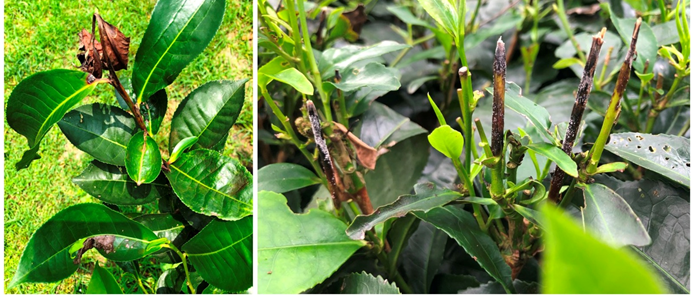
Nectria Dieback Causal Organism - Nectria spp. (N.cinnabarina (Tode ex Fr.) Fr. and Nectria sp.) Nectria sp._are mostly saprophytic wound parasites of the tea bush.The fungi generally infects grown tea plants and the immature tea plants below five years remain largely uninfected. Die back symptoms are observed in branches affected by _nectria infection [12]. The tea plant branches infected by the fungus shows gradual death from the tip or the specific point of infection backwards (Figure 1) [13]. Nectria sp. produces various kind of spores and dissemination of the disease propagules takes place as spores are carried by air currents. The imperfect stage of Nectria infection is seen as the affected stems show some minute, pink coloration in the form of a soft cushion like outgrowth. The perfect stage of infection also known as perithecia consists of small globules (about 0.2-0.4 mm) diameter which are produce singly or in groups intermingle with the conidia. _Nectria_infection in the plant can also occur within the natural or artificial openings on the plant stem.
Branch Canker
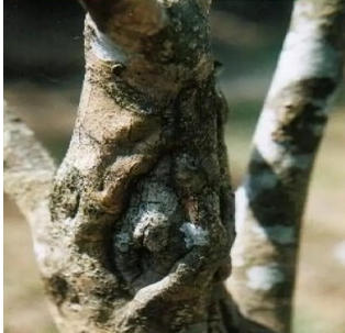
Causal Organism – Poria hypobrunnea Petch. Branch canker is a significant stem disease affecting the tea cultivation in Assam and other adjoining states of NE India. It significantly affects the physiological, phytochemical and quality properties of tea [14]. Poria hypobrunnea Petch is a wound parasite of the tea bush that causes significant loss in tea yield and initiates the infection as air-borne spores gain entrance into the tea plants through wounds, cuts and injuries especially in the thicker branches which are prone to small injury marks during heavy pruning, sun-scorch lesion, damages due to careless chopping, sawing, wrenching of branches and also due to falling of shade trees etc [15] .The disease can be identified by the presence of some partially buried lesions that are generally separated by a surrounding ring of tissue growth (Figure 2) [16]. The disease spreads below the branches affecting them one after another until it reaches the main stems [17]. It may take 8- 10 years for the pathogen to completely damage a fully grown tea bush while immature, developing bushes may be completely damaged in about 3 years [18]. The affected wood of the tea plant gradually turns yellowish, softens and gradually decays with the progression of the disease. Thin irregular brown lines can be seen in the wood. Infection sprayed through air borne spores called conidia.
Thread Blight Disease
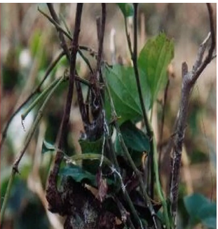
Causal Organism – Marasmius pulcher (B. & Br.) Petch. The infection is caused by a parasitic fungus belonging to the family Agaricaceae that generally affects the moribund and debilitated tea bushes in Assam and other adjoining states [19]. The disease affects young and mature tea plants causing significant crop losses. The most optimum conditions for smooth spread of this fungus are heavily shaded, damp and cool places. It has been reported that the fungus can complete its life cycle in about 57 days [20]. This fungus produces branching mycelial strands (threads) with chalky-white coloration on the stems. A crucial distinguishable feature of this disease is that as disease progresses, the chalky-white mycelial strands spread upwards along the length of the stem and reach the abaxial surface of the leaf blades (Figure 3). It was reported that the abaxial surface of older tea leaves are more prone to necrosis as compared younger and immature leaves because as the mycelial strands reach the under surface of leaves, the wider stomata of older leaves can be easily penetrated by the pathogen hypha as compared to the stomata of younger leaves. Disease spread mainly occurs via direct contact with infected plant or plant parts [21].
Pink Disease
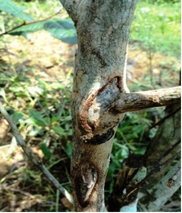
Causal Organism –Pellicularia salmonicolor (B. & Br. Rogers) and Corticium salmonicolor (B. & Br.) The causative organism of pink disease has resemblances withthe Black root rot fungi affecting tea plant which has been rarely found to attack young tea plantation in Cachar, Assam [21]. In Assam, this disease has been very commonly found on plants such as Crotoliaria anagyroids and Tephrosia candida. It forms a narrow layer of sticky white mycelial strands on the surface of stem of affected tea plant (Figure 4). In severe conditions, the fungal mycelium enters into the tissues and causes wilting of the plant [22]. Fungus fructification appear in shade trees of Crotoliaria and Tephrosis with thin rosy pink crust. The crust found to be on shaded side but sometimes surrounds the whole stem as well. Infected plants die following severe wilting.
Control measures of stem disease:
| SN | Disease | Causal Organism | Control Measures | Reference |
|---|---|---|---|---|
| 1 | Nectria Dieback | N.cinnabarina (Tode ex. Fr.) Fr. and Nectria sp. | Affected branches to be pruned atleast10 cm below die back limitduring the middle of November and end of December Use of Copper Fungicides (such as Copper Oxychloride) and Ovicides (such as Ethion) | Samah, Pandey, et al. [22] |
| 2 | Branch Canker | Poria hypobrunnea Petch. | Removal of dead wood at each pruning, protection from sun-scorch by providing adequate shade, smoothing off wounds using a knife.Thereafter, a layer of bitumen paint such as Indophalt can be applied, Avoidance of planting susceptible clones in areas with previous history of the disease Biological control using fungal antagonists such as Trichoderma sp. | Arulpragasam, et al. [23], Yao, et al. [24] |
| 3 | Thread Blight disease | Marasmius pulcher (B. & Br.) Petch. | Pruning, regular cleaning, field sanitation, minimization of over-dense shade trees to allow free air circulation. Application of 50% copper fungicides | Sarmah [21], Mouli [25] |
| 4 | Pink Disease | Pellicularia salmonicolor (B. & Br. Rogers) and Corticium salmonicolor (B. & Br.) | Diseased stems to be cut out and burnt; Field sanitation; Elimination of inoculum Spraying of 0.25% copper fungicides | Sarmah [21], Akrofi, et al. [26] and Chaliha [27] |
Root Diseases
It has been reported that root diseases affecting the tea plant are caused by parasites situated within the plant roots in the natural habitat. These root parasites pose a significant threat to the tea cultivation [28, 29]. The soil-borne fungi cause various root diseases in tea plants negatively affecting its yield and quality.
Primary Root Diseases
The Primary root diseases cause death of infected tea plants solely due to the successful fungal attack on the plant. These diseases can completely destroy fully mature plants. Wilting and dying up of foliage leaves remain as the most commonly visible symptoms of the disease. The withering of leaves proceeds slowly before eventual shedding of leaves [30]. In case of severe infections, a patch within a tea plant attacked by a primary root disease causing fungi can be identified by observing an arrangement of dead branches surrounding a center giving the appearance of a completely dead bush. These diseases infect the plants based on the plantation pattern and spacing between the plants [21].
Primary root diseases commonly occur due to the monoculture practice and also due to vegetation clearing for tea plantation. The mechanism of dissemination isvia soil or by root contact. When a healthy tea plant dies suddenly, the withered leaves remain attached to the plant for some time before eventual shedding [22]. Proper distinction between common, primary and secondary root disease is essential for prevention healthy tree plant excision from the field or spreading of an infection throughout the field [31].
Some important examples of primary root diseases in tea: Black Root Rot
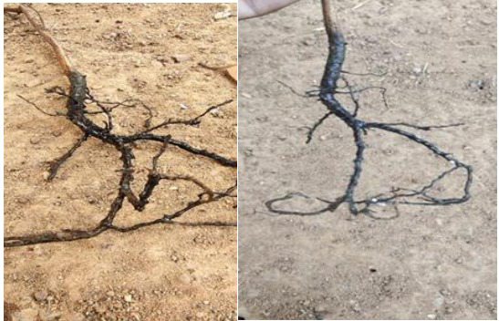
Causal Organism - Rosellinia arcuate Petch. Black root rot is an important type of root rot diseases in tea plant. The causal organism is Rosellinia arcuate Petch. The disease is considered as a serious root disease in Darjeeling region of North Bengal as well as in hilly regions of N.E. India [32]. This disease is characterized by woolly, black, non- uniform mycelia appearing on the root and collar portion of the plant after successful infection (Figure 5). ‘Star-like’ arrangement of white mycelium becomes prominent in the transition region between bark and wood. Some dash-like structures may sometimes be seen on the roots or collar [33]. The mycelial growthpersists and continues through upper soil surface particles and organic matter. The disease can spread faster under moist conditions from diseased plant to adjacent healthy plants within the field.
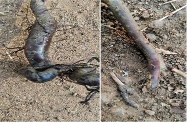
Red Root Rot
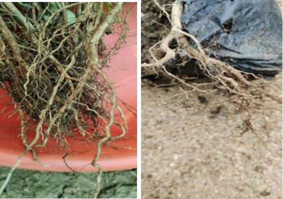
Causal Organism – Poria hypolateritia Berk It is one of the most common primary root diseases affecting tea as well as coffee plantations. The name of this disease has been derived from the red coloration of mycelial strands of the fungus visible in later stages of infection. As the disease spreads within the plant, the mycelia of the infecting fungus fuse together to form a sheet-like structure with dark mycelial strands [34]. The rhizomorphs of infected tea plants show the appearance of whitish conidial clumps. The infected plant roots when rinsed in running water and rubbed under sunlight, fungal mycelium can be seen with a reddish coloration (Figure 6). The fungal infection causes the roots to become light in weight and spongy in nature. The symptoms of successful infection become visible slowly after pathogen attack and thus the disease often remains undetected just after infection till symptoms become visible. Symptoms such as lack of thriftiness, yellow coloration of leaves and stoppage or slowness of growth are visible among immature and developing plants infected by this primary root disease. Most often, infection occurs via direct contact of infected plant propagules (inoculum) with the roots of healthy susceptible plant [35].
Brown Root Rot Disease
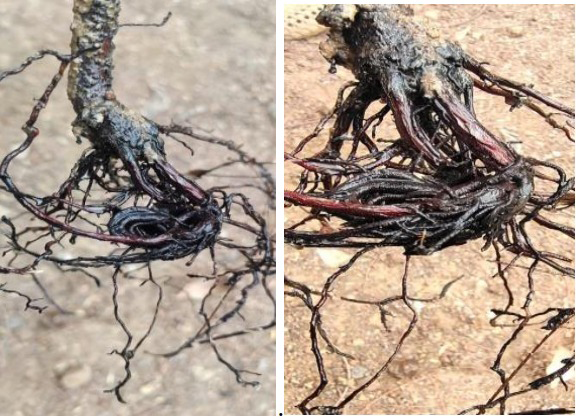
Causal Organism –Fomes lamoensis (Murr.) Sacc. and Trott The disease generally occurs in lower elevation areas under tea cultivationespecially in hills garden. It has been affecting the cultivation of tea since many decades [11]. It is found all types of soils throughout N.E. India but more common on sandy ones. The disease has been a major obstacle for tea cultivation in the districts under Barak Valley area of Assam that contributes about 7.64 % tea production in Assam [36]. This disease occurs in tea plants from about
3 years upwards but younger plants attacked easily and destroyed withina few months. Infection rapidly spreads as the roots of plants come in contact with remains of diseased plants in the soil [37]. Plants die suddenly and their dead leaves remain attached for sometimes. The disease slowly affects the plant and the mycelium produced by the fungus on the outer side are unable to spread through the soil over a distance. The primary root disease spreads from one plant to another through contact between the plant roots under the soil as the pathogen always enters through the plant roots. The external surface of the root contains fungal hyphae and mycelium that show the presence of soil particles adhered to it as the mycelium acts as a bridge to bind the soil particles with the roots of an infected plant (Figure 7) [38]. The mycelium isvisible as wool-like structures between the soil particles.24In severe cases, the mycelium may form a sheet-like structure on the stem with a blackish surface. Fructification is rarely found on diseased plants. It is bracket shaped, 3-4 inches wide and about half an inch thick, and a very hard. When mature and dry, it is purplish to dark brown in color and often in black concentric zones on the upper surface.
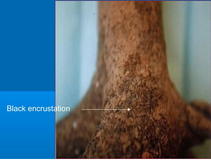
Charcoal Stump Root Rot
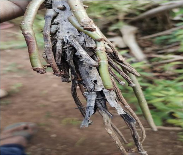
Causal Organism – Ustulina zonata (Lev.) Sacc The causative organism of charcoal stump root rot Ustulina zonata (Lev.) Sacc. has been reported to be one of the most destructive primary root diseases causing organisms [11]. The disease is prevalent in all type of soilsthroughout the tea growing regions of North-East India. The fungus attacks above three-year-old tea plantsand cause sudden death of tea bushes but the withered leaves remain attached for few days. On the other hand, sometimes in older bushes one side may be dead while other side remains looking healthy for few months [39, 40]. The fungus develops a characteristic fructification with white at early stage and later to charcoal-like, black brittle encrustation, showed wavy on the surface. The infected tea plant roots often show whitish or black-cushion like mycelial growth underneath the root bark (Figure 8). This fungus spreads through the roots, diseased woody plant or by wind borne fungal spores.
Purple Root Rot
Causal Organism – Helicobasidium compactum (Boedijin) Boedijin This is one of the less prevalent primary root diseases of tea plant. The causal organism for this disease I Helicobasidium compactum which can be found on all soils about 1-8 years of tea plant [41]. The disease however, is not common [21]. The characteristic detectable symptoms of this disease develop at the collar region of the plant which may extent up to 12-15 cm upwards as a deep brown to purple brown velvety pad. It may encircle the collar region completely or may develop on side alone. Infected plant when dug out reveals a pinkish coloration in the wood. Deep brown to purple brown mycelial cord can be noticed along the collar downwards even by naked eyes.
Tarry Root Rot
Causal Organism –Hypoxylon asarcodes (Theiss.) Mill Tarry root rot disease is prevalent in the tea growing areas where soils having sand and gravel. It is more common in West Bengal but rarely found in Assam. This disease mainly affects mature bushes and the affected plants show withering of leaves. There is no prominent external symptom shown by the affected bushes. Tea plants infected by this fungus show smooth, black crust-type structures that develops on the plant stem just above the ground level (Figure 9). Infection spreads through wind-borne spores or by contact with diseased and infected plant material.
Secondary Root Diseases
These diseases work by weakening the susceptible plant which has already been damaged or injured due to various biotic or abiotic factors. The causes of secondary infection may be a mechanical injury to the plant, drought condition leading to abiotic stress, lack of proper drainage systems in the field or hail storm or lightning. It has also been observed that presence of clayey soil is also one of the important factors behind occurrence of secondary root disease [42]. The following are some of the common secondary root diseases of tea.
Diplodia Root Disease
Causal Organism – Botryodiplodia theobromae Pat. It is one of the most common secondary root diseases affecting the tea production in North-East India. The fungusneeds a high temperature above a threshold level to cause successful infection in plants. The disease causes severe depletion of root starch reserves [43]. The most common reason for incidence of this disease is extensive harvesting practices following high demand resulting in mechanical injuries. Prolonged drought and a severe attack of pest and diseases may also enhance the chances of disease incidence. One of the most common symptoms of the Diplodia Root disease is that the external surface of infected roots shows a layer of blackish hairy cushion-like structures that can be designated as a diagnostic character of this disease.
This disease also causes the plant to look weaker and unhealthy with withered leaves. Due to this disease, formation of new shoot and leaves slow down. New shoots formed after pruning may also show visible die back symptom where death of tissues occur from the infected tip point backwards.
Violet Root Rot Causal Organism – Sphaerostilbe repens B & Br It is also another very common secondary root disease throughout North-East India. It mainly occurs in water logged and stagnant water conditions. The symptoms of this disease can be easily identified as the yellowing of infected leaves occur. As the infection intensifies and spreads throughout the plant, the bushes begin to die in a specific fashion the leaves drop off in their fresh state without any gradual withering and dying. Mycelial growth occurs on the infected roots in the form of dark violet spots or patches (Figure 10). The vinegar-like pungent smell of infected roots have been reported as another diagnostic feature of this disease. It is found most commonly on heavy soils particularly in case of flooding, back feeding of drains, collapsing or blockage of drains. It has also been observed that poor soil aeration also favors incidence of the disease.
Control Measures of Root Diseases
Diseases affecting tea plant roots are commonly seen when the plants are grown on forest land after clearing. The diseases are generally transmitted to young and healthy tea plants whenever the newly developed tea plant roots come in contact with pathogen inoculum or diseases plant remains in the field. As pathogen attack begins, it can spread to the nearby regions by root-contact or through airborne spores.
To control the root diseases of tea, there are no suitable fungicides available in the market. There is no well-known method to directly detect the disease in the roots until either the whole or some part of the tea plant dies suddenly [44]. Therefore, the first step to contain spread of root diseases was to isolate the diseased patch by digging a trench at least 3 inch deep and 1 inch wide all around bushes to uproot a complete ring of apparently healthy surrounds at the same time. Though this method produced good result, labour intensive and frequent inspection is required to keep the trenches clean. Otherwise, the disease can spread to the surrounding healthy plants [45]. In case of fields with root disease incidence, all diseased plant remains should be removed and all the wood material should be extracted from under the soil. Green crop should be grown in the tea cultivation area for two years before usual re-plantation. On the other hand, to avoid the entry of root diseases in plantations, certain preventive measures are considered pragmatic and essential. One of the crucial steps is that plants killed by primary root diseases must be completely cleared and field sanitation should be done in such a way that disease propagules are fully removed [46]. For secondary root diseases, field sanitation and removal of diseased plant materials are the best possible ways to avoid disease incidence.
Chemical control of root diseases is gaining popularity worldwide. The application of soil fumigant (Vapam and Shell DD) gave excellent control of primary root diseases such as brown and red root diseases of tea [47] 1:14 (cost : benefit ratio) has been reported with soil fumigation in comparison to other conventional treatments. Methyl bromide [48], Mancozeb or Carbendazim were found useful to controlling root diseases of tea. Antagonistic potential of several fungal species of Trichoderma and bacteria such as Pseudomonas, Bacillus against primary root diseases were reported under_in_ _vitro_conditions [49, 50, 51, 52, 53, 54].
The paper highlights some of the frequently occurring stem and root diseases of tea thatare responsible for significant setbacks in the overall tea production output of the state. Information on the causal organisms, mechanism of infection, disease cycle and possible control measures can be a reference aid for future research and exploration in this field leading towards the upliftment of the tea production and livelihood of the tea planters of Assam & North East India.
Conclusion and Future prospects
Due to prolonged monoculture practice and long- life cycle, tea has becomehighly susceptible to attack by several infectious pathogens causing different types of diseases responsible for huge crop losses. Tea diseases were scientifically described from the work Kumhar, et al. [55]. In North East India several workers such as Tunstall, et al. [56], Roy, et al. [57] described the diseases affecting tea. Gadd described diseases of tea occurring in Sri Lanka. In China Tzongmaochen, et al.. Hamaya, et al.; Tang, et al. [58] have described the various fungi and bacteria associated with the diseases of tea plants. There are about
209 genera of tea encompassing 389 species as reported by Agnihothrudu. Out of these, about190 occur in NE India. Among the fungal pathogens, the higher fungi, namely the Ascomycetes, Basidiomycetes and Deuteromycetes_comprised 73 genera. According to Li, et al. [59], Leaf diseases are the most major setback to tea cultivations worldwide. Chandra Mouli [60] stated that among the major pathogens, 37% caused various leaf diseases, 36% stem diseases and 27 % root diseases. Control of tea disease is very serious concern among the tea growers. Several workers were involved in the control of leaf diseases such as Sarmah [21]; Ordish [61]; Eden [32]; Soeb, et al. [62] and for stem and root diseases such as Sarmah [21], Satyanarayana [37]. Now a days biological control approached of root diseases is gaining popularity. Sarmah, et al. performed a comprehensive case study on the various pathways of biologically controlling tea diseases in North-East India. Baby, et al. [60], Mishra, et al. [63], Kumar, et al. Kumar, et al. [64] and Morang, et al. [40] reported the antagonistic potential of several fungal species of _Trichoderma and bacteria Pseudomonas, Bacillus against primary root disease in in vitro. A significant recent study endorsed by Department of Science and Technology (DST); Govt. of India was performed by Hazarika, et al. [65] which identified 35 (out of a total 88) isolated endophytic actinobacteria having antagonistic action against various fungal pathogens associated with diseases of tea. For biological control, detailed field evaluation is needed and efforts should be made to harness the benefits of other fungal and bacteria antagonists against the tea root disease pathogens [66, 67, 68, 69, 70, 71, 72, 73, 74, 75, 76, 77, 78, 79].
From the above study it is found that, stem and root diseases in tea is very common which can directly affect the productivity of this economically important crop. Though there are several methods that have been adopted to control the diseases in tea, keeping in view of environmental concerns more research needs to be done in order to develop an integrated disease management (IDM) approach so that the three important components of a disease triangle viz. Host, Pathogen and Environment can be suitably balanced for sustainable agricultural practices.
References
-
Kundu R, Mondal M, Garai S, Banerjee H, Ghosh D, et al. (2020) Efficacy of herbicides on weed control, rhizospheric micro-organisms, soil properties and leaf qualities in tea plantation. Indian Journal of Weed Science 52(2): 160.
-
Wang C, Han J, Pu Y, Wang X (2022) Tea (_Camellia_ _sinensis_) A review of nutritional composition, potential applications, and omics research. Applied Sciences 12(12): 5874.
-
Ye J, Wang Y, Hong L, Jia X, Kang J, et al. (2022) Improvement of soil acidification in tea plantations by long-term use of organic fertilizers and its effect on tea yield and quality. Frontiers in Plant Science 13.
-
Hamaya E (1981) Diseases of tea plant in Japan and their control. Reviews of plant protection Research 14: 96- 111.
-
Bhowmik SK (2011) Ethnicity and isolation: Marginalization of tea plantation workers. Race/ Ethnicity: Multidisciplinary Global Contexts 4(2): 235- 253.
-
Roy NC, Biswas D (2021) History of tea industry in India. _Human Resource Management in the Indian Tea_ _Industry_ pp: 12-21.
-
Parida BR, Bar S, Roberts G, Mandal SP, Pandey AC, et al. (2021) Improvement in air quality and its impact on land surface temperature in major urban areas across India during the first lockdown of the pandemic. Environmental Research 199: 111280.
-
Hazarika SN, Saikia K, Thakur D (2022) Characterization and selection of endophytic actinobacteria for growth and disease management of tea (_Camellia sinensis_ L.). Frontiers in Plant Science 13.
-
Piyasena KN, Hettiarachchi L, Edirisinghe E, Abayarathne A, Jayasinghe, W (2023) Quantification of dynamic changes of sugars during the aeration/ oxidation period of Black tea processing: A Sri Lankan study. Food and Humanity 1: 8-12.
-
Watt G, Mann HH (1903) The Pests and Blights of the Tea Plant.Government Printing Press, Calcutta, India 15: 429.
-
Petch T (1923) The diseases of tea bush. Mac Millan, London, UK, pp: 220.
-
Rattan PS, Pawsay RG (1981) Death of tea in Malawi caused by Pseudophaeolusbaudonie. Tropical Pest Management 27: 225-229.
-
Kumar BSD, Bezbaruah B (1997) Plant growth promotion and fungal pest control through an antibiotic and siderophore producing fluorescent Pseudomonas strain from tea (_Camellia sinensis_ (L) O. Kuntze) plantations. Ind J Exp Biol 35(3): 289-292.
-
Rattan PS (1992) Pest and diseases control. In: Wilson KC, Centiford MN (Eds.), Chapman and Hall, UK, pp: 331- 352.
-
Morang P, Devi SP, Doley SN (2023) Integrated approach to management of Brown root rot disease of tea (_Camellia_ _sinensis_ (L)O.Kuntze). Current Agriculture Research Journal 11(2): 468-483.
-
Borah A, Hazarika SN, Thakur D (2022) Potentiality of actinobacteria to combat against biotic and abiotic stresses in tea [_Camellia sinensis_ (L) O. Kuntze]. Journal of Applied Microbiology 133(4): 2314-2330.
-
Satyanarayana G, Barua GCS (1983) Leaf and stem diseases of tea in N.E. India with reference to recent advances in control measures. J Plantation Crops (Supplement) 11: 27-31.
-
More SJ, Bampidis V, Benford D, Bragard C, Halldorsson TI, et al. (2023) Re‐evaluation of the existing health‐based guidance values for copper and exposure assessment from all sources. EFSA Journal 21(1).
-
Sarkar S, Kabir SE (2016) A Field survey of sucking tea pests and their control measures in a few tea gardens of terai region, West Bengal, India. International Journal of Science and Research (IJSR) 5(3): 1343-1345.
-
Chen M (2021) The tea plant leaf cuticle: From plant protection to tea quality. Frontiers in Plant Science pp: 12.
-
Sarmah KC (1960) Diseases of tea and associated crops in N.E. India. ITA Memorandum 26: 44-46.
-
Pandey AK, Sinniah GD, Babu A, Tanti A (2021) How the global tea industry copes with fungal diseases – Challenges and opportunities. Plant Disease 105(7): 1868-1879.
-
Arulpragasum PV (1987) An in expensive and effective method for the control of red root disease of tea. S.L. J Tea Sci 56(1): 5-11.
-
Yao H, Su H, Ma J, Zheng J, He W, et al. (2023) Widely targeted volatileomic analysis reveals the typical aroma formation of Xinyang Black tea during fermentation. Food Research International 164: 112387.
-
Mouli BC (19990 Global status of tea diseases and management strategies. In N.K. Jain (ed). Global Advances in Tea Science. Aravali Books International (P) Ltd. New Delhi, India, pp: 647-658.
-
Akrofi AY, Amoako-Atta I, Assuah M, Kumi-Asare E (2014) Pink Disease Caused by Erythriciumsalmonicolor (Berk. & Broome) Burdsall: An Epidemiological Assessment of its Potential Effect on Cocoa Production in Ghana. J. Plant Pathol. Microb 5: 215.
-
Chaliha C, Kalita E (2020) Blister blight disease of tea: An Enigma. Diagnostics of Plant Diseases. Intechopen.
-
Butler EJ (1918) Fungal and Disease in Plants. Thacker Spinck & Co pp: 547.
-
Butler EJ, Bisby GR (1937) The fungi of India. Monogram Council Agricultural Research India II and XVII pp:237.
-
Mouli BC, Kumar PR (1997) Comparative evaluation of fungicides spray schedules against blister blight (ExobasidiumvexansMassee.) disease of tea. Pestology 21(2): 19-21.
-
Goodchild NA (1952) Stem diseases. Ann. Report Tea Research Institute of East Africa pp: 24
-
Eden T (1947) The effect of hard plucking- with special reference to blister blight. Tea Quarterly 19: 105-109.
-
Ram S, Panika S (2021) Identification of Tea Root Rots Disease Through Image Recognization in Conventional Neural Network. International Journal of Innovative Research in Technology 8(7): 445-450.
-
Mouli BC, Kumar RP (1988) Fungi occurring on tea and Grevilliarobusta in the plantation of South India. Ind. Phytopathol 41(3): 73-88.
-
Tompong S, Kunasakdakul K (2014) Causal Agent, Symptoms and Environmental Factors of Root Rot Disease of Organic Assam Tea in Mae Teang District, Chiang Mai Province. International Journal of Agricultural Technology 10(3): 767-777.
-
Mouli BC, Parthiban M (1992) Thorny stem blight disease of tea. Newsletter UPASI Tea Scientific Department Bulletin 44: 14-16.
-
Satyanarayana G (1980) Benefit evaluation of soil fumigation in root rot control in tea in NE India. Two and a Bud 27: 63-64.
-
Banerjee B (1993) Tea production and processing. Oxford and IBH Publishing Company Pvt. Ltd. Pp: 243- 248.
-
Morang P, Dutta B (2012) Growth promotion and Bi- control approaches of Brown root rot disease of tea by pseudomonas Aeruginosa. Journal of Plant Pathology & Microbiology 3(5).
-
Morang P, Kumar BS, Dutta BK (2011) _In vitro_ evaluation of antagonistic potential of Bacillus cereus isolated from Tea Rhizosphere soil in Barak Valley region of Assam. Assam University Journal of Science and Technology, Biological and Environmental Sciences. An International journal 8(1): 65-70.
-
Satyanarayana G, Barua GCS, Baruah KC (1975) Uncommon diseases. Two and a Bud 24(1): 30.
-
Yatoo AM, Ali MN, Baba ZA, Hassan B (2021) Sustainable management of diseases and pests in crops by vermicompost and vermicompost tea - A review. Agronomy for Sustainable Development 41(1).
-
Venktaram CS (1927) Control of red and brown root rot diseases in tea. Planters Chronicle 67: 163-165.
-
Bertus HL (1974) Fungicidal control of Camellia die back. J Hort Sci 49: 167-169.
-
Venkataram CS (1920) A reassessment of Diplodia root disease of tea plants. UPASI Scientific Department Tea Section 19: 18-28.
-
Saha A, Dasgupta S, Mandal P, Saha D (2005) Reduction in disease in young tea plants against Curvulariaeragrostidis by biotic and abiotic elicitors. In: Chakraborty U, Chakraborty BN (Eds.), Stress Biology, Narosa Publishing House, New Delhi, India, pp: 238-242.
-
Shanmuganathan N (1964) Recent development in the control of poria root disease. Tea Quality 35: 22-30.
-
Tunstall AC, Sarmah KC (1947) Black rot of tea in N.E. India (CorticuminvisumPetch and CorticumtheaeBernard). Memorandum No. 13, Indian Tea Association, Tocklai Experimental Station 1: 26.
-
Mouli BC (1979) Efficacy of protectant, eradicant and systemic fungicides in tea blister blight control in South India. Proceedings of plantation Crops Symposium pp: 205-211.
-
Dutta BK, Debnath S, Pradhan SK, Gurung RM (1992) Role of systemic fungicides in the management of blister blight in Darjeeling. Proceedings of 31st Tocklai Conference pp: 153-162.
-
Mouli BC, Kumar RP (1995) Hexaconazole, a novel fungicide in tea blister blight management. UPASI Tea Scientific Department Bulletin 48: 59-66.
-
Naglot A, Goswami S, Rahman I, Shrimali D, Yadav KK, et al. (2015) Antagonistic potential of native Trichodermaviridestrain against potent tea fungal pathogens in north east India. The Plant Pathology Journal 31(3): 278-289.
-
Jeyaraman M, Robert PS (2018) Bio efficacy of Indigenous biological agents and selected fungicides against branch canker disease of (Macrophomatheicola) tea under field level. BMC Plant Biology 18(1).
-
Sun Y, Wu F, Guo H, Li R, Yao J, et al. (2023) Tea Disease Net: Multi-scale self-attentive tea disease detection. Frontiers in Plant Science pp: 14.
-
Kumhar KC, Babu A, Nisha SN (2022) Management of tea (_Camellia sinensis_) diseases with application of microbes: A review. _Innovare Journal of Agricultural Sciences_.
-
Tunstall AC (1940) Notes on root diseases of tea in N.E. India.Memorandum No. 8 ITA, Tocklai Experimental Station.
-
Roy S, Barooah AK, Ahmed KZ, Baruah RD, Prasad AK, et al. (2020) Impact of climate change on tea pest status in Northeast India and effective plans for mitigation. Acta Ecologica Sinica 40(6): 432-442.
-
Tang Z, Zhu J, Song Q, Daly P, Kong L, et al. (2024) Identification and pathogenicity of Fusarium spp. associated with tea wilt in Zhejiang province, China. BMC Microbiology 24(1).
-
Li H, Shi H, Du A, Mao Y, Fan K, et al. (2022) Symptom recognition of disease and insect damage based on mask R-CNN, wavelet transform, and F-rnet. Frontiers in Plant Science 13.
-
Baby UI, Chandra Mouli B (1969) Biological antagonism of Trchoderma and Gliocladiumspp. against certain primary root pathogens of tea. Journal of Plantation Crops 24: 249-255.
-
Ordish G (1952) Untaken Harvest. Constable et Comp. Ltd. London, UK. pp:171.
-
Soeb MJ, Jubayer MF, Tarin TA, Al Mamun MR, Ruhad FM, et al. (2023) Tea leaf disease detection and identification based on YOLOv7 (YOLO-T). Scientific Reports 13(1).
-
Mishra AK, Dutta S, Kumar BSD (2005) Effect of fluorescent Pseudomonas strains on crop enhancement and suppression of root diseases of tea. In: Asian Conference on Emerging Trends in Plant-Microbe Interactions. Pp: 275-283.
-
Kumar BSD, Dutta S, Mishra AK (2004) Biocontrol of brown rot disease and improved crop production of tea by plant growth promoting rhizobacteria. Proceedings of International Conference on O-CHA (Tea) Culture and Science (ICOS 2004)Shizuoka, Japan pp: 98-101.
-
Hazarika LK, Puzari KC, Wahab S (2001) Biological control of tea pests. Biocontrol Potential and its Exploitation in Sustainable Agriculture pp: 159-180.
-
Agnohothrudu V (1964) Some aspects of Mycological investigation at Tocklai. Two and a Bud 10(4): 27-31.
-
Baxter CW (1974) Studies on the twig blight canker and die back of Camellia. American Camellia Year Book 2: 63- 75.
-
Corbu VM, Gheorghe-Barbu I, Dumbravă AS, Vrâncianu CO, Șesan TE (2023) Current insights in fungal importance—A comprehensive review. Microorganisms 11(6): 1384.
-
Das SC, Barua KC (1990) Scope of bio control of tea pests and diseases in tea plantations. Tea Research- Global Perspective Proceedings of the International Conference on R&D in Tea pp: 119-125.
-
Dutta BK, Borthakur B (1991) Some aspects of disease control in tea. Field management in Tea, Tea Research Association, Jorhat pp: 145-155.
-
Naglot A, Goswami S, Rahman I, Shrimali D, Yadav KK, et al. (2015) Antagonistic potential of native Trichodermaviridestrain against potent tea fungal pathogens in north east India. The Plant Pathology Journal 31(3): 278-289.
-
Mouli BC, Parthiban M (1991) Black root disease of tea. UPASI Scientific Department Tea Section Bulletin 44: 14- 16.
-
Niranjan D, Kumari B, Raghav YS, Mishra P, Al Khatib A, et al. (2022) Modeling And Forecasting Of Tea Production In India. Journal of Animal and Plant Sciences pp: 32.
-
Sarmah R, Bhattacharyya SN, Barooah A (2020) Microbial biocides-Viable alternatives to chemicals for tea disease management. _Journal of Biological Control_ _34_(2): 144-152.
-
Satyanarayana G (1972) Annual Scientific Report. Tea Research Association Tocklai Experimental Station 1: 98.
-
Subba MK (19360 Pink disease.Tea Scientific Department UPASI pp: 25.
-
Tunstall AC, Bose SC (1920) The fungal diseases of the tea leaf. Quart J of ITA pp: 152-154.
-
Tunstall AC (1930) Some observation on tea roots. The Indian Tea AssociationQuarterly Journal pp: 75-78.
-
Venkataram CS (1961) Application of nickel chloride to tea plants (_Camellia sinensis_) and control of blister blight. Current Science 30: 57-58.
- Enhancement of Vegetative Growth and Fruit Yield in Cucumber (Cucumis sativus L.) via Spiritual Blessing (Biofield) Energy Intervention
- Production of Açaí (Euterpe oleracea Mart.) under Different Agroforestry System Management Intensities in Amazonian Floodplain (Varzea) Forests
- Coffee and the Production Region: What is the Secret to the Expression "Quality"?
- Experiential Agripreneurship Training in Sub-Saharan Africa: Integrating a Business Incubator into Postgraduate Livestock Education at the University of Buea
- Advances in Agricultural High-Quality Development
- Linking Compost Residue to ABAGE in Plants - a Short Note