Micromorphology and Histochemistry of Vegetative Organs from Nicandra Physalodes (L.) Gaertn (Solanaceae)
Nicandra physalodes (L.) Gaertn is a sub-shrub used in folk medicine for various purposes, as it is also considered a toxic plant. Due to its ethnobotanical importance, a study of vegetative organs (leaves, stem, root) of this species was carried out, for its micromorphological and histochemical characterization, with the objective of finding parameters to support the taxonomy and quality control of their ethnodrugs. Paradermic and cross sections were carried out on fresh samples of leaves, stem and root, from the plant collected in the municipality of Bananeiras, State of Paraíba, Brazil, as well as histochemical tests with specific conductors, following the usual techniques in botanical anatomy. It was observed that N. physalodes has an amphistomatic leaf blade, anisocytic and anomocytic stomata, 1-serial epidermis, sinuous anticlinical cell walls, dorsiventral mesophyll, 1-serial palisade parenchyma and the 3-4-serial spongy border; biconvex midrib, bicolateral, arched; semicircular petiole with lateral, adaxial projections, 5-6-seriate collenchyma and vascular system similar to the midrib, 2 central bundles and 2 accessories in the lateral projections. The stem is irregularly pentagonal, epidermis 1-seriate, angular collenchyma 3-4-seriate, prismatic crystals are observed in the cortex and medullary parenchyma, and the vascular system is amphiphilic siphonostelic. The root is circular, with the endoderm 1-serial, siphonostelic vascular system. Histochemical tests showed a positive reaction for lipids, lignin, starch, alkaloids, phenolic compounds and proteins in the rib, petiole and stem. The anatomical character set of the leaf epidermis, stem and root together with the histochemical data are distinctive for N. physalodes, who support her taxonomy and the quality control of her ethnodrugs.
Introduction
Solanaceae Juss. It covers about 92 genera and 2000 species, widely distributed across continents, with its main center of diversity being South America [1]. The family has species of great economic importance, mainly used in human nutrition, such for example as tomatoes (Solanum lycopersicum L.), potatoes (Solanum tuberosum L.), eggplant (Solanum melongena L.), and species of the genus Capsicum L. (peppers). In addition, it has species of pharmaceutical importance and also species with narcotic effects such as Nicotiana tabacum L., and used in urban ornamentation, among which Petunia Juss., Solanum L. and Physalis L. [2].
Nicandra Adans is a monotypic genus of Solanaceae, whose species, Nicandra physalodes (L.) Gaertn. This species is a sub-shrub, annual, with petiolate leaves and irregularly lobed limbus, the flowers are pentamerous, lilac to blue corolla, and the fruit is a spherical berry. The species originates from the Andean region of South America, and is distributed in tropical areas [1], including Brazil, where it is widely distributed in the Northeast and Southeast regions [3].
Popularly known in Brazil as “quintilho”, the species is used in ethnomedicine as a tranquilizer and fly poison, from the decoction of the whole plant [4, 5]. Phytochemical studies of the plant revealed the occurrence of groups of steroids and phenolic amides with pharmacological effects, among them, the protection of liver cells against damage induced by oxidative stress [6]. Biological studies reveal that the species has a diuretic effect, in addition to anti-inflammatory, antimicrobial and antidiabetic activities Gupta A [7].
Plant anatomy is an important taxonomic tool, helping to characterize and distinguish between species [8]. According to Judd WS [2], the anatomical characters provide subsidies for the construction of hypotheses of phylogenetic relationships and, in addition, the analysis of the chemical compounds of the plant are also useful for the determination of phylogenetic relationships between families and other taxonomic groups. Although N. physalodes is recognized as a species of importance in folk medicine, there are few studies addressing the anatomy of the species, restricting themselves to the studies by Metcalfe CR [9] who described the epidermis and mesophyll, focusing on leaf anatomy [10].
Given the ethnomedicinal importance of the species and the limited information available on the micromorphology of the vegetative organs of N. physalodes, a micromorphological and histochemical study of the vegetative organs (leaves, stems and roots) of N. physalodes was carried out in order to find characters distinctive micromorphological features that can act as an additional tool to the taxonomy of the species, in addition to providing subsidy to the quality control of its ethnodrugs.
Material and Methods
Plant Material
Fertile samples of N. physalodes were collected in September 2019, in the municipality of Bananeiras, Paraíba, Brazil (6°43’36.7”S and 35°37’43.2”W). Part of the material collected was herborized, following the usual techniques in botanical taxonomy described by Bridson D [11], with specimens incorporated into the collection of the Herbarium Lauro Pires Xavier (JPB) of the Federal University of Paraíba, and another part fixed in a 50% FAA solution [12], for 48 hours and later stored in 70% ethyl alcohol for further anatomical studies.
Anatomical Study
Paradermal sections were made on both surfaces of the leaf blade, and transverse sections were made on leaves (petiole, midrib, mesophyll, and leaf margin), stem and root, freehand, with the aid of cutting blades on styrofoam support, following the usual techniques in plant anatomy described by Kraus JE [13]. The sections were clarified in sodium hypochlorite solution (10%), washed three times in distilled water, for five minutes, then neutralized with acetic acid (0.1%) and washed again in distilled water. The paradermic sections were stained with safranin (1%), while the cross sections were stained in safrablue, and finally mounted between slides and coverslips with glycerin (50%), analyzed and photographed under an optical microscope (Leica DM 750), fitted with a video camera (Leica ICC50 HD). The terminology of the epidermal patterns and stomata types follows [14].
Both surfaces of the dehydrated leaflet samples (approximately 1 cm2) were examined after attaching them to aluminum stubs with double-faced adhesive tape, metallizing with gold (a ca. 24 nm thick layer), and scanned using JEOL JSM-5600 scanning electron microscopy (SEM) at 15 kV. The terminology of the epicuticular wax follows [15, 16, 17].
Histochemical Tests
Cross sections were performed on fresh samples of the leaf (midrib and petiole), stem and root. The samples were treated with specific reagents to detect the presence of the following substances: lipids with Sudan IV [18]; proteins with Xilidine Ponceau [19]; starch with Lugol [12]; alkaloids with Wagner Reagent [20]; lignified elements with Phloroglucine Acid [12], and non-structural phenolic compounds with Ferric Chloride [12].
Results and Discussion
Epidermis
Scanning Electron Microscopy (SEM): Scanning electron microscopy (SEM) showed some types of epicuticular waxes on the leaf surface of Nicandra physalodes. Among them there are the granules type (Figure 1a, orange arrow), the granules are irregular crystalloids, generally round with and isodiametric. Smooth layers (Figure 1a, blue arrow), also observed are continuous covering waxes, which do not show prominence because they measure less than 1 micrometer. While the crusts (Figure 1b, green arrow), which are also continuous covering waxes, however with greater thickness, reaching more than 1 micrometer and slight prominence. Mev also allowed the identification of fissured layers (Figure 1a, red arrow), this type of wax is thicker and crusty, with conspicuous fractures. In addition to these, membrane platelets (Figure 1c, yellow arrow) and visibly open and closed stomata (Figure 1d, purple arrow) were also identified. Membrane platelets are flat, membranous, usually interconnected crystalloids.
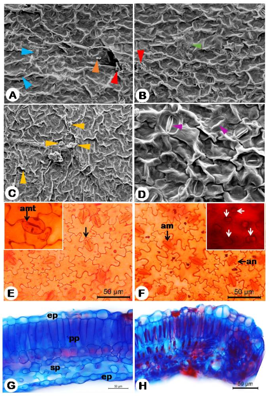
Figure 1: Leaf epidermis of Nicandra phisalodes observed by scanning electron microscopy (SEM) and light microscopy (LM). A-D: Epicuticular waxes: A. Epidermal surface showing granules (orange arrow), smooth layers covering stomata(blue arrow) and fissured layers (red arrow); B. crusts (green arrow) and fissured layers (red arrow); C. membrane platelets (yellow arrow); D. Detail of the epidermis highlighting open and closed stomata (purple arrow); E. adaxial surface showing anomotetracytic stomata (amt), F. Abaxial surface evidencing anisocytic (an), anomocytic (am) and prismatic crystals (arrow); G. Dorsiventral heterogeneous mesophyll showing epidermis ep), paliçadic parenchyma (pp), spongy parenchyma (sp); H. Rounded board fledged leaf to the abaxial face.
Ecologically, epicuticular waxes perform protective functions for plants, acting as a barrier against abiotic and biotic factors, such as against light, perspiration and pathogens [21]. However, waxes can be explored from another perspective, such as their taxonomic value, as emphasized by Barthlott W [16, 17], who considers waxes as an element of great systematic significance, even took care to classify types and terminologies for waxes that are characteristic of specific botanical families. The fissured layers and crusts, recorded here in N. physalodes, are commonly found in succulent species (Asclepiadaceae, Cactaceae), characterizing themselves as a taxonomically important wax for the recognition of species of this group [16, 17].
Although these waxes are generally found in these families, they can also occur in other groups of angiosperms, as is being referred directly here to Nicandra and also to Solanaceae. Studies on epicuticular waxes in Solanaceae are mostly about their chemical composition, which has also been used as taxonomic support, which are chemotaxonomic studies. In this research bias, we can highlight the work Khan Z [22] who researched the chemical composition of epicuticular waxes of the Solanum nigrum complex and concluded that based on the constituents of the waxes, a multivariate analysis indicated distances between the morphotypes and were interpreted as distinct species, while other morphotypes were closer, which still indicates that they are a variety or subspecies of Solanum nigrum. Waxes are therefore important for taxonomic support studies, whether with morphological or chemical approaches. Thus, the results reported here for the first time for Nicandra physalodes add additional information for the recognition of the species and Solanaceae.
In a paradermal section, the leaf epidermis of N. physalodes is glabrous and has sinuous anticlinal cell walls on both surfaces (Figures 1e-f). In cross-section, the epidermis is uniseriate on both surfaces (Figures 1g-h), with rectangular and rounded cells, being larger on the adaxial side and covered by a thin cuticle. The sinuous pattern of the cell walls observed on both surfaces of N. physalodes coincides with that previously recorded for the species by Nurit-Silva K [10], being the most frequent pattern in Solanaceae [8], and observed in other genera of the family, such as Nicotiana L. [23], Capsicum [24], and Solanum [25, 26].
The leaf blade is amphistomatic (Figures 1a-b), with anisocytic, anomocytic and anomotetracytic stomata on both surfaces, occurring at the level of the epidermal cells. The amphistomatic pattern with anisocytic and anomocytic stomata on both surfaces was previously observed for the species by Ferreira EA [9]and Nurit-Silva K [10], this stomata distribution pattern being common, but not the only one, in Solanaceae species, such as Nicotiana glauca [23], S. polytrichum [25], S. cappsicoides All [27]. Nurit- Silva K [10] também registram a presença de estômatos anisociticos e anomociticos em N. physalodes, entretanto o tipo anomotetracítico é citado aqui pela primeira vez para espécie.
Mesophyll and Leaf Margin: The leaf, in cross section, presents a dorsiventral mesophyll (Figure: 1g-h), with a palisade parenchyma uniseriate and the 3-4-seriate of spongy parenchyma, with cells of irregular sizes. According to Metcalfe CR [8], the dorsiventral mesophyll with palisade parenchyma is a common pattern in the Solanaceae family, previously observed in N. physalodes by Ferreira EA [9, 10], and in other genus of family [22, 26, 28, 29, 30, 31, 33] The leaf margin has a rounded edge, slightly revolute (Figure 1h). Maple anatomy had not been previously studied for N. physalodes by Ferreira EA [9] and Nurit-Silva K [10]. The pattern observed for the species coincides with that observed for Nicotiana glauca by Nurit-Silva K [23].
Midrib: The midrib had a biconvex contour (Figure 2a), in cross-section, with uniseriate epidermis, followed by angular collenchyma, 3-4-seriate (Figure 2b), and the fundamental parenchyma consisting of rounded cells. The outline of the midrib observed here corroborates that recorded for N. physalodes by Nurit-Silva K [10], as well as for other Solanaceae species, such as Solanum lycocarpum [29], Physalis angulata [10], Solanum paniculatum L. and S. rhytidoandrum Sendtn [23]. The contour pattern of the plano- convex type is also recorded in species of the family, as in Nicotiana glauca [23]. The angular collenchyma observed in the midrib, petiole and stem of N. physalodes was previously recorded for another genus of Solanaceae, such as Nicotiana [23], Solanum L. [31, 32], and Hyoscyamus L [34].
The vascular system is of the bicollateral type (Figure 2c), with a single central bundle, in the shape of an arch. Idioblasts of prismatic crystals were observed in the fundamental parenchyma and in the vascular system (Figure 2d). The bicollateral vascular system is a common feature of species in the Solanaceae family [8], and was previously observed in N. physalodes by Nurit-Silva K [10]. Prismatic crystals are cited here for the first time in N. physalodes, not being observed in previous studies for the species [9, 10].
Histochemical tests revealed the presence of a lipid cuticle in the epidermis (Figure 2e) In the vascular system, lignified xylem was observed, while in the fundamental parenchyma the presence of alkaloids (Figure 2f), Proteins in the parenchymal and perivascular region (Figure 2g), phenolic compounds and starch grains (Figure 2h) was registered in the endoderm.
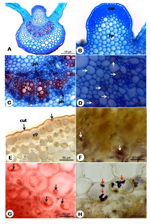
Figure 2: Anatomy and histochemical of the main ribs of Nicandra Physalodes. A. Overview of the ribs showing biconvex format; B. Detail of adaxial convexity by highlighting the angular collenchyma (Col); C. Detail of the bouncing vascular system highlighting external phloem (ph), xylem (xy) and internal phloem (ph). D. Detail of the vascular region highlighting prismatic crystals (arrows). E-H. Histochemical Tests: E. Positive reaction for lipids showing orange cuticle (arrow); F. Vascular region showing alkaloids; G.Proteins being evidenced in the parenchyma and vascular region (arrows); H. Endoderm showing starch grains (arrow).
Petiole: The petiole, in cross-section, is adaxially winged and has a semicircular outline (Figure 3a). The cortical region presents the same characteristics observed in the main vein (Figure 3b). The bicollateral vascular system has four bundles, two of which are larger central, in the shape of a semi-arc, and two smaller, rounded accessories, in the lateral wings (Figure 3c). Idioblasts containing prismatic crystals occur in both outer and inner phloem (Figure 3d). The winged petiole is a taxonomic character of N. physalodes, corroborating what was registered by Nurit-Silva K [10], including the organization of the vascular system. In Nicotiana glauca, Nurit-Silva K [23]
observed the occurrence of a central beam in the form of an open arch, while in Physalis angulata three central beams in the form of an arch are observed [10]. In both species smaller bundles are observed at the ends of the petiole, as observed in N. physalodes. Histochemical tests showed the presence of a lipid cuticle in the epidermis, lignified xylem in the vascular bundle (Figure 3e), proteins in the parenchyma and xylem (Figure 3f), in addition to phenolic compounds, alkaloids (Figure 3g) and starch grains (Figure 3h) in the parenchyma and endoderm.
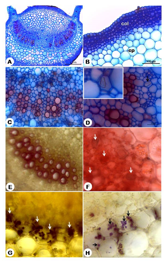
Figure 3: Anatomy and histochemistry of the petiole of Nicandra physalodes. A. General view of petiole showing winged biconvex shape; B. detail of the cortical region highlighting the angular collenchyma (col) and cortical parenchyma (pc); C. Detail of the vascular system showing phloem (ph) and xylem (xy); D. Detail of the parenchyma and phloem region showing prismatic crystals (cp)arrow. E-H. Histochemical tests: E. Positive reaction for acid floruglucine, showing lignified xylem; F. Proteins in the vascular region (arrow); G. Positive reaction to alkaloids in endoderm; H. Starch grains in the endodermis.
Stem: The stem, in cross-section, has an irregularly pentagonal outline (Figure 4a). The uniseriate epidermis is formed by tabular cells, covered by a thin cuticle (Figure 4b), and presents sparse, simple, biseriate and stipitate glandular trichomes (Figure 4c). In the wider portions of the sinuosities of the stem, the cortex was observed with 3-4 strata of angular collenchyma, similar to that of the petiole, followed by a fundamental parenchym. In the deeper portions of the sinuosities, the cortex is filled only by 2-5 strata of fundamental parenchyma, where crystalline sand idioblasts and prismatic crystals are observed (Figure 4c). The irregularly pentagonal outline of the stem observed in N. physalodes differs from the circular pattern observed in other groups of Solanaceae, such as in the genera Cestrum L. and Sessea Ruiz, et al. [35], Datura L. [36], and Physalis [37]; and the quadrangular pattern observed in Capsicum and Solanum species [36]. Thus, the outline of the irregularly pentagonal stem of N. physalodes presents taxonomic potential for the recognition of the species.
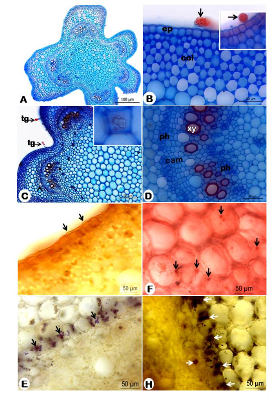
Figure 4: Anatomy and histochemistry of the stem of Nicandra physalodes. A. Overview of the stem showing irregular grooved pentagonal shape; B. Detail of the cortical region showing uniseriate epidermis (ep), glandular trichome (tg) and angular collenchyma (col), C. Wide view showing glandular trichomes (gt) in the epidermis, cortical region, vascular bundles (vb) and prismatic crystals; D. Detail of the vascular system, shown external phloem (ph) cambium (cam), xylem (xy) and internal phloem (ph). E-H Histochemical tests: E. Positive reaction for lipids, showing cuticle (arrow); F; Positive reaction for proteins (arrows); G. Starch grains in the endodermis; H. Positive reaction for alkaloids.
The vascular system is of the siphonostelic-amphiphloic type (Figure 4d), formed by five free bundles, organized close to the projections (Figure 2d). The medullary parenchyma is well developed, formed by irregularly rounded cells and occupying most of the stem (Figure 2c). The vascular organization of the stem of the siphonostelic-amphiphloic type observed in N. physalodes, was previously recorded in other species of Solanaceae, as in Solanum torvum Sw. [32] and in species of the Cestreae tribe occurring in Argentina [35]. Prismatic crystals were observed in the cortex and medullary parenchyma (Figure 2c). Idioblasts of the prismatic crystal type observed in this study in the midrib, petiole and stem of N. physalodes, had not been previously reported for the species [10], which is an additional character observed here, with taxonomic potential for recognition of the species. In Solanaceae, crystalline sand idioblasts are commonly observed [23, 31, 33], and more rarely drusen [28, 32]). Histochemical tests showed lipid cuticle in the epidermis (Figure 4e), lignified xylem in the vascular bundle, proteins (Figure 4f) and phenolic compounds in the cortical region, in addition to starch grains (Figure 4g) and alkaloids (Figure 4g) in the fundamental parenchyma and endoderm.
Root: The root in the primary structure, in cross-section, has a circular outline (Figure 5a). The circular contour observed in the root of N. physalodes corroborates with that registered in other Solanaceae species, such as in S. caavurana [31] and S. torvum [32]. Adjacent to the uniseriate epidermis (Figure 5b), the 4-5-stratified cortical parenchyma is observed, followed by the uniseriate endodermis (Figure 5b).
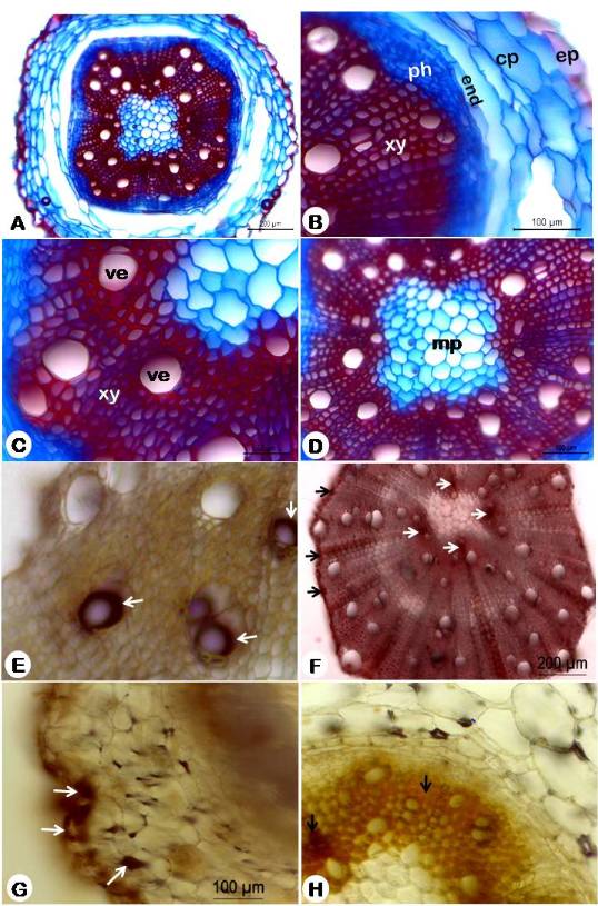
Figure 5: Anatomy and histochemistry of Nicandra physalodes root. A. Overview of root showing circular shape; B. Detail of the cortical and vascular region showing uniseriate epidermis (ep), cortical parenchyma (pc), endoderm (end), xylem (phloem) and xylem (xy); C. Detail of the xylem showing vessel elements (ev); D. Detail of the central region showing reduced pith (mp); E-H Histochemical tests: E. Test with acidic phloruglucine showing elements of lignified xylem; F. Proteins being evidenced in the perivascular region of the xylem, xylem rays and points close to the medulla (arrow); G. Phenolic compounds highlighted in the root epidermis and cortical parenchyma (arrow); H. Positive reaction for alkaloids in the root xylem (arrow).
The vascular system has a siphonostelic organization, with a single circular vascular bundle (Figure 5a). The phloem is obliterated towards the cortical portion and the xylem is well developed in the central region, showing rays and vessel elements (Figure 5c). The vascular system with siphonostelic organization observed in N. physalodes differs from that registered in other species of Solanaceae. Protostelic vascular system formed by a massive cylinder of xylem (Figure 5c) in the central portion, without the occurrence of medullary parenchyma, is a common feature in Solanum species, with tetrarch edges in S. rhytidoandrum [23] and S. torvum [32], and hexarch edges in S. paniculatum [23] and S. caarvurana [31]. The medullary portion is reduced, formed by smaller parenchyma cells, close to the xylem, larger and more rounded in the medullary portion (Figure 5d).
Histochemical tests showed lignified xylem elements (Figure 5e) and starch grains and protein in the perivascular region of the root (Figure 5f). Phenolic compounds were found sparsely in the epidermis and part of the cortical region (Figure 5g), while alkaloids were identified only in the xylem region of the root (Figure 5h). The presence of starch grains in the cortical parenchyma of the roots is a characteristic of the Solanaceae family [8], also observed in Solanum caavurana [31] S.paniculatum and S. rhytidoandrum [23].
Conclusion
Nicandra physalodes has characteristics common to Solanaceae species, such as the sinuous contour of the anticlinal epidermal walls, anisocytic and anomocytic stomata, uniseriate epidermis, biconvex main vein, angular collenchyma and collateral vascular system. However, a set of characters such as anomotetracytic stomata, adaxially winged petiole, irregular pentagonal contour in the stem, organization of the siphonostelic vascular system in the root, and the occurrence of prismatic crystal-type idioblasts, present taxonomic potential for species identification and distinction. from other Solanaceae genera. Histochemical tests reveal the presence of common metabolites in Solanaceae species, especially alkaloids, a substance of great importance in the family. In this way, the micromorphological study of the vegetative organs of N. physalodes revealed a set of useful characters to its taxonomy, contributing to the quality control of its ethnodrugs.
Acknowledgment
The Federal University of Paraíba (UFPB) and the National Council for Scientific and Technological Development (CNPq) for the scholarships granted to the authors.
Conflict of Interest
The authors declare no competing interests.
References
-
Davenport LJ (2004) Genera Solanacearum: the genera of Solanaceae illustrated, arranged according to a new system by Armando T. Hunziker Syst Bot 29(1): 221-222.
-
Judd WS, Campell CS, Kellogg EA, Stevens PF, Donoghue MJ (2009) Sistemática vegetal: um enfoque filogenético. Artmed, Porto Alegre in 3rd (Edn.).
-
Flora e Funga do Brazil.
-
Agra MF, Baracho GS, Nurit-Silva K, Basílio IJL, Coelho VPM (2007) Medicinal and poisonous diversity of the flora of “Cariri Paraibano”, Brazil. J Ethnopharmacol 111(2): 383-395.
-
Agra MF, Nurit-Silva K, Basílio IJL, Freitas PF, Barbosa- Filho JM (2008). Survey of medicinal plants used in the region Northeast of Brazil. Rev Bras Farmacogn 18(3): 472-508.
-
Wang L (2018) Withanolides isolated from Nicandra physaloides protect liver cells against oxidative stress- induced damage. J Funct Foods 40: 93-101.
-
Gupta A, Singh P, Trivedi N, Jha KK, Kumar S, Singh B (2014) A review on pharmacognostical and pharmacological activities of plant Nicandra physalodes. Pharma Res 11(1): 42-47.
-
Metcalfe CR, Chalk L (1951) Anatomy of the dicotyledons: leaves, stem and wood in relation to taxonomy, with notes on economic uses. Clarendon Press, Oxford 26(3): 294.
-
Ferreira EA, Procópio SO, Silva EAM, Silva AA, Rufino RJN (2002) Estudos anatômicos de folhas de plantas daninhas: I-Nicandra physaloides, Solanum viarum, Solanum americanum e Raphanus raphanistrum. Planta Daninha 20(2): 159-167.
-
Nurit-Silva K, Agra MF (2005) Estudo farmacobotânico comparativo entre Nicandra physalodes e Physalis angulata (Solanaceae). Rev Bras Farmacogn 15(4): 344- 351.
-
Bridson D, Forman L (1992) The herbarium handbook. Royal Botanic Gardens, Kew.
-
Johansen DA (1940) Plant microtechnique. McGraw-Hill, New York.
-
Kraus JE, Arduin M (1997) Manual básico de métodos em morfologia vegetal. Universidade Federal do Rio de Janeiro, Rio de Janeiro.
-
Dilcher DL (1974) Approaches to the identification of angiosperm leaf remains. Bot Rev 40: 1-157.
-
Barthlott W (1994) Epicuticular wax ultrastructure and systematics. In: Behnke HD, Mabry TJ (Eds.), Caryophyllales. Springer, Berlin, pp: 75-86.
-
Barthlott W, Neinhuis C, Cutler D, Ditsch F, Meusel I, et al. (1998) Classification and terminology of plant epicuticular waxes. Biol J Linn Soc 126(3): 237-260.
-
Barthlott, W, Neinhuis, C, Cutler, D, Ditsch, F, Meusel, I, et al. (1998) Classification and terminology of plant epicuticular waxes. Botanical journal of the Linnean society 126: 237-260.
-
Pearse AGE (1972) Histochemistry: theoretical and applied. The Williams & Wilkins Company, Baltimore.
-
Vidal BC (1970) Dichroism in collagen bundles stained with xylidine-Ponceau 2R. Ann Histochim 15(4): 289- 296.
-
Furr M, Mahlberg PG (1981) Histochemical analyses of laticifers and glandular trichomes in Cannabis sativa. J Nat Prod 44(2): 153-159.
-
Sharma, P, Kothari SL, Rathore M, Gour V (2018). Properties, variations, roles, and potential applications of epicuticular wax: a review. Turkish Journal of Botany 42(2): 135-149.
-
Khan, Z, Ahmad M, Kashmiri MA, Yasmin S, Asghar MN, et al. (2010) Epicuticular waxes from Solanum nigrum complex: Chemotaxonomic implications. Asian Journal of Chemistry 22(4): 2919.
-
Nurit-Silva K, Agra MF, Baracho GS, Basílio IJL (2007) Estudo farmacobotânico de folhas de Nicotiana glauca (Solanaceae). Lat Am J Pharm 26(3): 499-506.
-
Zhigila DA, Sawa FBJ, Aluko TA, Oladele FA, Rahaman AA (2015) Leaf epidermal anatomy in five varieties of Capsicum annuum L. Solanaceae. Am J Exp Agric 5(4): 392-399.
-
Nurit‐Silva K, Agra MF (2011) Leaf epidermal characters of Solanum sect. Polytrichum (Solanaceae) as taxonomic evidence. Microsc Res Tech 74(12): 1186-1191.
-
Sampaio VS, Araújo ND, Agra MF (2014) Characters of leaf epidermis in Solanum (clade Brevantherum) species from Atlantic forest of northeastern Brazil. S Afr J Bot 94: 108-113.
-
Dharman AK, Anilkumar M (2018) Pharmacognostic studies in Solanum capsicoides all. J Pharmacogn Phytochem 7(4): 397-410.
-
Reis C, Sajo MG, Stehmann JR (2002) Leaf structure and taxonomy of Petunia and Calibrachoa (Solanaceae). Braz Arch Biol Technol 45(1): 59-66.
-
Elias SR, Assis RM, Stacciarini-Seraphin E, Rezende MH (2003) Anatomia foliar em plantas jovens de Solanum lycocarpum A. St.-Hil. (Solanaceae). Revista Brasil Bot 26(2): 169-174.
-
Nurit-Silva K, Costa-Silva R, Basílio IJ, Agra MF (2012) Leaf epidermal characters of Brazilian species of Solanum section Torva as taxonomic evidence. Botany 90(9): 806-814.
-
Nurit-Silva K, Agra MF (2009) Estudo morfoanatômico de órgãos Vegetativos de Solanum caavurana Vell. (Solanaceae). Lat Am J Pharm 28(5): 675-681.
-
Nurit-Silva K, Costa-Silva R, Coelho VP, Agra MF (2011) A pharmacobotanical study of vegetative organs of Solanum torvum. Rev Bras Farmacogn 21(4): 568-574.
-
Araújo ND, Coelho VPM, Agra MF (2010) Estudo farmacobotânico comparativo de folhas de Solanum crinitum La., Solanum gomphodes Dunal e Solanum lycocarpum A. St.-Hil, Solanaceae. Rev Bras Farmacogn 20(5): 666-674.
-
Satıl F, Aslan M, Erdoğan E, Polat R, Selvi S (2015) Comparative anatomical studies on some species of Hyoscyamus L. (Solanaceae) growing in Turkey. Bangladesh J Bot 44(1): 37-43.
-
Liscovsky IJ, Cosa MT (2005) Anatomía comparativa de hoja y tallo en los representantes de Cestreae G. Don (Solanaceae) de Argentina. Gayana Bot 62(1): 33-43.
-
EI-Nagdy G, Yossef FA, Abou-Bakr MHA, Ahmed AM (2005) Morphological and anatomical studies on the stem of some Solanaceae genera. J Plant Prod 30(11): 6669-6686.
-
Săvulescu E, Hoza G (2011) Anatomy study of Physalis peruviana L. species (Solanaceae). Lucrări Științifice USAMV București. Seria B, Horticultură 55: 643-646.
- Evaluation of Comparative Morphological and Phytochemical Studies on the Seeds Extracts of Cocos nucifera (L.) and Elaeis guineensis Jacq. (Arecaceae)
- Aborted Spores in Argentine Ferns
- Ficus middletonii Chantaras. – A New Distributional Record For Central India
- A Study on the Phytochemical, Antioxidant and Antimicrobial Activities of Ficus glumosa Delile (Moraceae)
- Paclobutrazol (PBZ) and its Metabolic Function in Agriculture: A Review
- Decalepis arayalpathra: Ethnobotany, Scientific Interventions and Prospects