The Wnt Pathway Components are Upregulated in Different Molecular Subtypes of Invasive Ductal Carcinoma Patients of Breast
Wnt/β-catenin pathway is a key pathway associated with the development of breast cancer. Hence, the aim of this study was to investigate the expression of Wnt pathway components in primary invasive ductal carcinoma (IDC) patients of breast and explore their relation with clinico-pathological parameters as well as the prognostic significance. The study comprised of 160 IDC patients classified into different molecular subtypes and who have undergone surgery as the primary treatment. The expression of important pathway components- Fzd7, β-catenin and cyclin D1 was detected by immunehistochemical staining and analyzed using H-score method. Statistical analysis was carried out using SPSS and p≤0.05 were considered significant. Fzd7 was significantly over-expressed in breast tumours with smaller size and metastatic lymph nodes. The expression of Fzd7, β-catenin and cyclin D1 were associated with grade 2 tumours than grade 3 tumours. Upon correlating with the hormone receptors, the molecules were found to be positively related to the receptor positivity. Also, the patients comprising Luminal subtypes exhibited higher expression of the molecules than the triple negative breast cancer (TNBC) subtype. Finally, Fzd7 and cyclin D1 emerged as the significant prognosticator of overall survival (OS) in patients with grade 3 tumours and TNBC, respectively. Wnt pathway components are up regulated during the early stages of the breast cancer development with a loss during the disease advancement. Althoughthe pathway failed to predict the prognosis in total breast cancer patients, in patient subgroups having grade 3 tumours and TNBC, Fzd7 and cyclin D1 were found to be significant predictors of OS.
Introduction
Breast cancer has thrived to become the most common cancer in women with the second leading cause of cancer mortality worldwide [1]. The clinical parameters and pathological markers (estrogen receptor [ER], progesterone receptor [PR] and human epidermal growth factor 2 [Her2]), are routinely used by the clinicians to stratify patients for prognostic predictions and to select treatments [2]. However, inspite of various treatment modalities, breast cancer rate keeps on increasing due to the complexity of the disease. This is due to the association of a number of pathways in the development and progression of breast cancer [3]. Wnt/β-catenin signalling pathway is one of such pathways which is actively engaged in various stages of development including adult tissue organization [4]. The binding of the Wnt ligand to its receptor Frizzled (Fzd) and co-receptors at the cell membrane initiates several downstream pathways. Activation of β-catenin- dependent canonical pathway results into cytoplasmic accumulation of β-catenin and thereby its nuclear translocation. This subsequently transcribes the target genes of the pathway resulting in carcinogenesis [5]. The role of aberrant Wnt signaling is most prominent in colorectal cancer, but it is also observed in many cancer types such as non-small cell lung cancer, prostate cancer, breast cancer and so on [6]. However, the role of Wnt signaling in breast cancer is controversial with few reports showing Wnt pathway mutations in only rare cases [7]. Hence in the present study, we aimed to determine the incidence of the Wnt pathway in primary breast cancer patients by analyzing the expression of important pathway components. We studied the expression and sub- cellular localization of the Wnt ligand receptor- Frizzled7 (Fzd7), the principle protein of the pathway- β-catenin and the activated target gene- cyclin D1 in invasive ductal carcinoma (IDC) patients of breast. Further, the protein expression of these molecules was correlated with different clinicopathological parameters, hormone receptors and molecular subtypes. Also, the prognostic significance of these molecules was studied in the breast cancer patients.
Materials & Methods
Patients
A total of 160 untreated histologically confirmed breast cancer patients with Invasive Ductal Carcinoma (IDC) type registered at Gujarat Cancer & Research Institute from March 2014 to December 2015 were enrolled in the present study. The written consent forms were obtained from all the patients prior to treatment administration and the study was approved by the Institute’s Ethics Committee Board. Detailed clinical and demographic history of the patients was obtained from the case files maintained at the Medical Record Department of the institute. Histopathological details such as tumour size, lymph node status, disease stage, BR score, histological grade and status of ER, PR, Her2 were evaluated and reported on routine basis by the pathologists of our institute. The patients were classified into different molecular subtypes based on the expression of hormone receptors and Her 2 in breast tumours, where Luminal A subtype comprised patients with ER+, PR+/-, and Her2-, Luminal B subtype comprised of patients with ER+, PR+/-, and Her2+, Her2 positive subtype were patients having ER-, PR-, and Her2+ while those who were negative for all the three receptors were classified as TNBC patients. Detailed clinico-pathological characteristics of the enrolled patients are enlisted in Table 1. Complete follow- up details of 69% (111/160) patients were obtained, who were included in overall survival (OS) analysis. Amongst these, 3% (2/111) patients had persistent disease and hence only 68% (109/160) patients were included for the analysis of relapse free survival (RFS).
| Variables | N (%) | ||||
|---|---|---|---|---|---|
| Total patients | 160 (100) | ||||
| Age (years) | |||||
| ≤50 | 81 (51) | ||||
| >50 | 79 (49) | ||||
| Menopausal Status | |||||
| Pre-menopausal | 56 (35) | ||||
| Post-menopausal | 104 (65) | ||||
| Tumour size | |||||
| T1 (≤20 mm) | 21 (13) | ||||
| T2 (20-50 mm) | 113 (71) | ||||
| T3 (>50 mm) | 17 (11) | ||||
| T4 (Extension to chest wall and/or skin) | 09 (05) | ||||
| Node involvement | |||||
| Negative | 65 (41) | ||||
| Positive | 95 (59) | ||||
| TNM Stage | |||||
| I | 12 (08) | ||||
| II | 86 (54) | ||||
| III | 61 (38) | ||||
| IV | 01 (06) | ||||
| Tumor Grade | |||||
| Grade 1 | 13 (08) | ||||
| Grade 2 | 103 (64) | ||||
| Grade 3 | 44 (28) | ||||
| ER | |||||
| Negative | 72 (45) | ||||
| Positive | 88 (55) | ||||
| PR | |||||
| Negative | 100 (62) | ||||
| Positive | 60 (38) | ||||
| Her2-neu | |||||
| Negative | 91 (57) | ||||
| Positive | 69 (43) | ||||
| Molecular subtype | |||||
| Luminal A | 52 (33) | ||||
| Luminal B | 36 (22) | ||||
| Her2-positive | 35 (22) | ||||
| TNBC | 37 (23) | ||||
| Treatment administered | |||||
| S | 11 (07) | ||||
| S+CT | 45 (28) | ||||
| S+CT+RT | 27 (17) | ||||
| S+CT+HT | 35 (22) | ||||
| S+CT+RT+HT | 42 (26) |
Table 1: Clinicopathological characteristics of IDC patients
Immunohistochemistry
For immunohistochemical (IHC) study, formalin-fixed paraffin embedded tissue blocks of the enrolled patients were retrieved from the tissue repository of our institute’s Pathology Department. The blocks were cut into 4 μm sections and placed on 3-amino propyl triethoxy silane (APES)-coated slides. IHC staining was performed for the detection of Fzd7, β-catenin and cyclin D1 using HRP/DAB (ABC) Detection IHC kit (Abcam) according to manufacturer’s protocol. Briefly, antigen retrieval treatment was given by heating the sections in 10 mM sodium citrate buffer (pH-6.0) in a pressure cooker. Then after, sections were incubated overnight at 40 C with respective primary antibodies procured commercially, after appropriate dilutions: Fzd7 (Aviva Systems Biology, USA; 1:100), β-catenin (Genetex Inc, USA; 1:300) and cyclin D1 (Biogenex, USA; 1:50). The stained sections were mounted with DPX and observed under the light microscope. A section with intense staining for a given marker was used as a positive control, whereas negative control was obtained by omission of primary antibody.
Assessment of Fzd7, β-Catenin and Cyclin D1 Expression
The stained sections were evaluated immunohistochemically by semi-quantitative method. The scoring was done independently in a blinded fashion on the basis of staining intensity and percentage of positive cells. The staining intensity was scored on a scale of 0-3 where 0 indicated no staining obtained, 1+ for weakly stained cells, 2+ for moderately stained cells and 3+ for strong intense staining of the cells. The extent of staining was expressed by percentage of positive cells (0- 100%) by 10% intervals. The final histoscore (H-score) was calculated by multiplying the staining positivity score with the staining intensity score of each section, ranging from 0 to 300. The mean value of the H-score for each of the studied protein (i.e. Fzd7, β-catenin, cyclin D1) was set as the cut-off value. The calculated mean for Fzd7 score was 130, β-catenin was 169 and cyclin D1 was 162. The H-score values above this cut-off were considered as the over-expression/upregulation of the protein and the values below the cut-off were considered as low expression.
Statistical Analysis
The data was analyzed statistically using SPSS Inc.
version 23 software. The correlation between the
expression of studied parameters and various clinicopathological characteristics of breast cancer
patients was determined by two-tailed chi square test (χ2)
and spearman’s correlation. Survival analysis was
performed using Kaplan-Meier survival function and the
differences in survival were tested for statistical
$$ \text{significance using log-rank statistic. } p \leq 0.05 \text{ was} $$
considered to be statistically significant.
Results
Incidence of Fzd7, β-Catenin and Cyclin D1
The immunostaining pattern of Fzd7 protein was intensely membranous, that of the β-catenin protein was found to be cytoplasmic whereas for cyclin D1 protein, it was in the nucleus of the breast tumour cells (Figure 1). The immunoreactivity of Fzd7, β-catenin and cyclin D1 was detected in 92% (148/160), 96% (154/160) and 96% (154/160) patients, respectively. According to the cut-off value as described previously, 48% (77/160), 56% (89/160) and 50% (80/160) patients exhibited overexpression of Fzd7, β-catenin and cyclin D1, respectively.
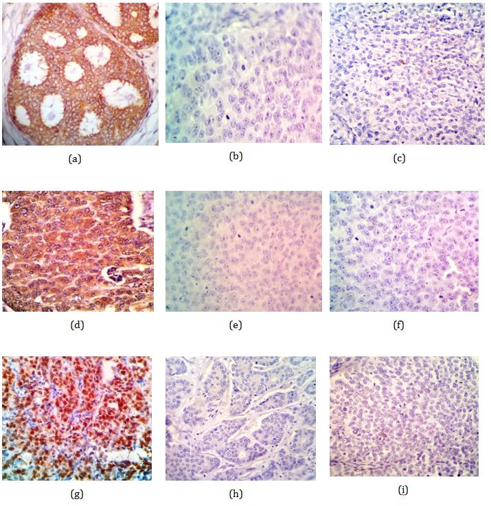
- showed significant upregulation in patients having lymph node metastasis (p=0.043). Moreover, a trend was observed for higher expression of Fzd7 with various tumour sizes (p=0.061) (Table 2). Furthermore, ER+ patients exhibited significant over-expression of Fzd7
- (p=0.002), β-catenin (p=0.010) and cyclin D1 (p<0.001) as compared to ER– patients. Similarly, PR+ patients showed significant higher expression of Fzd7 (p=0.003) and cyclin
- D1 (p<0.001) than PR– patients. However, in case of βcatenin over-expression, only a trend was observed in PR+ patients (p=0.065). On the other hand, no correlations were observed for any of the protein expression with
- Her2 status of the tumours.
Table 2: Correlation of Fzd7, β-catenin and cyclin D1 expression with pathological features
Variables N Fzd7 expression β-catenin expression Cyclin D1 expression Low High Low High Low High N (%) N (%) N (%) N (%) N (%) N (%) Tumor size T1 21 08 (38) 13 (62) 11 (52) 10 (48) 06 (29) 15 (71) T2 113 58 (51) 55 (49) 46 (41) 67 (59) 60 (53) 53 (47) T3 17 11 (65) 06 (35) 11 (65) 06 (35) 11 (65) 06 (35) T4 09 06 (67) 03 (33) 03 (33) 06 (67) 03 (33) 06 (67) χ2=3.521 r=-0.144 p=0.061 χ2=4.452 r=-0.012 p=0.883 χ2=6.761 r=-0.074 p=0.141 Nodal status Negative 65 40 (62) 25 (38) 28 (43) 37 (57) 36 (55) 29 (45) Positive 95 43 (45) 52 (55) 43 (45) 52 (55) 44 (46) 51 (54) χ2=4.095 r=0.16 p=0.043 χ2=0.075 r=-0.022 p=0.786 χ2=1.27 r=0.089 p=0.263 Tumor grade Grade 2 103 48 (47) 55 (53) 39 (38) 64 (62) 48 (47) 55 (53) Grade 3 44 29 (66) 15 (34) 26 (59) 18 (41) 29 (66) 15 (34) χ2=4.607 r=-0.17 p=0.030 χ2=5.632 r=-0.196 p=0.018 χ2=4.607 r=-0.177 p=0.032 Tumor grade Grade 1 13 06 (46) 07 (54) 06 (46) 00 (54) 03 (23) 10 (77) Grade 3 44 29 (66) 15 (34) 26 (59) 18 (41) 29 (66) 15 (34) χ2=1.653 r=-0.17 p=0.205 χ2=0.682 r=-0.109 p=0.418 χ2=7.477 r=-0.362 p=0.006 ER status Negative 72 47 (65) 25 (35) 40 (56) 32 (44) 53 (74) 19 (26) Positive 88 36 (41) 52 (59) 31 (35) 57 (65) 27 (31) 61 (69) χ2=4.607 r=-0.17 p=0.030 χ2=6.63 r=0.204 p=0.010 χ2=29.192 r=0.427 p<0.001 PR status Negative 100 61 (61) 39 (39) 50 (50) 50 (50) 64 (64) 36 (36) Positive 60 22 (37) 38 (63) 21 (35) 39 (65) 16 (27) 44 (73) χ2=8.894 r=0.236 p=0.003 χ2=3.418 r=0.146 p=0.065 χ2=20.907 r=0.361 p<0.001 Correlation of Fzd7, β-catenin and cyclin D1 with molecular subtypes: The expression of Fzd7 (p=0.007), β-catenin (p=0.008) and cyclin D1 (p<0.001) were significantly associated with the molecular subtypes of the patients. Hence, upon correlating with the individual subtype it was observed that, the upregulation of Fzd7 (p=0.015, p=0.041), β-catenin (p=0.011_, p_=0.024) and cyclin D1 (p<0.001, p=0.006) were significantly higher in
- patients with Luminal A subtype as well as Luminal B subtype when compared to TNBC patients, respectively (Table 3). Further, Fzd7 (p=0.003) and cyclin D1 (p=0.005) were over-expressed in Luminal B subtype when compared to Her2-positive subtype. However, only cyclin D1 expression was significantly upregulated in Luminal A subtype when compared to Her2-positive subtype (p<0.001) (Table 3).
Table 3: Correlation of Fzd7, β-catenin and cyclin D1 expression with molecular subtypes.
Variables N Fzd7 expression β-catenin expression Cyclin D1 expression Low High Low High Low High N (%) N (%) N (%) N (%) N (%) N (%) Luminal A 54 24 (44) 30 (56) 19 (35) 35 (65) 13 (24) 41 (76) Her2- positive 35 21 (60) 14 (40) 17 (49) 18 (51) 26 (74) 9 (26) χ2=2.081 r=-0.155 p=0.153 χ2=1.695 r=-0.14 p=0.197 χ2=20.54 r=-0.486 p<0.001 Luminal A 54 24 (44) 30 (56) 19 (35) 35 (65) 13 (24) 41 (76) TNBC 37 26 (70) 11 (30) 23 (62) 14 (38) 27 (73) 10 (27) χ2=5.915 r=-0.255 p=0.015 χ2=6.43 r=-0.266 p=0.011 χ2=21.31 r=-0.484 p<0.001 Luminal B 34 13 (36) 23 (64) 13 (36) 23 (64) 14 (39) 22 (61) TNBC 37 26 (70) 11 (30) 23 (62) 14 (38) 27 (73) 10 (27) χ2=8.557 r=-0.342 p=0.003 χ2=4.954 r=-0.261 p=0.026 χ2=8.61 r=-0.343 p=0.003 Luminal B 36 13 (36) 23 (64) 13 (36) 23 (64) 14 (39) 22 (61)
- catenin and cyclin D1 failed to discriminate better or worse disease outcome. Further, patients were stratified into various subgroups according to clinico-pathological parameters and it was observed that in patients with grade 3 tumours, low expression of Fzd7 showed a trend of increased incidence of death as compared to the high expression of Fzd7 (Log rank= 3.606, df=1, p=0.058)
- (Figure
- 2a,
- Table
- 4).
- Additionally, although not statistically significant, in TNBC patients, low expression of cyclin D1 showed an increased incidence of death as compared to the high expression of cyclin D1 (Log rank=
- 2.724, df=1, p=0.099) (Figure 2b, Table 4).
Table 4: Overall survival analysis of Fzd7 and Cyclin D1 in patient subgroups.
N Alive Dead Log rank df p N (%) N (%) Fzd7 protein expression Patients with grade 3 tumors (N=29) Low 19 13 (68) 06 (32) 3.606 1 0.058 High 10 10 (100) 00 (00) Cyclin D1 protein expression TNBC patients (N=25) Low 17 15 (88) 02 (12) 2.724 1 0.099 High 08 05 (62) 03 (38)
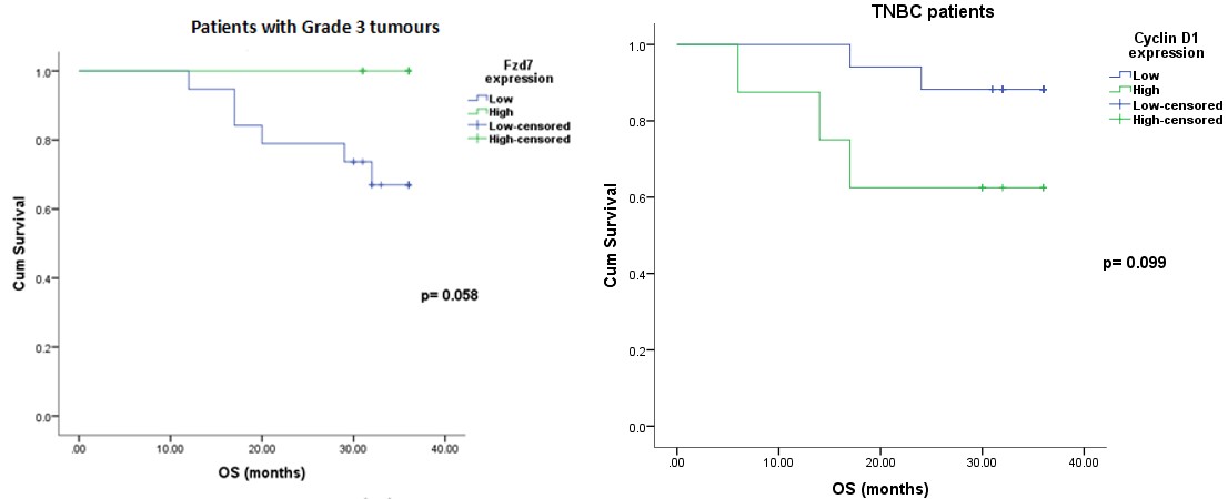
(a) (b) Figure 2: (a) Overall survival curve of breast cancer patients with grade 3 tumours in relation to Fzd7 protein expression. (b) Overall survival curve of TNBC patients in relation to cyclin D1 protein expression.
Wnt/β-catenin signalling pathway has various functions in embryonic development as well as normal homeostasis [8, 9, 10]. However, dysregulation of the pathway leads to its aberrant activation and these results in tumorigenesis [11]. Hence, the present study aimed to analyse the expression of Wnt pathway components in breast cancer patients. The immunoreactivity of Fzd7, β-catenin and cyclin D1 were found in cell membrane, cell cytoplasm and nucleus of 48%, 56% and 50% of breast cancer patients, respectively. Similarly, Corda et al. also reported membranous localization of Fzd7 [12]. Wang, et al. showed cytoplasmic as well as nuclear expression of β- catenin while Lee et al. found the normal membranous β- catenin expression with the loss of membranous staining for abnormal β-catenin expression [13, 14]. Thus the observed only cytoplasmic staining pattern in current study indicated the probability of loss of membranous pattern implying that the Wnt/ β-catenin pathway is most likely activated in breast cancers [15]. Consequently, as observed in our study, cyclin D1 has been reported to be localized in the nucleus with an overexpression in about 50% of human breast cancers [16]. In addition, Peurala et al. has also reported nuclear expression of cyclin D1 in 60% of breast cancer patients [17]. Further, the proteins were correlated with clinicopathological parameters, and we observed that high expression of Fzd7, β-catenin and cyclin D1 proteins were associated with smaller tumor size and node negative tumour cells. Although, there was a difference between high and low β-catenin and cyclin D1 proteins the data failed to reach statistical significance, but overall it indicates that there was a loss of protein expression with advanced disease. Additionally, higher expression of Fzd7, β-catenin and cyclin D1 were also significantly associated with tumor differentiation that is higher expression in grade 2 breast tumors than that of grade 3 tumors. Likewise, the expression was also high in grade 1 tumors than in grade 3 tumors, but difference was statistically significant for only cyclin D1. Hence the current data strengthen the above finding that there was an association of loss of the three proteins with the disease progression. In agreement with our results, Peurala, et al has observerd high cyclin D1 to be significantly expressed in lower grade of breast tumors [17]. Likewise Shapiro, et al. in non small cell lung carcinoma observed that β-catenin expression is increased in stage 1 patients [18]. On the other hand, contradictory to our results, He et al reported significant correlation of β-catenin and cyclin D1 over expression with the differentiation grade, clinical stage and lymph node status of breast cancer patients [19]. Apart from this, Zengel, et al_._ failed to find any correlation of clinic pathological parameters with β-catenin or cyclin D1 in breast cancer patients [20]. It is well established that in patients with breast cancer, hormone receptors and Her2 status is used for treatment decision. Corda, et al. observed over expression of Fzd6 in ER+ breast cancers [12]. Zwijsen, et al. showed an association of cyclin D1 over expression with the estrogen response element in breast cancer tissues. The authors hypothesized that the effects of estrogen on breast tumors may be exerted via the cyclin D1 pathway, and that ER-α may promote breast cancer occurrence through the induction of cyclin D1 [21]. On the other hand, Luo, et al. suggested that ER pathway activation may activate β-catenin, which may further activate the Wnt signaling pathway and result in the increased expression of cyclin D1 [22]. The present study too observed significant association of Fzd7, β-catenin and cyclin D1 with ER+ and PR+ status indicating that ER pathway activates Fzd7 and β-catenin resulting in activation of cyclin D1 and thereby Wnt signaling pathway. Contradictorily, some studies reported higher cyclin D1 expressions to be associated with ER-, PR- and Her2- breast tumours [20, 23]. Moreover, β-catenin gene has been identified to correlate with the ER expression in breast cancer cell lines and patient derived metastatic breast cancer samples [23]. On the other hand, Zengel, et al. did not observe any correlation between β-catenin and ER/PR or Her2 expression [20]. In addition, we analyzed the expression of Fzd7, β- catenin and cyclin D1 in different molecular subtypes of breast cancer patients, where it was observed that all the three proteins were significantly higher in Luminal subtypes as compared to Her2-positive and TNBC subtypes. The probable reason for this could be that Wnt pathway is related to estrogen and Her2 signaling in breast tumors, leading to enhanced malignant properties. In literature, there are a number of contradictory reports for the activation of Wnt pathway in the molecular subtypes of breast cancer. Fzd7 over-expression is inversely associated with the high levels of Her2 in breast cancer cell lines, while there was no difference in β- catenin expression amongst the four molecular subtypes of breast cancer [24, 25]. On the other hand, still another study shows β-catenin to be preferentially expressed in triple-negative/basal-like breast carcinomas [26]. Also, in accordance to our study, cyclin D1 is shown to have a positive correlation with hormone receptors as well as non-basal breast carcinomas and non-TNBC phenotype [27]. In univariate survival analysis, low Fzd7 expression was found to be significantly associated with reduced overall survival in patients with grade 3 tumours. Additionally, in the present study, low expression of cyclin D1 showed a trend of association with decreased overall survival of TNBC patients. Similarly, Guo, et al. has also shown patients with positive cyclin D1 expression to exhibit a longer survival time compared with those with negative cyclin D1 expression [28]. However, the significance of cyclin D1 as a prognostic factor in breast cancer is controversial with studies reporting both positive and negative findings [29]. Cyclin D1 expression was not of prognostic value in early stage breast cancer patients [30]. Contradictorily, higher expression of cyclin D1 was associated with better prognosis in breast cancer patients with Her2 over expression [20, 31]. In conclusion, the over-expression of the Wnt pathway components is likely to have a role in early stages of breast cancer development leading to the cancer progression. The pathway molecules are also associated with the surface receptors with predominance in non-TNBC subtype. Also, Fzd7 and cyclin D1 emerged as a significant prognosticator in patient subgroups of poorly differentiated tumours and TNBC subtype. However, further studies with a large sample size in each of the molecular subtype of patients are required for an exact correlation of the pathway components.
Conflicts of interest
There are no conflicts of interest in the present study.
Acknowledgement
The financial support for the present study was received from Gujarat Cancer Society and Gujarat Cancer & Research Institute.
References
-
Downs-Holmes C, Silverman P (2011) Breast cancer: overview & updates. Nurse Pract 36 (12): 20-26.
-
Prat A, Cheang MC, Martín M, Martín M, Parker JS, et al. (2012) Prognostic significance of progesterone receptor-positive tumor cells within immunohistochemically defined luminal A breast cancer. J Clin Oncol 31(2): 203-209.
-
Kamdje AH, Etet PF, Vecchio L, Muller JM, Krampera M, et al. (2014). Signaling pathways in breast cancer: therapeutic targeting of the microenvironment. Cell Signal 26(12): 2843-2856.
-
Klemm F, Bleckmann A, Siam L, Chuang HN, Rietkotter E, et al. (2011) β-catenin-independent WNT signaling in basal-like breast cancer and brain metastasis. Carcinogenesis 32(3): 434-442.
-
MacDonald BT, Tamai K, He X (2009) Wnt/β-catenin signaling: components, mechanisms, and diseases. Dev Cell 17(1): 9-26.
-
Zhan T, Rindtorff N, Boutros M (2017) Wnt signaling in cancer. Oncogene 36(11): 1461-1473.
-
Polakis P (2012) Wnt signaling in cancer. Cold Spring Harb Perspect Biol 4(5): a008052.
-
Teo JL, Kahn M (2010) The Wnt signaling pathway in cellular proliferation and differentiation: a tale of two coactivators. Adv Drug Deliv Rev 62(12): 1149-1155.
-
Kim W, Kim M, Jho EH (2013) Wnt/β-catenin signalling: from plasma membrane to nucleus. Biochem J 450(1): 9-21.
-
Wang J, Sinha T, Wynshaw-Boris A (2012) Wnt signaling in mammalian development: lessons from mouse genetics. Cold Spring Harb Perspect Biol 4(5): a007963.
-
Kazi MM, Trivedi TI, Kobawala TP, Ghosh NR (2016) The Potential of Wnt Signaling Pathway in Cancer: A Focus on Breast Cancer. Cancer Transl Med 2(2): 55- 60.
-
Corda G, Sala G, Lattanzio R, Iezzi M, Sallese M, et al. (2017) Functional and prognostic significance of the genomic amplification of frizzled 6 (FZD6) in breast cancer. J Pathol 241(3): 350-361.
-
Wang Z, Zhang H, Hou J, Niu J, Ma Z, et al. (2015) Clinical implications of β-catenin protein expression in breast cancer. Int J Clin Exp Pathol 8(11): 14989- 14994.
-
Lee WA (2005) Prognostic Significance of Abnormal β-catenin Expression in Breast Carcinoma. Korean J Pathol 39: 114-119.
-
Reya T, Duncan AW, Ailles L, Domen J (2003) A role for Wnt signalling in self-renewal of haematopoietic stem cells. Nature 423(6938): 409-414.
-
Eeckhoute J, Carroll JS, Geistlinger TR, Torres-Arzayus MI, Brown M (2006) A cell-type-specific transcriptional network required for estrogen regulation of cyclin D1 and cell cycle progression in breast cancer. Genes Dev 20(18): 2513-2526.
-
Peurala E, Koivunen P, Haapasaari KM, Bloigu R, Jukkola-Vuorinen A (2013) The prognostic significance and value of cyclin D1, CDK4 and p16 in human breast cancer. Breast Cancer Research 15(1): R5.
-
Shapiro M, Akiri G, Chin C, Wisnivesky JP, Beasley MB, et al. (2013) Wnt pathway activation predicts increased risk of tumor recurrence in patients with stage I non-small cell lung cancer. Ann Surg 257(3): 548-554.
-
He Y, Liu Z, Qiao C, Xu M, Yu J, et al. (2014) Expression and significance of Wnt signaling components and their target genes in breast carcinoma. Mol Med Rep 9(1): 137-143.
-
Zengel B, Vardar E, Postaci H, Keçeciler S, Alacacioğlu A, et al. (2011) β-Catenin Stability, Cyclin D1 and Frizzled Proteins Expression in Human Breast Cancer and Their Relation with the Prognosis. Turkiye Klinikleri J Med Sci 31(2): 350-357.
-
Zwijsen R, Wientjens E, Klompmaker R, van der Sman J, Bernards R, et al. (1997) CDK-independent activation of estrogen receptor by cyclin D1. Cell 88(3): 405-415.
-
Luo J, Chen YL, Xu H (2010) Expression of β -catenin, cyclin D1 and ERα in breast cancer. 17: 1502-1504.
-
Lamb R, Ablett MP, Spence K_,_ Landberg G, Sims A, et al. (2013) Wnt pathway activity in breast cancer sub- types and stem-like cells. PloS one 8(7): e67811.
-
Yang L, Wu X, Wang Y, Zhang K, Wu J, et al. (2011) FZD7 has a critical role in cell proliferation in triple negative breast cancer. Oncogene 30(43): 4437- 4446.
-
Li S, Li S, Sun Y, Li L (2014) The expression of β- catenin in different subtypes of breast cancer and its clinical significance. Tumor Biology 35(8): 7693- 7698.
-
Geyer FC, Lacroix-Triki M, Savage K, Arnedos M, Lambros MB, et al. (2011) β -Catenin pathway activation in breast cancer is associated with triple- negative phenotype but not with CTNNB1 mutation. Mod Pathol 24(2): 209-231.
-
Boström P, Söderström M, Palokangas T, Vahlberg T, Collan Y, et al. (2009) Analysis of cyclins A, B1, D1 and E in breast cancer in relation to tumour grade and other prognostic factors. BMC Research Notes 2: 140.
-
Guo L, Yilamu D, Sun L, Liu S, Ma F (2015) Association among the expression of β-catenin, cyclin D1and estrogen receptor-β in human breast cancer. Exp Ther Med 10(4): 1423-1428.
-
Taneja P, Maglic D, Kai F, Zhu S, Kendig RD, et al. (2010) Classical and novel prognostic markers for breast cancer and their clinical significance. Clin Med Insights Oncol 4: 15-34.
-
Michalides RJ, Hageman PH, Van Tinteren H, Houben L, Wientjens E, et al. (1996) A clinicopathological study on overexpression of cyclin D1 and of p53 in a series of 248 patients with operable breast cancer. Br J Cancer 73(6): 728-734.
-
Lee A, Park WC, Yim HW, Lee MA, Park G, et al. (2007) Expression of c-erbB2, cyclin D1 and estrogen receptor and their clinical implications in the invasive ductal carcinoma of the breast. Jpn J Clin Oncol 37(9): 708-714.
- Cancer Diagnosis from RNA Sequence of Blood Cells by Using AI
- Field Cancerization in Oral Cavity, Case Report and Review of Literature. Oncologic Program Salud Integral Hospital, Managua, Nicaragua
- Identification of B Lymphocytes in Cancer Patient’s Blood
- A Case Report of a Breast Cancer Patient Developing Pneumonitis as a Result of Abemaciclib Therapy
- Immune Checkpoint Therapeutics for Today’s Fight and Beyond
- The Amalgamated Sophomore-Gonadoblastoma