Transdermal Delivery Offer for Low Dose Acetylsalicylic Acid
The use of Acetyl Salicylic Acid (ASA) in the secondary prevention of cardio vascular diseases is well known. Orally administered ASA is rapidly metabolized to its principal metabolite Salicylic Acid (SA) in the gastrointestinal fluids, during absorption in the intestine and liver. ASA, and not its hydrolysis product SA, which exhibits antithrombotic activity. Transdermal delivery of acetylsalicylic acid is a safe and convenient alternative, especially during long-term use. The goal of present study was to design transdermal delivery system, using Eudragit E100 weak organic acid and plasticizer. The influence of different dissolution medium for preparation of model systems, the type and amount of cross-linking agents and plasticizer on the release of acetylsalicylic acid was investigated. Matrix systems were prepared by solvent casting method. The dissolution study was carried out in water for 72 hours by disk assembly method paddle and in-vitro release of acetylsalicylic acid from loaded model systems was studied. Quantitative determination of released acetylsalicylic acid by UV spectroscopy at wavelength of 265 nm was made. Significantly sustained release show systems prepared by using water as dissolution medium, MA, like cross linking agent, PEG400 like plasticizer
Introduction
ASA in low dose irreversibly blocks the formation of thromboxane A2 in platelets, producing an inhibitory effect on platelet aggregation. Orally administered aspirin requires high and frequent dosing because it undergoes extensive metabolism in the intestine and liver into salicylic acid. Metabolite does not possess antiplatelet activity. The major side effects of per orally administered ASA are gastric irritation and ulceration due to inhibition of cyclooxygenase [1]. The plasma concentration of АSА decays with a half-life of 15–20 min. [2]. Very frequent oral dosing with aspirin tablet (every 2-3 hrs) is not practical. The low bioavailability of transdermally administered ASA and the avoidance of direct contact with COX-1 expressed on gastric mucosal cells may provide a safer means of inhibiting platelet function. ( [1, 3]. Transdermal delivery will retain the inhibitory effect of aspirin on platelet COX-2 and minimize that on vascular COX-1, thus continuous low-dose aspirin therapy can be used without the reported risks on GI tract [4, 5]; In vitro studies showed that the skin acts like reservoir for ASA. About 15% of the administered dose is absorbed over 48 hours after a single application [6]. Aspirin is rapidly deacetylated in aqueous conditions and is possible only salicylic acid was absorbed. Preventing the deacetylation includes aspirin in propylene glycol and alcohol like vehicle and penetration enhancer and applied on the human skin in vivo. They established that ASA applied daily induced a dose-dependent inhibition of platelet cyclooxygenase. In another study ASA 84 mg and 120 mg was included in a transdermal patch with surface area 50 cm2 and also found to induce marked suppression of platelet cyclo-oxygenase [7]. Aim of this study is to prepare model for transdermal system on base of Eudragit E100, weak organic acid malic or citric and plasticizer, releasing low-dose acetylsalicylic acid in order to achieved extended antithrombotic activity [8]. Structure of the obtained model system was proven, by method of IR spectroscopy and by determination of the specific viscosity of dilute solutions of the basic components. Preliminary in vitro studies to clarify influence of process and biopharmaceutical factors on rate and degree of release of ASA from model systems. ASA like a candidate for the transdermal drug delivery has low molecular weight 180.157 g/mol, low melting point i.e. 135°C., an elimination plasma half-life of 15-20 min, log P (lipophilic character) value of 1.426., causes gastric irritation and ulceration due to inhibition of cyclooxygenase [9], is used for a long period, undergoes extensive first pass metabolism.
Methods and Materials
Materials
Acetylsalicylic acid (ASA), Triethylcitrate (TEC), Triacetin (TA), Dibutylphtalate (DBF), polyethylene glycol 400 (PEG400), Malic acid (MA), Citric acid (CA), Ethanol, Acetone and Isopropanol were purchased from Sigma Aldrich, Germany, Eudragit E 100 (EE100) was purchased from Evonik industries, Germany.
Methods
1st Method: The cross linking agent in water was dissolved. To this solution ASA previously dissolved in ethanol was added to solution of cross linking agent. Next step, EE 100 to the acidic solution was added and finally the plasticizer was added. Stirred on a magnet stirrer Kika®Labortechnik RH basic, Germany to obtain a uniform solution for 20 minutes. 2nd Method: The polymer was dissolved in ethanol: acetone: isopropanol mixture for a period of 90 min. Then the solution of ASA in ethanol was added slowly in the polymeric solution and stirred on the magnetic stirrer to obtain a uniform solution for 10 minutes. DBF, TEC, TA or PEG400 were added and continue stirring for 10 min. Finally ethanol solution of cross linking agent was added and stirred for 20 min. 6.0 grams of mixtures prepared by 1st or 2nd method on the silicon forms having surface area of 28 cm2 were poured and dried at the room temperature. Systems prepared by 2nd method were dried covered with inverted funnel to control solvent evaporation. After drying patches were weighed and cut into 2x2 cm2 patches for dissolution study. Drug incorporated for each 2x2 cm2 patch on base of weight after drying and lose of solvent was calculated.
Results and Discussion
IR spectrum interpretation
IR spectrums of EE100, MA, CA, ASA and a mixture of them by using method I and II technique were taken, as the solvent is evaporated. [Figure 1] demonstrates the IR spectra of (a) EE100, (b) MA, (c) EE100/MA obtained by method 2 and (d) EE100/MA obtained by method 1. As can be seen from the comparison of spectra b, c and d the absorption band at 1716 cm-1, which corresponds to the stretching vibrations υc=o in free MA, shifts to 1722cm-1 in case of system obtained by method 2 (c) and to 1727 cm-1, system obtained by method I (d). The absorption band at 1720 cm-1, which corresponds to the stretching vibrations υc=o in free CA, shifts to 1723cm-1 in case of system obtained by method 2 and to 1733 cm-1, system obtained by method I (not shown on figure 1). From the comparison of spectra a, c and d the absorption band at 2951 cm-1 which corresponds to the stretching vibrations υNH3+ in free EE100, shifts to 2983 cm-1 in case of system obtained by method 2 (c) and to 2954 cm-1, system obtained by method 1(d). The absorption band at 2951 cm-1 which corresponds to the stretching vibrations υNH3+ in free EE100, shifts to 2989 cm-1 in case of system EE100/CA obtained by method 2 and to 2954 cm-1, system obtained by method 1(not shown on fig.1). Coates, J, Yong-Cheng Ning, Norman, B. [9, 10, 11] the high-frequency shift to the stretching vibration bands υc=o and υNH3+ in the IR spectra of complex between EE100 and MA/CA obtained by method I is explained as follows. The formation of chemical bond between the functional groups of interacting substances increases the energy required for the excitation of stretching vibration in a complexed functional group. The stretching vibrations υc=o and υNH3+ in the region of higher frequencies in case of complex EE/MA and EE/CA obtained by method I but in the IR spectra of complexes obtained by method II is negligibly low.
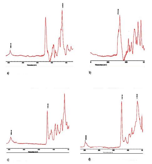
Presence of water in the systems obtained by method I, and pH 3.5 suppose that ionic bond is formed between the protonated amine groups of the EE 100 and the ionized carboxyl groups of MA / CA. In case of systems obtained by method II the absence of water in the system protonation of the amino groups and the ionization of the carboxyl groups are not observed, therefore, suggests that after evaporation of solvent have a physical mixture of them. ASA has a carboxylic group that is possible to interact in case of method II preparation with protonated tertiary amino groups of EE 100. This interaction by IR spectroscopy can be proven in this case. From a chemical point of view, at pH 3.5 and pKa of ASA 3.5, shows that 50% of ASA is ionized, pKa of malic acid 3.4 and 5.20, shows that 55% of first and 30% of second carboxylic groups are ionized and pKa of citric acid 3.13, 4.76 and 6.39, shows that 60% of primary, 40% of secondary and 25% tertiary carboxylic groups are ionized. EE100 is 99.9 % protonated. This means that the primary carboxyl groups certainly displace the ASA from ionic bonds with the protonated amino groups of the EE 100. Secondary carboxyl groups of MA react with protonated amino groups of the EE 100 when ionized ASA is exhausted. For CA this applies to secondary and tertiary carboxyl groups. Therefore assume that the non-ionized ASA remains physically included into the system. Ionized ASA is chemically bound with some of the protonated amino groups of EE100. With the rest protonated amino groups ionic bond secondary carboxyl groups of MA and secondary and tertiary carboxyl groups of CA formed. To confirm the results specific viscosity of dilute solutions of EE 100 and MA, CA and ASA were determined. Dependences of specific viscosity ηsp of mixed solutions of EE 100 with MA, CA and ASA at a constant concentration of a EE100 on the z = [EE]/[MA], z = [EE]/[CA] and z = [EE]/[ASA] mass/mass ratio, where [MA] is the mass concentration of malic acid, [CA] is the mass concentration of citric acid, [ASA] is the mass concentration of acetylsalicylic acid and [EE] = const = 0.01 g/dL was shown on figure 1. At low EE100 concentrations, the system forms true solutions without gelation.
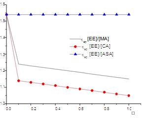
The decrease in value of the specific viscosity (Figure 2■) when adding a solution of malic acid about the formation of a complex between the protonated tertiary amino groups of the polymer, and the ionized carboxylic groups of MA give information. Difference between of the value of specific viscosity compared with the value of system with MA is due to a participating third carboxyl groups of CA and the engagement of a third chain of EE100 thereof, which results in the formation of a folded conformation and decrease the value of the specific viscosity (Figure 2●). By addition of increasing concentrations of ASA to the solution of EE, change in the specific viscosity is not observed (Figure 2▲). Valid reason for that is the presence of only one carboxyl group in the molecule of the ASA, which cross-linking of the polymer chains of EE 100 does not allow.
Biopharmaceutical characterization of model systems
Determination of λmax
A 10mg of ASA was accurately weighed and was first dissolved in 35ml methanol solution. The solution was then diluted using water to 100 ml. UV spectrum was recorded in the wavelength range 200-600 nm.
λmax was observed at 265 nm In-vitro release of ASA loaded model systems by paddle over disk method
• Dissolution medium - purified water (Ph.Eur.) Volume
of solvent - 900 ml.
- Stirring speed - 50 rpm/min. Temperature - 32
- C ±0, 5.
- Samples were taken every hour for the first 6 hours and at and 12, 24, 48 and 72 hours thereafter. Quantitative determination of released ASA by UV spectroscopy
- Spectrophotometric measurement at wavelength of 265 nm.
In-vitro release of ASA loaded model systems by paddle over disk method
- Dissolution medium - purified water (Ph.Eur.) Volume of solvent - 900 ml.
- Stirring speed - 50 rpm/min. Temperature - 32
- C ± 0, 5.
- Samples were taken every hour for the first 6 hours and at and 12, 24, 48 and 72 hours thereafter. Quantitative determination of released ASA by UV spectroscopy
- Spectrophotometric measurement at wavelength of 265 nm.
Influence of different dissolution medium for preparation of model systems on the release of ASA from the model system
Investigated was the release of ASA systems prepared by method I and method II as described above. For the purpose a concentration of 5% MA, like cross linking agent, plasticizer DBF and concentration of ASA 10% were chosen. Results were shown at Figure 3.
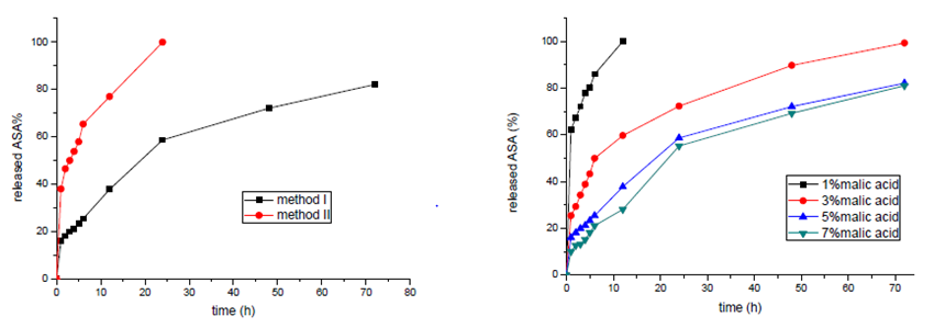
The results show sustained release for the systems prepared by two methods. Significantly sustained release was observed when the systems were prepared by using water as a dissolution medium. In dissolution medium water and pH 3, 5 tertiary amino groups in molecules of EE100 were protonated and carboxylic groups in molecules of MA were ionized. This allows cross linking of EE100 molecules with MA. The network sustained release up to 82% for 72 hours allows. In dissolution medium organic solvent there are not enough protonated molecules EE 100 and ionized molecules of MA. ASA is released from the model system completely for 24 hours.
Influence of concentration of the cross linking agent on the release of ASA from the model system
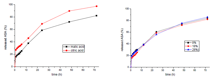
In order to observe the influence of the concentration of the cross linking agent on the release of ASA 4 different concentrations of MA were varied – 1%, 3%, 5%, 7% (Figure 4). Like plasticizer DBF and concentration of ASA 10% were chosen. With increase of the concentration of acid in the system, release of ASA is delayed. At concentration 1% malic acid, 62% of included ASA at first hour is released. At concentration 5% and 7 % release profiles are not significantly different. We accept that the concentration of malic acid 5% is optimal and allows release of 82% for 72 hours. In order to observe the influence of the type of the cross linking agent on the release of ASA, model systems with MA and CA in a concentration of 5% were prepared and the release of ASA, for a period of 72 hours was investigated. Systems with CA show a release of 97%, and systems with MA 82% for 72 hours (Figure 5). High percentage released ASA explained by the participation of the third carboxyl group at the cross linking of the molecules of EE 100 with CA. We assume that steric obstruction allows the rapid release of ASA from the resulting net.
Influence of the concentration of plasticizer on the release of ASA from the model system
Concentration of the plasticizer does not have a significant effect on the release of ASA from the model system prepared with 8, 18 and 28% of DBF (Figure 6). We assume that the concentration of the plasticizer has a significant effect on the mechanical properties of the system, such as elasticity, elongation and tensile strength. These studies may be subject to subsequent work if the systems demonstrate potential for administration.
Influence of type of a plasticizer on the release of ASA from the model system
In order to observe the influence of type of plasticizer on the release of ASA with DBF, TA, TEC and PEG 400 like plasticizers were prepared system. 5% of MA like cross linking agent was used. Systems with PEG and DBF show release respectively 80 and 82%, while systems with TEC and TA - 89 and 90 respectively (Figure 7).
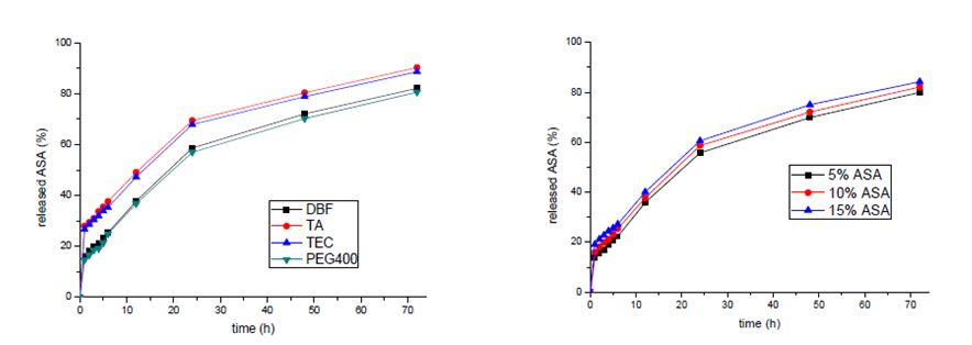
Influence of loading of the model system with ASA on release from them
Increasing of the amount of ASA does not change significantly the rate and degree of release of the ASA (figure 8). Concentration gradient is not the main factor that determining release of the active substance. Influence has structural, mechanical and physicochemical properties of the system.
Conclusions
In this work an attempt was made to formulate and evaluate TDDS for sustained release ASA by solvent casting method. Using IR spectroscopy and viscometer, it has been shown that EE100 form complexes with MA or CA in aqueous solutions. The complexation is realized via bonding between dissociated COO-groups of the organic acids and NH3+ groups of the EE100. Non-ionized ASA remains physically included into the system. Significantly sustained release show systems prepared by using water as dissolution medium, concentration of 5% and MA, like cross linking agent, 18% PEG400 like plasticizer. Results provide convincing evidence for the feasibility of transdermal low-dose aspirin patch. Further work is required to establish the utility of such transdermal drug delivery system through in vitro testing of skin absorption using static Franz-type diffusion cells.
References
-
Keimowitz RM (1996) Transdermal aspirin and gastric ulcer healing after coronary artery stent placement. Circulation 94: 3002.
-
Roth GJ, Stanford N, Majerus PW (1975) Acetylation of prostaglandin synthase by aspirin.
-
Keimowitz R, Pulvermacher G, Mayo G, Fitzgerald D (1993) Transdermal modification of platelet function. A dermal aspirin preparation selectively inhibits platelet cyclooxygenase and preserves prostacyclin biosynthesis. Circulation 88: 556-561.
-
Derry S, Loke Y (2000) Risk of gastrointestinal hemorrhage with long term use of aspirin: meta- analysis. British Medical Journal 321: 1183-1187.
-
Krishna DR, Srinivas G, Srinivas A (2000) Transdermal aspirin: influence of platelet aggregation and serum lipid peroxides. Indian Journal Pharmaceutical Science 62(3): 200-204.
-
Bronaugh RL, Collier SW (1993) In vitro methods for measuring skin permeation. Skin Permeation – Fundamentals and Applications. Allured Publishing Corporation, Wheaton, Illinois_,_ 93-111.
-
McAdam B, Keimowitz R, Maher M, FitzGerald D (1996) Transdermal modification of platelet function: an aspirin patch system results in marked suppression of platelet cyclooxygenase. Journal of Pharmacology and Experimental Therapeutics 277(2): 559-564.
-
Fowler P (1979) Role of NSAIDs in Rheumatic Arthritis, Clinics in rheumatic disease 5: 427.
-
Antithrombotic Trialists’ Collaboration (2002) Collaborative meta-analysis of randomized trials of antiplatelet therapy for prevention of death, myocardial infarction and stroke in high risk patients. BMJ 324(7329): 71-86.
-
Coates J (2000) Interpretation of infrared spectra, a practical approach in Encyclopedia of analytical chemistry. RA Meyers John Wiley & Sons Ltd 10815- 10837.
-
Ning YC (2011) Interpretation of organic spectra. John Wiley & Sons.
-
Norman B (1969) Colthup. Interpretaion of infrared spectra, American Chemical Society.
- Acido Labile or Gastro Irritant Apis and Enteric Release in Galenic Practice: An Overview
- A Study on Knowledge, Attitude and Practice of Hand Hygiene among Healthcare Professionals at a Tertiary Care Hospital, India
- Influence of Inoculum Concentration on In Vivo Incubation Period of Emmia lacerata, Pathogenesis and Management of Wilt in Pepper (Capsicum annuum L.)
- Vanilla’s Chemistry
- Marine Anti-Cancer Compounds and Adverse Effects of Global Warming on Oceans: An Overview
- Serological Investigation of Chikungunya Virus Antibody among Malaria-Suspected Febrile Patients in Some Healthcare Facilities in Rivers State