Cytotoxic and Anti-Cervical Cancer Effects of the Camellia sinensis Byproducts from Various Kenyan Factories
Cervical cancer is a significant global health challenge and necessitates the search for innovative and patient-friendly therapies due to the limitations and challenges posed by conventional treatments. This study explored the largely overlooked potential of Camellia sinensis processing wastes as a natural source for developing effective and safe anticancer compounds. Tea waste samples from Kenyan processing facilities demonstrated concentration-dependent cytotoxic effects on normal epithelial cells (Vero), with the fluff sample from Chelal displaying superior efficacy. Median inhibitory concentration (IC50) values highlighted the potency of the samples, especially the fluff sample from Chelal (IC50: 483.99μg/ml). Selectivity indices revealed favorable safety profiles, indicating selective potential anticancer effects against cervical cancer cells. This study contributes substantively to understanding tea waste’s anticancer potential, providing valuable insights for drug development. The study offers a foundation for continued research into sustainable and impactful anticancer drug discovery, emphasizing specific results that pinpoint promising tea waste samples and their potential applications in cervical cancer therapy and anticancer drug development
Introduction
Recent statistics reveal that there were approximately 19.3million new cases of cancer and an astonishing 10 million deaths attributed to cancer globally in 2020 alone [1]. Amongst this overwhelming burden, cervical cancer stands out as one of its principal contributors, warranting not only attention but also innovative approaches for effective management [2, 3]. In 2020 alone, there were 604,127 reported cases of cervical cancer, with an associated death toll reaching 341,831 [4]. The multifaceted challenges of cervical cancer span various aspects, from delayed diagnoses, especially in resource-limited settings, to its often aggressive nature, which poses substantial obstacles within healthcare systems worldwide, with even more tremendous toll on the less-developed countries of sub-Saharan Africa [5].
Moreover, conventional treatment modalities such as surgery, radiation, and chemotherapy may impose physical and emotional strain on patients [6, 7]. These treatments also demonstrate limitations in terms of efficacy, especially in advanced disease stages [6].
Furthermore, the challenges of managing and treating cervical cancer intensify due to its high incidence rates in low- to middle-income countries where access to healthcare resources is limited [4, 8, 9, 10]. Given these complexities and recognizing a need for more patient-friendly yet effective therapeutic approaches, we must explore alternative therapies. Camellia sinensis processing waste, often disregarded, presents a glimmering hope to diversify our therapeutic arsenal against cervical cancer. This quest not only presents opportunities for superior results but also promises diminished side effects and heightened accessibility, especially in areas grappling with healthcare disparities [10, 11, 12].
Natural compounds exhibiting potential anticancer properties are garnering prominence [13, 14, 15, 16]. Camellia sinensis, commonly identified as tea, has attracted immense research attention, transcending its conventional role as a beverage due to its rich polyphenols, catechins, flavonoids, and other bioactive elements [17, 18, 19]. Various health benefits, such as antioxidant effects, anti-inflammatory actions, and even the potential for cancer prevention or suppression, have been associated with tea consumption [18, 20]. Epigallocatechin gallate (EGCG), a polyphenol found in tea, has captured particular interest for its demonstrated potential in suppressing the proliferation of cancer cells by inducing apoptosis and inhibiting angiogenesis [18, 20, 21].
Tea dust stems and twigs, among other tea wastes, harbour a notable density of bioactive compounds [22]. The investigation into potential tea waste adheres to sustainability principles and reveals an overlooked resource for novel pharmaceutical compounds to combat cervical cancer. Previous reports indicate green tea extracts possess cytotoxic effects on various cancer cell lines [18, 19]. Research shows that EGCG has the potential to inhibit HeLa cell growth; however, there is a dearth of empirical data to appraise the anti-cervical cancer efficacy of Camellia sinensis byproducts. Moreover, focusing on isolated compounds or specific types of tea creates the existing research gaps, neglecting the broader spectrum of bioactive molecules found in byproducts.
The present study aimed to bridge the existing gaps in the literature, offering a comprehensive understanding of tea byproducts’ potential therapeutic applications for cervical cancer. The range of bioactive compounds in Camellia sinensis presents a versatile strategy against tumorigenesis by targeting multiple pathways involved in cancer development. We can potentially develop potent anticancer therapies with precise targets by comprehending the molecular mechanisms and pinpointing specific bioactive compounds that induce cytotoxic effects on HeLa cells. Delving deeper into these natural compounds’ complex biochemistry may uncover novel cancer prevention and treatment strategies.
Materials and Methods
Test Samples and their Preparation
The tea waste samples, which included fluffs, cyclones, cyclone fluffs, dry-offs, and fluff-dry mouths, were systematically obtained from various tea processing facilities in tea-growing regions of Kenya. They were placed in clean, brown-hued glass bottles and taken to the Kenya Medical Research Institute (KEMRI), Nairobi, where they were oven- dried (103°C) until attaining a consistent weight. After that, an electric blending device (Moulinex AR 1043, China) was employed to mill the dried samples finely. The stock concentrations of the powdered samples were prepared in dimethyl sulphoxide and diluted two-fold to achieve the working concentrations, which ranged from 7.81 to 1000 μg/ ml.
Cell Culture and Cytotoxicity Assay
The previously described cell viability assay method was adopted in this study [23]. Briefly, Cervical cancer (HeLa) and normal mammalian epithelial (Vero CCL-81) cell lines were cultured in Eagle’s Minimum Essential Medium (EMEM) supplemented with Earle’s saline, an antibiotic- antimycotic mixture (penicillin 100 U/mL, streptomycin 100 mg/mL, amphotericin B 0.25mg/ml) and 10% fetal bovine serum (Sigma, USA) [24]. In a cell culture incubator, the cell cultures were inoculated in 96-well plates and maintained in a humidified atmosphere saturated with 5% CO2 at 37°C.
The cells were washed with phosphate-buffered saline (PBS) washing and then exposed to a culture medium with the test samples at various working concentrations (7.81 to 1000μg/ml) or without. After a 72-hour incubation period, the culture medium was aspirated, and 150ml of a 5μg/ml MTT solution in PBS (pH 7.2) was added to each well, followed by incubation for 4 hours at 37°C in a humidified atmosphere saturated with 5% CO2. Post-incubation, 750ml of dimethyl sulfoxide was added to each well, and the plates were shaken gently for 15 minutes to solubilize the formazan dye. After that, the absorbance values of each well were obtained spectro photometrically at 570nm using a Quant Universal Microplate Spectrophotometer (Bio-Tek Instruments Inc, USA). The percentage of cytotoxicity was calculated using the following formula (Eq. 1) [25].
% 1 100
The absorbance of treated cells
Cytotoxicity
The absorbance of control cells
$$ v = 1 - \left(\frac {\text {The absorbance of treated cells}}{\text {The absorbance of control cells}}\right) \times 1 $$ (Eq.1)
Selectivity Index
The degree of selectivity for the compounds was gauged by their Selectivity Index (SI) [24]. A high SI value (>2) signifies selective toxicity against cancer cells, while an SI value <2 indicates general toxicity with potential cytotoxicity in normal cells. Each SI value was determined using the formula presented in Equation 2:
81 CC value for Vero CCL cells SI IC value for cancer cells − =
50 (Eq.2)
50
Data Analysis
This study gathered quantitative data, tabulated it, and organized it in an Excel spreadsheet (Microsoft 365). The organized dataset was exported to Minitab version 21.4 for rigorous analysis. Descriptive statistics were performed, and the results were presented as mean± standard deviation (SD) from triplicate experiments. Subsequently, inferential statistical techniques were executed using One-Way Analysis of Variance (ANOVA) followed by Tukey’s post hoc analysis to assess the statistical significance of variations among means and pairwise comparisons and segregation of means. Statistical significance was established at a threshold of P<0.05. Ethical Approval This study received ethical approval from the Ethical Review Committee of Jomo Kenyatta University of Agriculture and Technology (JKUAT-ERC) under the reference number BPS/HSB/411-1433/2020 before its commencement.
Results
Cytotoxic Effects of The Selected Tea Waste Samples
The cytotoxic effects of the selected tea waste samples were investigated in normal mammalian epithelial cells derived from the green monkey kidney (Vero cell line). The results showed significant concentration-dependent increases in cytotoxicity among Vero cells treated with the studied tea waste samples (P<0.05). It was further observed that the fluff sample from the Chelal factory caused significantly higher percentage cytotoxicity at concentrations of 7.81µg/ml, 15.63µg/ml, 500µg/ml and 1000 µg/ml, respectively P<0.05 (Figure 1). Likewise, the percentage cytotoxicity caused by the cyclone sample from the Mudete factory at 125µg/ml and 250µg/ml were significantly higher than those of all the other samples P<0.05; (Figure 1). The results further showed that the median cytotoxic concentration (CC50) of the fluff sample from Chelal (586.13µg/ml) was lower than those of all the other samples (Figure 1). Conversely, the CC50 of the cyclone sample from the Gitambo factory (901.80µg/ml) was higher than those of all the other samples (Figure 1).
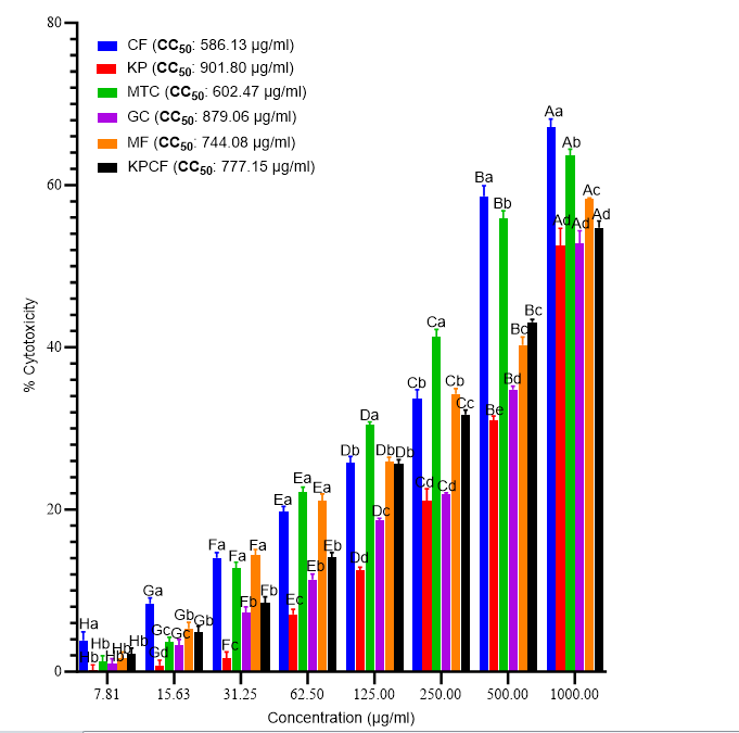
Figure 1: Cytotoxic effects of the selected tea waste samples from various factories. Values are presented as X SD ± ; Bars with dissimilar upper-case alphabets across concentrations and lower-case alphabets within the same concentration are significantly different (P<0.05; One-Way ANOVA with Tukey’s post hoc). CF: Chelal fluff; GC: Gitambo Cyclone; GF: Gitambo Fluff; IC: Itumbe Cyclone; KP: Kaptumo Fluff; KPCF: Kaptumo cyclone fluff. CC50: Median Cytotoxic concentration.
Antiproliferative Effects of The Selected Tea Waste Samples from Various Factories
Inhibition of Cervical Cancer Cell Growth: The results revealed a positive significant dose-dependent increase in percentage inhibition of cervical cancer cell growth P<0.05; (Figure 2). Notably, the percentage inhibitions recorded by the fluff sample from the Chelal factory were significantly higher than those of all the other samples at all concentrations, except at concentrations of 62.5µg/ml, 125µg/ml and 250µg/
ml (P<0.05). It was observed that at these concentrations (62.5µg/ml, 125µg/ml, and 250µg/ml), the cyclone from the Mudete factory caused significantly higher inhibitions of cervical cancer cell growth than all the other tested samples (P<0.05; Figure 2). However, no significant differences among the percentage inhibitions of cervical cell growth were observed among the fluff samples collected from the Kaptumo and Momul factories, cyclone samples from the Mudete and Gitambo factories, and cyclone fluff from the Kaptumo factory P>0.05 (Figure 2). Furthermore, each sample’s median inhibitory concentration (IC50) was determined to appraise its anticancer capacity. The results showed that the fluff sample from Chelal had lower IC50 (483.99µg/ml) than all the examined samples (Figure 2). It was observed that the IC50 values of all the other samples were more than 500µg/ml, with the cyclone sample from Gitambo recording the highest (836.55µg/ml) (Figure 2).
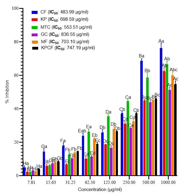
Figure 2: Antiproliferative effects of the selected tea waste samples from various factories against cervical cancer cells (HeLa). Values are presented as X + ; Bars with dissimilar upper- case alphabets across concentrations and lower-case alphabets within the same concentration are significantly different (P<0.05; One-Way ANOVA with Tukey’s post hoc). CF: Chelal fluff; GC: Gitambo Cyclone; GF: Gitambo Fluff; IC: Itumbe Cyclone; KP: Kaptumo Fluff; KPCF: Kaptumo cyclone fluff. IC50: Median inhibition concentration.
Selectivity Indices: As the results in Table 1 show, the selectivity indices of the fluff samples from Kaptumo and Chelal factories were higher than those obtained from other factories. Notably, the selectivity of doxorubicin was higher than that of all the tea waste samples in this study (Table 1).
| Selectivity Index | |
|---|---|
| CF | 1.21 |
| KP | 1.29 |
| MTC | 1.09 |
| GC | 1.05 |
| MF | 1.06 |
| KPCF | 1.04 |
| Doxorubicin | 2.62 |
Table 1: Selectivity Indices of the selected tea waste samples against cervical cancer cells (HeLa). CF: Chelal fluff; GC: Gitamb
Discussion
The need for potent and safe anticancer agents is imperative, considering the challenges experienced by the conventionally used chemotherapeutic agents [6, 26]. Therefore, analysing plant-derived materials, such as C. sinensis processing byproducts, may help identify active compounds suitable as lead substances for semi-synthetic drug development and direct pharmaceutical application [27, 28]. Numerous novel anticancer drugs have originated from natural products, with ongoing research leading to the continual discovery of new compounds [14, 29]. These cytotoxic natural products have the potential to complement conventional chemotherapeutic drugs, enhancing efficacy or reducing toxicity, thereby offering significant promise in the treatment of cancers [16].
The investigation into the cytotoxic and anti-cervical cancer effects of C. sinensis processing effluents presents compelling insights that hold significance for environmental waste management and the potential development of anticancer drugs. This study revealed concentration- dependent responses to various tea waste samples, revealing distinctive patterns in cytotoxicity and anti-cervical cancer effects. The higher cytotoxicity induced by the Chelal factory fluff sample and the Mudete factory cyclone sample provides concrete evidence of bioactive phytochemicals with diverse biological activities in tea waste [27, 30]. This suggests that these wastes probably contain cytotoxic alkaloids, terpenoids, anthraquinones, and others, which can induce cell death [31, 32]. Determining median cytotoxic concentration (CC50) values further refines this understanding, indicating the concentrations at which the toxicity becomes prominent. The lower CC50 of the fluff sample from Chelal implies a higher toxicity level than other samples, in line with previous research [33, 34, 35], warranting attention to its potential environmental and health implications. Moreover, research has shown that test samples with lower CC50 values may possess anticancer activity [36, 37].
The study unraveled a positive dose-dependent increase in the Inhibition of cervical cancer cell growth across all samples. The superior inhibitory effects of the fluff sample from the Chelal factory, as evidenced by higher percentage inhibitions, and the intriguing potency of the cyclone sample from the Mudete factory at specific concentrations signify the potential of these tea waste components in hindering cancer cell proliferation. Determining median inhibitory concentration (IC50) values refines this insight, with the lower IC50 of the fluff sample from Chelal indicating its considerable potency in inhibiting cervical cancer cell growth. Previous research shows that C. sinensis contains antioxidant polyphenols, such as catechins and gallic acid, with anticancer efficacy [27, 30]. Therefore, it is suggestive that upon processing, these phytochemicals accompany the tea wastes, ultimately contributing to the observed effects. Besides, the variations in cytotoxicity towards normal and cancer cells are attributed to the differential concentrations of the associated phytochemicals in the various studied samples.
The computation of Selectivity Indices is crucial as it offers insights into the safety profiles of the samples on normal versus cancer cells [38]. Research shows that SI values 1 ≥ denote selective toxicity towards cancer cells while sparing normal cells, indicating anticancer potential [39]. Thus, the recorded SI of the fluff samples from the Chelal, Gitambo, and Kaptumo factories, cyclone samples from the Gitambo and Itumbe factories, and cyclone fluff from the Kaptumo factory indicate a favorable safety profile. This implies that these waste components may contain specific anticancer-associated phytochemicals [13, 40], which exhibit a selective effect, individually or synergistically, on cancer cells compared to normal cells. This selective cytotoxicity is a crucial aspect in anticancer drug research, as it suggests a potential avenue for developing drugs that target cancer cells while sparing normal cells [38, 39]. While limited studies directly address tea wastes cytotoxic and anti-cervical cancer effects, the present findings contribute substantively to the current body of knowledge. The concentration-dependent responses and nuanced anti-cervical cancer effects observed in some tea waste samples provide valuable data for anticancer drug research.
Identifying tea waste components with cytotoxic and anti-cervical cancer effects directs attention to potential candidates for drug development, considering the observed potential of this often neglected resource. The distinct patterns observed in some tea waste samples offer a roadmap for researchers to explore and isolate bioactive compounds with anticancer properties [28, 41]. These compounds could be the foundation for developing targeted and efficacious anticancer drugs [42, 43]. Employing proper tea waste management procedures, including proper storage, may prevent leakage, loss, or degradation of the bioactive phytochemicals, thereby increasing their potency and safety [44, 45, 46].
Moreover, the nuanced understanding of concentration- dependent responses and the safety profiles of tea waste components are critical considerations in drug development. The lower CC50 and IC50 values, coupled with higher Selectivity Indices, suggest that these components can integrated into drug formulations with improved efficacy and safety profiles [22, 44, 45, 47]. The observed variations among tea waste samples underscore the importance of targeted investigations, allowing researchers to identify and harness the unique properties of specific waste streams for drug development.
This initial study advances our understanding of the cytotoxic and anti-cervical cancer effects of tea waste and offers valuable insights for anticancer drug research. Additionally, the findings of this study pave the way for continued interdisciplinary research, bridging environmental science and medicinal chemistry for sustainable and impactful anticancer drug development.
Conclusions and Recommendations
This study underscores the potential of tea-processing effluents to serve as natural sources of cytotoxic and anti- cervical cancer lead molecules for future anticancer drug development. The concentration-dependent responses, safety profiles indicated by Selectivity Indices and nuanced variations among waste samples contribute valuable insights, emphasizing the importance of targeted investigations for identifying bioactive compounds. The findings also highlight the significance of proper waste management to maintain the potency and safety of these compounds. Moreover, this study bridges environmental science and medicinal chemistry, advancing our understanding of tea waste’s potential as a resource for sustainable and impactful anticancer drug discovery.
Going forward, conducting in-depth analyses of specific bioactive compounds of tea waste samples to understand their mechanism of action and potential synergies is crucial. Additionally, exploring optimal extraction methods to maximize the yield and stability of these compounds is imperative for drug development. Further investigations should focus on the feasibility of incorporating tea waste-derived bioactive compounds into drug formulations, considering potential synergistic effects with existing anticancer drugs. Long-term studies assessing the environmental impact of tea waste and its sustainable utilization in drug development are also warranted. Collaboration between environmental scientists and pharmaceutical researchers is encouraged to foster interdisciplinary approaches for comprehensive understanding and utilization of tea waste in anticancer drug development.
Acknowledgments
We appreciate the technical support offered by the laboratory technologists at the Kenya Medical Research Institute, Centre for Traditional Medicine, and Drug Research (CTMDR), especially Ms. Sally Kamau, during our experiments.
Authors Contributions
Thaddeus Mangenya conceived the research idea, with contributions from Daniel Kariuki, Johnson Kinyua, Martin Obanda, and Simon Ochanda. Thaddeus Mangenya conducted the experiments, analyzed the data, and drafted the manuscript.
Gervason Moriasi provided the reagents and optimized the experimental design and study methods.
Daniel Kariuki, Johnson Kinyua, Martin Obanda and Simon Ochanda supervised the study. All authors reviewed and approved the final draft for submission and publication.
Availability of Data and Materials
All data is presented within the manuscript; however, the authors may provide any additional information upon reasonable request.
Competing Interests
The authors declare that no known or perceived competing interests/conflicts of interest regarding this publication exist.
Funding
This study did not receive formal funding from private, public, or not-for-profit research granting agencies.
References
-
Siegel RL, Miller KD, Wagle NS, Jemal A (2023) Cancer statistics, 2023. CA Cancer J Clin 73(1): 17-48.
-
Dy GW, Gore JL, Forouzanfar MH, Naghavi M, Fitzmaurice C (2017) Global Burden of Urologic Cancers, 1990–2013. Eur Urol 71(3): 437-446.
-
Jianrong Z, Jianfu L, Shan X, Qihua H, RebeccA J, et al. (2021) Global burden of lung cancer: implications from current evidence. Ann Cancer Epidemiol 5: 4-4.
-
World Health Organisation (2023) cervical cancer. Cervical cancer Fact Sheet.
-
GBD 2019 Ageing Collaborators (2022) Global, regional, and national burden of diseases and injuries for adults 70 years and older: systematic analysis for the Global Burden of Disease 2019 Study. BMJ 376: e068208.
-
Zugazagoitia J, Guedes C, Ponce S, Ferrer I, Molina P (2016) Current Challenges in Cancer Treatment. Clin Ther 38(7): 1551-1566.
-
Hegde P, Chen D (2020) Top 10 Challenges in Cancer Immunotherapy. Immunity 52(1): 17-35.
-
Lei J, Ploner A, Elfström K, Jiangrong W (2020) HPV Vaccination and the Risk of Invasive Cervical Cancer. New England Journal of Medicine 383(14): 1340-1348.
-
Fokom J, Schmeler K (2019) Conservative management of cervical cancer: Status and obstetrical implications. Best Pract Res Clin Obstet Gynaecol 55: 79-92.
-
Stelzle D, Tanaka LF, Lee KK, Ahmadaye I, Iacopo B, et al. (2021) Estimates of the global burden of cervical cancer associated with HIV. Lancet Glob Health 9(2): 161-169.
-
Leng A, Jing J, Nicholas S, Wang J (2019) Geographical disparities in treatment and health care costs for end- of-life cancer patients in China: A retrospective study 11 Medical and Health Sciences 1117 Public Health and Health Services. BMC Cancer 19(1): 39.
-
Jemal A, Bray F, Forman D, Meg O, Jacques F, et al. (2012) Cancer burden in Africa and opportunities for prevention. Cancer 118(18): 4372-4384.
-
Singh S, Sharma B, Kanwar SS, Kumar A (2016) Lead phytochemicals for anticancer drug development. Front Plant Sci 7: 1667.
-
Shah U, Shah R, Acharya S, Acharya N (2013) Novel anticancer agents from plant sources. Chin J Nat Med 11(1): 16-23.
-
Greenwell M, Rahman P (2015) Medicinal Plants: Their Use in Anticancer Treatment. Int J Pharm Sci Res 6(10): 4103-4112.
-
Prakash O, Kumar A, Kumar P, Ajeet A (2013) Anticancer Potential of Plants and Natural Products: A Review. Am J Pharmacol Sci 1(6): 104-115.
-
Pradhan S, Dubey RC (2020) Proximate Analysis, Total Phenolic Content, Antioxidant Activity and Anti-Diabetic Property of Camellia sinensis and Camellia Assamica. Biochem Cell Arch 20(1): 15-23.
-
Zhang L, Ho C, Zhou J, Santos JS, Armstrong L, et al. (2019) Chemistry and Biological Activities of Processed Camellia sinensis Teas: A Comprehensive Review. Compr Rev Food Sci Food Saf 18(5): 1474-1495.
-
Gonçalves D, Windson I, Cristina P, Arruda F, Maria G (2021) Processing, chemical signature, and food industry applications of Camellia sinensis teas: An overview. Food Chem X 12: 100160.
-
Esghaei M, Ghaffari H, Esboei B, Tapeh Z, Salim F, et al. (2018) Evaluation of anticancer activity of Camellia sinensis in the Caco-2 colorectal cancer cell line. Asian Pacific Journal of Cancer Prevention 19(6): 1697-1701.
-
Paiva L, Rego C, Lima E, Marcone M, Baptista J (2021) Comparative analysis of the polyphenols, caffeine, and antioxidant activities of green tea, white tea, and flowers from azorean Camellia sinensis varieties affected by different harvested and processing conditions. Antioxidants 10(2): 1-16.
-
Abraham AM, Alnemari RM, Brußler J, Keck C (2021) Improved antioxidant capacity of black tea waste utilizing plantcrystals. Molecules 26(3): 592.
-
Meerloo J, Kaspers L, Cloos J (2011) Cell Sensitivity Assays: The MTT Assay. Methods Mol Biol 731: 237-245.
-
Fulya T, Ali K, Gul O, Melikoglu G, Anil S, et al. (2016) In vitro anticancer and cytotoxic activities of some plant extracts on HeLa and Vero cell lines. JBUON 21(3): 720- 725.
-
Bibi Y, Nisa S, Zia M, Waheed A, Ahmed S, et al. (2012) In vitro cytotoxic activity of Aesculus indica against breast adenocarcinoma cell line (MCF-7) and phytochemical analysis. Pak J Pharm Sci 25(1): 183-187.
-
Price J, Ndom P, Atenguena E, Mambou N, Robert W (2012) Cancer care challenges in developing countries. Cancer 118(14): 3627-3635.
-
Filippini T, Malavolti M, Borrelli F, Angelo A, Susan J, et al. (2020) green tea (Camellia sinensis) for the prevention of cancer. Cochrane Database of Systematic Reviews 3(3): CD005004.
-
Naeem A, Hu P, Yang M, Jing Z, Yali L, et al. (2022) Natural Products as Anticancer Agents: Current Status and Future Perspectives. Molecules 27(23): 8367.
-
Xu J, Mao W (2016) Overview of Research and Development for Anticancer Drugs. J Cancer Ther 7(10): 762-772.
-
Zhao T, Li C, Wang S, Song X (2022) Green Tea (Camellia sinensis): A Review of Its Phytochemistry, Pharmacology, and Toxicology. Molecules 27(12): 3909.
-
Fozia A, Victor K, Armelle T, Matthias H, Andreas K, et al. (2019) Cytotoxic flavonoids from two Lonchocarpus species. Nat Prod Res 33(18): 2609-2617.
-
Canga I, Vita P, Oliveira A, Castro M, Pinho C (2022) In Vitro Cytotoxic Activity of African Plants: A Review. Molecules 27(15): 4989.
-
Hamzeloo M, Khalaj A, Malekmohammadi M (2015) Cytotoxic Activity and Apoptosis Induction of Hypericum scabrum L. Iran Red Crescent Med J 17(10): e19453
-
Ahmed EM, Khalid A, Penech F, Mesaik MA (2018) Anti Prostate Cancer, Antioxidant Activity and Cytotoxicity of Some Sudanese Medicinal Plants 1(1): 1-5.
-
Steenkamp V, Gouws MC (2006) Cytotoxicity of six South African medicinal plant extracts used in the treatment of cancer. South African Journal of Botany 72(4): 630-633.
-
Odira HO, Mitema SO, Mapenay IM, Moriasi GA (2022) Anti-inflammatory, Analgesic, and Cytotoxic Effects of The Phytexponent: A Polyherbal Formulation. J Evid Based Integr Med 27.
-
Suchada J, Madaka F, Wunnakup T, Teeratad S, Chanamon P, et al. (2023) In vitro antioxidant, anti-inflammatory, and anticancer activities of mixture Thai medicinal plants. BMC Complement Med Ther 23(1): 43.
-
Radha A, Jawad KN (2021) Improvement of the Selectivity Index (SI) and cytotoxicity activity of doxorubicin drug by panax ginseng plant extract. Arch Razi Inst 76(3): 659-666.
-
Kissin I (2013) An early indicator of drug success: Top journal Selectivity Index. Drug Des Devel Ther 7: 93-98.
-
Raletsena MV, Mongalo NI (2023) Phytochemical analysis, in vitro antimicrobial, anticancer, anti-inflammatory, and antioxidant activity of extracts from Bulbine anguistifolia Poelln (Asphodelaceae). South African Journal of Botany 159: 588-595.
-
Huang M, Lu J, Ding J (2021) Natural Products in Cancer Therapy: Past, Present and Future. Nat Prod Bioprospect 11(1): 5-13.
-
Solowey E, Lichtenstein M, Sallon S, Paavilainen H, Solowey E, et al. (2014) Evaluating medicinal plants for anticancer activity. Scientific World Journal.
-
Lu J, Bao L, Chen X, Huang M, Wang Y (2012) Alkaloids isolated from natural herbs as anticancer agents. Evidence-based Complementary and Alternative Medicine.
-
Debnath B, Haldar D, Purkait MK (2021) Potential, and sustainable utilization of tea waste: A review on present status and future trends. J Environ Chem Eng 9(5): 106179.
-
Zahra A, Lim SK, Shin SJ, Yeon IJ (2022) Properties of Green Tea Waste as Cosmetics Ingredients and Rheology Enhancers. Applied Sciences (Switzerland) 12(24): 12871.
-
Gao P, Ogata Y (2020) CHAMU: An effective approach for improving the recycling of tea waste. In: IOP Conference Series: Materials Science and Engineering 711: 012024.
-
Athirojthanakij W, Rashidinejad A (2023) Delivery of Catechins from Green Tea Waste in Single- and Double-Layer Liposomes via Their Incorporation into a Functional Green Kiwifruit Juice. Molecules 28(2): 575.
- Acido Labile or Gastro Irritant Apis and Enteric Release in Galenic Practice: An Overview
- A Study on Knowledge, Attitude and Practice of Hand Hygiene among Healthcare Professionals at a Tertiary Care Hospital, India
- Influence of Inoculum Concentration on In Vivo Incubation Period of Emmia lacerata, Pathogenesis and Management of Wilt in Pepper (Capsicum annuum L.)
- Vanilla’s Chemistry
- Marine Anti-Cancer Compounds and Adverse Effects of Global Warming on Oceans: An Overview
- Serological Investigation of Chikungunya Virus Antibody among Malaria-Suspected Febrile Patients in Some Healthcare Facilities in Rivers State