Prenatal Diagnosis of Fetal Goiter: Case Report and Review of Literature
Pregnancy hyperthyroidism due to Plummerâs disease is an uncommon event. Therapy is based on propylthiouracil (PTU), starting from the first trimester of pregnancy. Among the complications of the antithyroid drugs (ATDs) there is the risk of hypothyroidism and fetal goiter, in addition to well-known possible teratogen effects. Maternal hormonal monitoring with a personalized pharmacological treatment and fetal ultrasound control, starting from the 18th week of pregnancy, are mandatory for the mother and fetus health and therefore for a successful pregnancy. In this paper well describe the case of a woman with toxic adenoma, in which the ultrasounds diagnosis of fetal goiter at the 29th gestational-week of pregnancy and the consequent suspension of the ongoing therapy with PTU, succeeded in the end of the pregnancy with the birth of a healthy fetus.
Introduction
The diagnosis of fetal goiter is relatively easier when the mother has a declared hypothyroidism than in patients with unknown medical history. In Europe it is estimated that hyperthyroidism is not recognized in about 1.7% of cases [1]; in the absence of predisposing factors for hyperthyroidism, it may be difficult to discriminate clinical signs of pregnancy with mild hyperthyroidism. The manifest hyperthyroidism of pregnancy is estimated in Europe with a prevalence of 0.2 to 0.8% [2, 3]: Basedow’s disease represents about 85% of cases, while a toxic nodule, recurring mainly in old age, is rare in pregnancy, representing about 5% of cases [4, 5] but some studies have also reported an increase in the number and size of nodules during pregnancy [6]. This article describes the case of a patient with known toxic nodule, treated with methimazole (MMI). From the 6th
week of gestation the patient switched to propylthiouracil (PTU) therapy. Subsequently the fetus has developed a goiter, diagnosed by ultrasound at the 29th gestational week. The therapy was suspended after the diagnosis, confirmed by nuclear magnetic resonance. Fetal goiter regressed in a few weeks before the delivery, performed by caesarean section.
Case Presentation
41-year-old patient, gravida3 para2.
A first pregnancy failed with a spontaneous abortion at the 7th week of amenorrhea; no clinical signs of hyperthyroidism reported.
A second pregnancy was complicated in the first trimester, by gravidic thyrotoxicosis: diagnosis of Plummer adenoma of the left lobe (diameter of 14mm), treated with propylthiouracil (PTU). Pregnancy ended with intra-partum fetal death at the 39th gestational week for shoulder dystocia; the newborn weighed 3700 grams. Two months after the delivery the therapy was changed and she started to take methimazole (MMI) 5mg once a day. Thyroid scintigraphic examination was proposed to consider a subsequent surgical therapy, that it was not performed.
A third pregnancy was complicated by hyperemesis and thyrotoxicosis, treated from the 8th week with PTU, 50 mg twice a day. At the 32nd weeks of pregnancy FT3 3.70 pg/ml, FT4 0.79 ng/ ml, TSH 2.36 mU/L. A caesarean section was performed at the 39th week of amenorrhea, with the delivery of a 4300 gr male infant. Two months after delivery: FT4 0.63 ng / ml, TSH 1.10 mU / L, Ab- TPO0.6 IU / ml, TRAB 0.10 IU / L ; so the patient switched to MMI 5 mg once a day. The thyroid ultrasound showed a 6 mm nodule in the left lobe with a barely evident colloid cyst component.
At the fourth pregnancy, in 2018, the patient had not undergone neither a scintigraphic examination nor a surgical consultation.
At the 6th week of amenorrhea, MMI was stopped and therapy with PTU 50 mg twice a day started.
At the 13th week, normal obstetric ultrasound. At the 16th week, FT4 0.58 ng / ml, TSH 1.93 mU / L, TRAB 0.10 IU/ L.
At the 20th week, FT4 0.44 ng / ml, TSH 2.5 mU /L, OGTT (91-157-168 mg / dL): gestational diabetes mellitus (GDM) was diagnosed; therapy with PTU 50 mg in the morning and 25 mg in the evening began.
At the 22nd week, morphological ultrasound was normal.
Apart from a compensated gestational diabetes and normal thyroid function, no other obstetric marks are reported in the 2nd, 4th and 7th month.
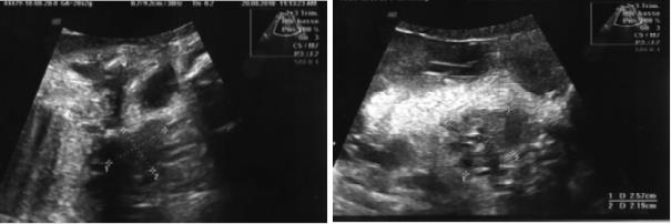
Ultrasonography at 29th week described: single fetus, biometrics at the 59th percentile with movements and regular heartbeat, normoinserted placentas with regular dimensions and echo structure, amniotic fluid of adequate volume, fetal morphology within the normal range except for a homogeneously increased volume of the thyroid (longitudinal diameter 20 mm x anterior-posterior 21 mm x lateral-lateral 25 mm), with increased peripheral vascularization to the echo-doppler (Figure 1).
Subsequent control with magnetic resonance imaging (MRI) confirmed fetal goiter. TRAB, anti-TPO antibodies, anti-TG antibodies in maternal blood were absent. Cordocentesis was performed for the evaluation of fetal thyroid function: TSH 66.27 mU / L, fT4 12.5 pmoli / L. PTU therapy was suspended.
At the 35th gestational week fetal ultrasonography reported a significant volume reduction of the thyroid.
At the 39th week and two days of gestational age, 68 days after PTU suspension, a caesarean section was performed. A female with an APGAR index at 1 minute of 9 and, at 5 minutes 10 was born, with an arterial pH of 7.36. Amniotic fluid meconium stained, weighed 3140 gr, length 50 cm, head circumference 32 cm. The neurological examination and the physical examination of the apparatus were normal. Screening of congenital metabolic diseases (cystic fibrosis, hyperphenylalaninemia, congenital hypothyroidism, congenital adrenal hyperplasia) was negative; audiological screening: bilateral pass. Ocular screening: red reflex normally evocable resulted bilaterally, without pharmacological distension. The electrocardiogram showed no anomalies; a color-doppler-echocardiography showed a slight shunt of the interatrial septum at the level of the oval fossa, and a modest tricuspid insufficiency with PAPs still within the age limits. At an ultrasound examination, performed on the second day, the thyroid had regular profiles, dimensions and eco-structure (the right thyroid lobe had an anteroposterior diameter of 7 mm and the left lobe 8 mm), with a normal vascular pattern. The ultrasound of the complete abdomen showed no pathologies. In the second day: TSH 4.38 mU / L, FT3 3.86 pg /mL, FT4 2.88 ng / ml. The little patient was discharged on the third day in good general conditions, breast feeded. At one month of life, we report normal thyroid function. The mother resumed the treatment with 5 mg MMI in the morning three months after delivery. At six months the child presented normal thyroid function and the mother was in good health.
Discussion
The fetal thyroid originates from the endoderm of the second pharyngeal pouch between the first and second pair of pharyngeal arches, during the first trimester of pregnancy while the functional development takes more time. The hormonal production of fetal thyroid begins during the 11th week of pregnancy but remains low until the 20th week [7]. Fetal thyroid is subjected to maternal hormonal activity, mediated by the placenta, which is impermeable to TSH, weakly permeable to thyroid hormones, and permeable to TRAB and TPO-Ab as well as to ATDs [8]. Functional thyroid disease is the second most common endocrine disorder after diabetes in pregnancy. Thyroid hormones play an essential role in the development and cerebral organization of the fetus with consequences on the future cognitive and behavioral capacities [9]. Maintaining a good and well-balanced maternal thyroid function with a healthy fetal development is the goal of an adequate obstetric assistance. In Europe, hyperthyroidism has a prevalence of 0.8% [3]: causes are various (Table 1), but, in about 80% of the cases, hyperthyroidism is due to Basedow - Graves’ disease; while Plummer’s disease it is relatively rare.
- Graves’ disease
- thyroiditis
- toxic adenoma
- toxic multinodular goiter
- gestational transient thyrotoxicosis
- hyperemesis gravidarum
- gestational trophoblastic disease
- TSH receptor mutation
- Iatrogenic –excessive levothyroxine intake-amiodarone -iodine
Table 1: Hyperthyroidism in pregnancy.
Complications of improperly treated hyperthyroidism can affect both, the mother and the fetus (Table 2).
| Maternal | Miscarriages, gestational hypertension, preeclampsia, congestive heart failure, Thyroid storm. |
| Obstetrical | Premature delivery, placental abruption, premature ruptures of membrane, gestational hypertension. |
| Fetal | Congenital malformations, hyperthyroidism, developmental dysplasia of the hip associated with first-trimester maternal hyperthyroidism, intrauterine growth restriction (IUGR), small for gestational age (SGA), prematurity, stillbirth. |
Table 2: Complications associated with hyperthyroidism in pregnancy [10].
the first check during pregnancy. The therapy of hyperthyroidism is based on ATDs of the thionamide group (MMI) and on thiouracil derivatives (PTU). All
ATDs pass the placental barrier and can block the fetal thyroid causing fetal hypothyroidism with goiter. In the first trimester of pregnancy the use of PTU is preferred: firstly because the placenta is less permeable to the PTU than to the MMI, and secondly because the PTU is quicker in blocking the peripheral conversion of T4 to T3, with less teratogenic effects on the fetus [5] (Table 3).
| MMI | Aplasia cutis, choanal atresia, esophageal atresia, omphalocele, urinary tract malformations, eye defects, ventral septal defects, dysmorphic facies, athelia, developmental delay. |
| PTU | Pre-auricular sinus/fistula and cysts, Urinary tract abnormalities in males. |
Table 3: Birth defect associated with ATD [10].
However, since PTU treatment can cause severe maternal hepatic cytolysis (in literature 1.8 out of 1000 treated pregnancy), requires close hematological controls to avoid the risk of acute drug induced hepatitis [11]. The patient in the described case, at the 6th week of gestation, moved from a 5 mg MMI therapy in the morning to a 50 mg PTU twice a day. In the 20th week the PTU was reduced to 50 mg plus 25 mg. When the mother is affected by hyperthyroidism in therapy with ATDs, especially with a positive TRAb at TPO-Ab pattern, the follow up of fetal status needs ultrasound examination. A careful evaluation of the size of the fetal thyroid starting from the 18th week of gestation with scheduled ultrasound scans is essential to recognize a thyroid dysfunction [12] and to arrange a prompt and adequate therapy; and maintaining the FT4 values at the upper limits of the standard reduces the risks of side effects on the fetus [10].
Fetal hypothyroidism due to overtreatment with ATDsis shown by an increase of the thyroid volume and of the peripheral gland vascularization [10], by delayed fetal growth, delayed bone maturation and polyhydramnios [13]. The fetal Magnetic Resonance Imaging (MRI) can confirm and sometimes improve the diagnosis but, when the doubt persists, cordocentesis is necessary to detect the fetal thyroid dysfunction. During the ultrasound examination at the 29th week of pregnancy the fetus, in this case, presented a goiter without other significant ultrasound signs; the following check with MRI confirmed the ultrasound data and cordocentesis was proposed to evaluate the fetal thyroid functional status, and allowed the diagnosis of a fetal hypothyroid goiter. Therapy with ATDs was suspended with a rapid improvement. The caesarean delivery at the end of the pregnancy brought to light a healthy newborn with mild residual thyroid dysfunction that normalized in the first month of life.
Conclusions
Diagnosis and treatment of hyperthyroidism in pregnancy represent a challenge that involves all the obstetricians, endocrinologists, pediatricians and anesthesiologists’ skills with the common goal of assuring a healthy development, delivery, and consequently maternal and newborn’s health status. Besides the diagnostic and therapeutic difficulties, should be considered also the problems caused by the patients’ low compliance to follow the medical instructions. This case shows how obstetric ultrasound performed in the third trimester of pregnancy was able to avoid fetal complications and the immediate suspension of PTU therapy allowed the birth of a healthy euthyroid newborn, without maternal complications [14].
Conflict of Interest
None of the authors received any support in the form of grants, equipment, or drugs.
Declarations
The patient agreed to share ultrasounds data, maintaining her anonymity.
References
-
Garmendia Madariaga A, Santos Palacios S, Guillén- Grima F, GalofréJC (2014) The incidence and prevalence of thyroid dysfunction in Europe: a meta- analysis. J Clin Endocrinol Metab 99(3): 923-931.
-
Taylor PN, Albrecht D, Scholz A, Gtierrez-Buey G, Lazarus JH, et al. (2018) Global epidemiology of hyperthyroidism and hypothyroidism. Nat Rev Endocrinol 14(5): 301-316.
-
De Leo S, Lee SY, Braverman LE (2016) Hyperthyroidism. Lancet 388(10047): 906-918.
-
KrassasGE, Poppe K, Glineor D (2010) Thyroid function and human reproductive health. Endocr Rev 31: 702-755.
-
Illouz F, Luton D, Polak M, BesaçonA, Bournaud C (2018) Graves’ disease and prengnancy Maladie de Basedow et grossesse . Annales d’Endocrinologie 79(6): 636-646.
-
Stagnaro-Green A (2015) Postpartum management of women begun on levothyroxine during pregnancy. Front Endocrinol 6: 183.
-
Polak M (2014) Human fetal thyroid function. Endocr Dev 26: 17-25.
-
Polak M, Luton D (2014) Fetal thyroidology. Best Pract Res Clin Endocrinol Metab 28(2): 161-173.
-
Besançon A, Beltrand J, Gac IL, Luton D, Polak M (2014) Management of neonates born to women with Graves’ disease: a cohort study. Eur J Endocrinol 170: 855-862.
-
Nguyen CT, Sasso EB, Barton L, Mestman J (2018) Graves’ hyperthyroidism in pregnancy: a clinical review. Clin Diabetes Endocrinol 4: 4.
-
Lo JC, Rivkees SA, Chandra M, Gonzalez JR, Korelitz JJ, et al. (2015) Gestational thyrotoxicosis, antithyroid drug use and neonatal outcomes within an integrated healthcare delivery system. Thyroid 25(6): 698-705.
-
De Groot L, Abalovich M, Alexander EK, Amino N, Barbour L, et al. (2012) Management of Thyroid dysfunction during pregnancy and postpartum: an Endocrine Society clinical practice guideline. J Clin Endocrinol Metab 97(8): 2543-2565.
-
Polak M (1998) Hyperthyroidism in early infancy: pathogenesis, clinical features and diagnosis with a focuson neonatalhyperthyroidism. Thyroid 8(12): 1171-1177.
-
King JR, Lachica R, Lee RH, Montoro M, Mestman J (2016) Diagnosis and Management of hyperthyroidism in pregnancy: a review. Obstet Gynecol Surv 71(11): 675-685.
- Understanding Pediatric Multiple Sclerosis: Clinical Presentation, Diagnostic Criteria, Therapeutic Advances, and Supportive Care Approaches
- Hemophilia in Children
- Xia-Gibbs Syndrome- A Case Report
- A Study to Assess Effectiveness of Play Therapy in Reducing Post-Operative Pain among Children Age 2 To 5 Year who have Undergone General Surgeries in Selected Pediatric Hospitals of Vadodara
- Preterm Birth: Scope of the Problem, Cost of Care, Potential Complications and Current Guidelines for Management
- Noradrenaline: Can we Use it to Manage Hemodynamic Instability among Neonatal Septic Shock at the NICU?