Pathogenetic Approach to Surgery in Children with Old Discosions of the Radial Head
The results of surgical treatment of 83 patients with dislocations of the radial head were analyzed. To recreate the annular ligament, the joint capsule itself and segments of the annular ligament were used, which is a more anatomical and physiological approach in relation to the elbow joint. The results were evaluated on a 5-point system, which included the functions of the joint and the axis of the upper limb. Good and excellent results when assessed after 6, 12 months (4-5 points) were obtained in 56 patients, which accounted for 57.4%, satisfactory results (3 points) were observed in 26 (31.3%).
Introduction
Elbow joint injuries in children, according to various authors, account for 40 to 50% of all injuries of the musculoskeletal system. And post-traumatic complications as a result of damage to the elbow joint take the first place and in 29.9% of cases lead to permanent disability of patients [1, 2]. In terms of the frequency of errors and complications, the difficulty of treating injuries in this area, they take the first place relative to other joints, which dictates the need for a more thorough and detailed study of these issues [3, 4, 5]. Unsatisfactory results in the treatment of elbow joint injuries still remain high, reach 21% and do not tend to decrease [2, 6]. Often, the treatment of injuries of the elbow joint, as concomitant injuries, is performed in the long term, when it is no longer possible to obtain good results [7, 8, 9, 10]. The scientific literature covering the tactics of treatment of chronic injuries of the glenohumeral joint, available to us, Clinical Note remains small. All this indicates the need for further research on more optimal options for the surgical restoration of the annular ligament in case of its damage.
Purpose
To describe the clinical, radiological, ultrasound features of chronic dislocations of the head of the radius and indications for surgery depending on the type of dislocation of the radius.
Materials and Methods
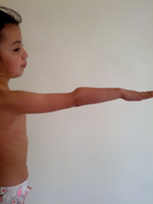
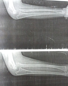
Our report is based on a study of treatment outcomes in 83 children who were hospitalized from 2015 to 2020 of these, isolated dislocations in 62 patients (74.6%), with Montage injuries in 21 patients (25.4%). There were 54 (65.1%) boys and 29 (34.9%) girls. Damage was localized on the right in 41, on the left in 39, and bilateral in 3 patients. By age: up to 5 years - 22 patients, 6-12 years - 46 patients, 13-18 years - 15 patients. According to the prescription of the injury: out of 62 patients with isolated dislocations, only one patient had a stale character, and the rest had all cases of an old character. Clinically: all patients had deformities of the elbow joint, in the form of protrusion of the head of the beam from under the skin (Fig. 1), 67 patients had flexion- extension contractures of the elbow joint and 72 patients had pronation-supination contractures of the forearm. X-ray: subluxation of the head of the radius in 16 patients and in 67 complete dislocation of the head of the beam. Dislocation of the head of the beam anteriorly was in 19 patients, antero- medial - in 48 patients. The axis of the radius was broken in 38 patients and the axis of the ulna was broken in 12 patients.
Surgical Interventions and Indications for them
1st Group of Patients a. Chronic dislocations of the head of the radius. Anterior- medial dislocations of the head of the beam are usually observed. They showed: the operation of open reduction of the head of the beam, plastic of the annular ligament according to the method of P.U. Urinbaev, Sh.N.
b. In not very neglected cases, it is possible to form a flap and fix it in the lateral region of the ulna to the remnants of the fibrously altered laterally posterior part of the annular ligament.
c. In very advanced cases: retention and fixation of the head of the beam with a bone suture through the channel of the ulna made with lavsan, lye.
2nd Group of Patients: Chronic Lesions of Monteggia a. In cases where the fracture of the ulna healed without deformity, but the dislocation of the ray head remained, the operation was performed by open reduction of the ray head, annular ligament plasty according to our method.
b. In case of fusion of the ulna fracture with angular deviation, osteotomy of the ulna, open reduction of the ray head, and annular ligament plasty according to our method are indicated. Use of the device according to indications.
3rd Group of Patients: Congenital Defects, Ulna Dysplasia and Dislocation of the Radial Head a. Osteotomy and lengthening of the ulna on the Ilizarov apparatus, open reduction of the head of the beam.
b. Reduction of the ray head on the device, open reduction of the ray head and replacement of the ulna defect with an autograft.
4th Group Patients: Removal of the chondroma, arthrosis of the joint, modeling of the head of the radius, reduction and reconstruction of the annular ligament.
Results
Analysis of the results of treatment was carried out using clinical and radiological methods. The results were evaluated according to a 5-point system, including the function of the joint and the axis of the upper limb (P.U.Urinbaev 1995). Good and excellent results when assessed after 6, 12 months (4-5 points) were obtained in 56 patients, which accounted for 57.4%, satisfactory results (3 points) were observed in 26 (31.3%). Recurrence in the form of subluxation was observed in 7 patients, they underwent a second operation.
Discussion
When performing an open reduction of the radial head in children with chronic trauma, we consider it appropriate to pay attention to the following points:
- Reconstruction of the annular ligament of the radius from the anterior capsule of the elbow joint;
- Excision of scar tissue from the radial notch of the ulna.
So, with changes in the humeroradial joint during surgery, it was shown that there was a layer of the joint capsule between the head of the radius and the articular end of the shoulder. Similar changes were observed in 80% or more of patients. Such changes are consecrated according to the new pathology of chronic dislocations of the ray head and substantiate the proposed tactics of surgical treatment of patients.
Clinical Observation
Patient Z., born in 2011 was admitted to the hospital with a diagnosis of chronic dislocation of the head of the right radius, more than a year old (Figure 1).
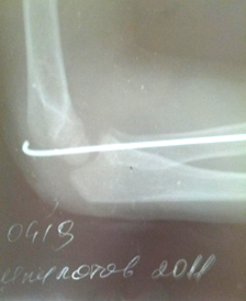
The operation was performed under general anesthesia, open reduction of the head of the right radius with restoration of the annular ligament from the joint capsule and transarticular fixation with a pin. The postoperative period proceeded without complications. After immobilization with a plaster splint in the supination position, the patient was discharged for outpatient treatment for 3 weeks. After 3 weeks, the pin was removed from the patient, the plaster splint was removed for 4 weeks, and a course of rehabilitation treatment was prescribed.
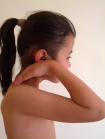
The anatomical and functional result of the treatment was studied 1 year after the end of treatment, while the patient had a limitation of movement in the elbow joint of 20 0 (range of motion - 120 0 ), valgus deviation of the forearm up to 5 0 , based on these data, the result was considered good (Figure 2).
Conclusion
The causes of chronic dislocations of the head of the beam are explained by diagnostic errors, untimely medical care due to late treatment of patients, when the parents of the patients considered the injury not serious. To recreate the annular ligament, we used the joint capsule itself and segments of the annular ligament, which is more anatomical and physiological in relation to the elbow joint. Surgical treatment of children with chronic dislocations of the radial head, reconstruction of the annular ligament, allowed us to achieve good and satisfactory functional results in 91.8% of cases, and anatomical results in 89.3% of cases.
Conflict of Interest: The author declares no conflict of interest.
References
-
Kapanji AI (2014) Upper limb. In: 6th (Edn.).
-
Meltsin II, Pavlov VA, Afaukov IV, Kotlubaev RS, Lyashchenko OA (2016) Injuries of the shoulder joint in children. Pediatric Surgery 20(1): 23-26.
-
Merkulov VN, Dergachev DA, Dorokhin AI (2014) Arthroplasty in the treatment of post-traumatic contractures and ankylosis of the elbow joint in children. Pediatric Surgery 4: 34-38.
-
Ovsyankin NA, Naumochkina NA, Pozdeeva NA (2014) Surgical treatment of patients with dislocations of the head of the beam and rotational contracture of the forearm with birth paralysis of the upper limb. Orthopedics Traumatology and Reconstructive Surgery of Children 2(1): 32-38.
-
Proshchenko YN (2015) Reasons for the development of instability in the distal radioulnar joint in children. Pediatric Surgery 1: 28-30.
-
Azizovich HT (2021) A Modern Approach to the Care of Victims with Combined Pelvic and Femoral Bone Injuries Based on the Severity of the Injury and the Severity of the Condition. Central Asian Journal of Medical and Natural Science 2(4): 156-159.
-
Goyal T, Arora SS, Banerjee S, Kandwal P (2015) Neglected Monteggia fracture dislocations in children: a systematic review. J Pediatr Orthop B 24(3): 191-199.
-
Tilyakov HA, Valiyev EY, Tilyakov AB, Tilyakov AB (2021) A new approach to surgical treatment of victims with pelvic and femoral fracture injuries, taking into account the severity of the condition and the severity of the injury. International Journal of Health & Medical Sciences 4(3): 338-346.
-
Tilyakov KA, Tilyakov AB, Valiyev EY, Tilyakov AB (2021) Apparatus for treatment of combined fractures of pelvic and femoral bones. Bull 897: 1-24.
- Understanding Pediatric Multiple Sclerosis: Clinical Presentation, Diagnostic Criteria, Therapeutic Advances, and Supportive Care Approaches
- Hemophilia in Children
- Xia-Gibbs Syndrome- A Case Report
- A Study to Assess Effectiveness of Play Therapy in Reducing Post-Operative Pain among Children Age 2 To 5 Year who have Undergone General Surgeries in Selected Pediatric Hospitals of Vadodara
- Preterm Birth: Scope of the Problem, Cost of Care, Potential Complications and Current Guidelines for Management
- Noradrenaline: Can we Use it to Manage Hemodynamic Instability among Neonatal Septic Shock at the NICU?