An Infantile Left Amyand Hernia: Case Report and Review of Literature
The incidence of incarceration in pediatric inguinal hernia is between 12 to 17% especially during infancy. It may contain small gut, omentum, cecum, appendix, or ovary and uterine tube, and appendix could be incarcerated on the right side (82%) mostly. Hypermobility of cecum which is a triggering factor of Left-sided Amyand’s hernia and an explanation of De Garengeot hernia occurs in 10-20% of population. Herein, a 9-month-infant with a left-sided incarcerated inguinal hernia containing both cecum and appendix was successfully operated through a groin incision. A hypermobile cecum is accused of the left-sided incarcerated Amyand’s hernia. Preservation or removal of the appendix is controversial which depends on the infection or trauma of hernia contents but the definitive repair of hernia is mandatory. Postoperative imaging should be done for exclusion of the possible underlying anomalies as hypermobile cecum, abnormal intestinal rotation, or situs inversus.
Introduction
Amyand’s hernia is an inguinal hernia which contains the appendix but it is rare in pediatrics, especially during infancy [1, 2]. The first surgeon who described this phenomenon in 1735, is Dr. Claudius Amyand, Sergeant to King George II of England. He reported a child whose age was 11-years old with an enterocutaneous fistula due to a perforated appendix by a pin within the hernial sac. The primary repair of hernia and appendectomy were done within half an hour [3]. Amyand hernia is usually diagnosed intraoperatively as it is rare and its presentation is as any inguinal hernia except in case of incarceration in which erythema and tenderness are the usual signs [2]. Herein, our case is a 9-month-old male had left incarcerated Amyand hernia.
A nine-month-old infant attended to our emergency complaining of vomiting, irreducible left inguinal hernia 8 hours ago. Examination showed normal vital signs, an irreducible left inguinal hernia, distended abdomen without peritonitis. Ultrasound showed intestine within the hernia sac and his laboratory investigations were normal. Dilatation of small intestine was detected in an abdominal x-ray. Manual reduction failed so groin exploration was considered.
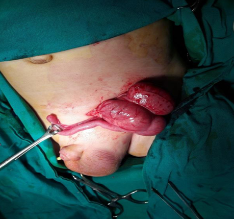
After induction of general anesthesia, a left inguinal transverse skin incision was done followed by opening the sac showing the congested appendix and cecum (Figure
1). The appendix was not removed as it was normal but reduction of the hernia contents into the abdominal cavity and left herniotomy were done. The patient had an uneventful postoperative course.
Retrospectively, a chest-abdominal X-ray detected normal gas distribution in the colon which was an indicator of a hypermobile cecum rather than of abnormal gut rotation. Two days postoperatively, he was stable and discharged. Over one year follow-up, no recurrence and or testicular atrophy were detected.
Discussion
The inguinal hernia has the appendix as a content is defined as Amyand’s hernia [4]. This study case is left Amyand’s hernia. Among general population in the literature, its incidence is 0.4–0.6 % and increases to be 1 % in pediatrics [4]. The appendix is mostly detected in the right hernia sac with the large gut either cecum and/or right colon. Left Amyand’s hernia is rare, and only reported in 13 pediatric cases [5, 6, 7, 8, 9, 10, 11]. The data of all 15 pediatric patients that have been reported in literature beside our case, with a left Amyand’s hernia in (Table 1).
| Pt. No | Author | Year | Gender | Age | Symptoms | Contents of hernia | Presence of appendicitis | Underlying condition | Surgical treatments | Outcome |
|---|---|---|---|---|---|---|---|---|---|---|
| 1 | Gupta S [1] | 2005 | Male | 9 months | Bilious vomiting, irreducible inguinal swelling | Appendix, cecum | (−) | Mobile cecum | herniorraphy | uneventful |
| 2 | Gupta N [5] | 2007 | Male | 11 months | Bilious vomiting, irreducible inguinal swelling | Appendix, cecum and terminal ileum | (+) | Mobile cecum | Herniorraphy, appendectomy | uneventful |
| 3 | Kaymakci A [6] | 2009 | N/A | N/A | N/A | N/A | (+) in one of three | Mobile cecum | Herniorrhaphy in three, appendectomy in one with appendicitis | uneventful |
| 4 | Kaymakci A [6] | 2009 | N/A | N/A | N/A | N/A | Mobile cecum | uneventful | ||
| 5 | Cankorkmaz L [7] | 2010 | Male | 4 months | Vomiting, irreducible inguinal swelling | Appendix | (−) | Mobile cecum in the one not known in the other | Herniorraphy, appendectomy | uneventful |
| 6 | Cankorkmaz L [7] | 2010 | Male | 2 months | Irreducible inguinal swelling | Appendix | (−) | Herniorraphy, appendectomy | uneventful | |
| 7 | Khan R [8] | 2011 | Male | 10 months | Scrotum swelling | Appendix, part of the cecum | (+) | Mobile cecum | Herniorrhaphy, appendectomy, another midline incision to explore malrotation, cecopexy Except for surgical site infection | Uneventful |
| 8 | Singh K [2] | 2011 | Male | 18 months | Bilious vomiting, fever, irreducible inguinal swelling | Appendix, cecum (perforation) | (−) | Mobile cecum | Herniorraphy, closure of cecal perforation | uneventful |
| 9 | Singh K [2] | 2011 | Male | 18 months | Scrotum swelling | Appendix, cecum, terminal ileum (serosal tear) | (−) | Not known | Herniorrhaphy, repair of serosal tear | uneventful |
| 10 | Pun A [9] | 2013 | Male | 18 months | Vomiting, irreducible inguinal swelling | Appendix, sigmoid colon | (+) | Mobile cecum | Herniorrhaphy, appendectomy | uneventful |
| 11 | Al-Mayoof [10] | 2014 | Male | 4 months | Vomiting, irreducible inguino- scrotal swelling | Appendix, cecum | (−) | Situs inversus | Herniorrhaphy, appendectomy | uneventful |
| 12 | Al-Mayoof [10] | 2014 | Male | 10 months | Bilious vomiting, fever, irreducible inguinal swelling | Appendix, cecum | (−) | Mobile cecum | Herniorrhaphy, appendectomy | uneventful |
| 13 | Fumiya Yoneyama, et al. [11] | 2014 | Male | 8 months | Bilious vomiting, irreducible inguinal swelling | Appendix, cecum (serosal tears), terminal ileum | (−) | Mobile cecum | Herniorrhaphy, repair of serosal tears | uneventful |
| 14 | Our case | 2022 | Male | 9 months | Bilious vomiting, irreducible inguinal swelling | Appendix, cecum | (−) | Mobile cecum | Herniorrhaphy | uneventful |
Table 1: The demographics and clinical outcomes of pediatric patients with left-sided Amyand’s hernia, including our case.
As regard the preoperative imaging studies, ultrasound was requested in three cases and our study case without detection of the contents except in our case, the gut was shown (data not illustrated). Detection of ileocecum and or appendix within left Amyand’s hernias has been reported in some adult patients using Contrast-enhanced computed tomography (CT) and contrast enema [12, 13, 14]. This preoperative radiography would be helpful for detection of left Amyand’s hernia and its associated intestinal anomalies, but it is not beneficial as the clinical examination is enough for undergoing emergency operation which is mandatory.
Intraoperative suspicion of the left sided ileocecum as a content of the sac should be considered by surgeons as mentioned in the majority of this review cases. Appendicitis is rare to be seen in Amyand’s hernia which is usually due to ischemia and or trauma of incarceration [15]. Appendectomy was done for 4 out of 15 pediatric patients having a left Amyand’s hernia and showed appendicitis (Table 1). The other eleven cases, involving our case, had a normal appendix (seven appendices were left; four were resected). According to literature, the presence of the appendix within the sac is not an indication of appendectomy except if it is inflamed. Removal of the normal appendix is controversial as follows: (1) the proposed surgical wound infection [4]; (2) the enlargement of wound to control the appendiceal base leading to the abdominal wall weakness and a possible recurrence [16]; and (3) loss of the benefit of the appendix for possible urinary diversion (Mitrofanoff appendicovesicostomy) [6].
One pediatric case with a left Amyand’s hernia underwent an intraoperative abdominal search for the possible anomalies [8], which revealed a mobile cecum. Recently, laparoscopy has an advantage over conventional approach in case of incarcerated inguinal hernia for reduction and visualization [17]. In literature, laparoscopy was successfully used for management of an adult right-sided Amyand’s hernia [18]. Hence, laparoscopy could be advantageous for left hernias in pediatrics, to confirm the abovementioned possible anomalies intraoperative.
Regarding to proposed detection of the abovementioned anomalies, no management is expected for situs inversus or mobile cecum whereas intestinal malrotation may need surgery as Ladd’s procedure if there is a history of obstruction due to possible volvulus.
Conclusion
The left-sided Amyand hernia is rare in pediatrics which could be managed in form of herniotomy and appendicectomy in case of infection and/or trauma with many advantages of laparoscopic management. The associated anomalies as a mobile cecum, intestinal malrotation, or situs inversus should be investigated through intra- or postoperative imaging studies.
References
-
Gupta S, Sharma R, Kaushik R (2005) Left sided Amyands hernia. Singapore Medical Journal 46(8): 424-425.
-
Singh K, Singh RR, Kaur S (2011) Amyands hernia. Journal of Indian Association of Pediatric Surgeons 16(4): 171-172.
-
Hutchinson R (1993) Amyands hernia. Journal of the Royal Society of Medicine 86(2): 104-105.
-
Michalinos A, Moris D, Vernadakis S (2014) Amyands hernia a review. Am J Surg 207(6): 989-995.
-
Gupta N, Wilkinson T, Wilkinson A, Akhtar M (2007) Left sided incarcerated Amyand’s hernia. Indian J Surg 69: 17-18.
-
Kaymakci A, Akilliouglu I, Akkoyun I, Guven S, Ozdemir A, et al. (2009) Amyand’s hernia a series of 30 cases in children. Hernia13 (6): 609-612.
-
Cankorkmaz L, Ozer H, Guney C, Atalar M, Arslan M, et al. (2010) Amyand’s hernia in the children: a single center experience. Surgery 147(1): 140-143.
-
Khan R, Wahab S, Ghani I (2011) Left-sided strangulated Amyand’s hernia presenting as testicular torsion in an infant. Hernia 15(1): 83-84.
-
Pun A, Khatri R (2013) Left sided Amyand’s hernia with sliding component. J Nepal Med Assoc 52(189): 285-287.
-
Al-Mayoof A, Al-Ani B (2014) Left-sided Amyand hernia: report of two cases with review of literatures. Eur J Pediatr Surg Rep 2(1): 63-66.
-
Yoneyama F, Tanaka H, Ono K, Sasaki T,Jimbo T, et al. (2015) An incarcerated appendix and the ileocecum within aleft inguinal hernia in an infant. Surgical Case Reports 1: 61.
-
Bo D, Mojin W, Wei Z, Lie Y, Zongguang Z, et al. (2014) Successful management of an incarcerated left-sided Amyand’s hernia in a 63-year-old male. Chin Med J 127(5): 980-981.
-
Kinoo S, Aboobakar M, Singh B (2013) Amyand’s hernia: a serendipitous diagnosis. Case Rep Surg 125095.
-
Unver M, Ozturk S, Karaman K, Turgut E (2013) Left sided Amyand’s hernia. World J Gastrointest Surg 5(10): 285-286.
-
Kevorkian N, Rennie C, Asarian A, Pappas P (2013) Left inguinal appendix in an HIV patient: a case report and review of the literature. Int J Surg Case Reports 4(3): 293-295.
-
Psarras K, Lalountas M, Baltatzis M, Pavlidis E, Tsitlakidis A, et al. (2011) Amyand’s hernia-a vermiform appendix presenting in an inguinal hernia: a case series. J Med Case Reports 5: 463.
-
Esposite C, Turial S, Alicchio F, Enders J, Castagnetti M, et al. (2013) Laparoscopic repair of incarcerated inguinal hernia. A safe and effective procedure to adopt in children. Hernia 17(2): 235-239.
-
Vermillion J, Abernathy S, Snyder S (1999) Laparoscopic reduction of Amyands hernia. Hernia 3: 159-160.
- Understanding Pediatric Multiple Sclerosis: Clinical Presentation, Diagnostic Criteria, Therapeutic Advances, and Supportive Care Approaches
- Hemophilia in Children
- Xia-Gibbs Syndrome- A Case Report
- A Study to Assess Effectiveness of Play Therapy in Reducing Post-Operative Pain among Children Age 2 To 5 Year who have Undergone General Surgeries in Selected Pediatric Hospitals of Vadodara
- Preterm Birth: Scope of the Problem, Cost of Care, Potential Complications and Current Guidelines for Management
- Noradrenaline: Can we Use it to Manage Hemodynamic Instability among Neonatal Septic Shock at the NICU?