Four Vessel Umbilical Cord - Three Arteries and One Vein
Here we report a case of a neonate with four-vessel umbilical cord containing three arteries and one vein associated with multiple congenital anomalies. This was due to the rare persistence of a small vitelline artery. Although the incidence of fourvessel umbilical cord is rare, its presence should mandate comprehensive workup for associated anomalies.
Introduction
There are numerous abnormalities of the umbilical cord known in the literature, related to morphology, placental insertion, number of vessels, and primary tumours, which can influence the perinatal outcome and may be associated with other fetal anomalies. Single umbilical artery being the most prevalent one, 4 vessels umbilical cord is found less frequently and more over less well known [1]. There are few case reports of 4 vessel cord in literature that had 2 veins and 2 arteries [2, 3]. Here we report a case with 4 vessel cord with 3 arteries and 1 vein associated with multiple congenital anomalies.
Case Report
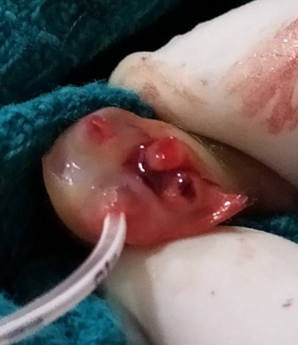
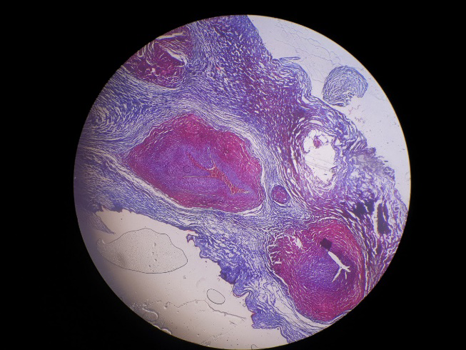
26-year-old primi mother, at 37 weeks of gestation delivered a male baby weighing 2.54 kg by normal vaginal delivery. Antenatal period was uneventful and antenatal scans done outside were normal. Baby had a normal transition at birth with Apgar scores of 7 and 8 at 1 and 5 min. The baby developed central cyanosis and tachypnea at 32 hours of life and was referred to us for further management. At admission, baby had a soft systolic murmur and central cyanosis with preductal and postductal saturation of 72% and 68% without any dysmorphism. The baby was started on prostaglandin infusion suspecting critical congenital heart disease. While we are about to insert an umbilical venous catheter to our surprise we noticed 4 umbilical vessels. The pathological examination of the umbilical cord showed three arteries and one vein. The placenta could not be examined as it was discarded. Cardiac evaluation done by doing an echo showed dextroposed transposition of great vessels with an intact septum and single coronary artery. The baby also had left-side urinary tract dilatation (UTD-P1) due to ureteropelvic obstruction. USG cranium was normal. The baby was initially stabilized by doing a PDA stenting and taken up for corrective surgery on day 5 for the arterial switch. However baby succumbed during the perioperative period.
Discussion
Umbilical arteries arise as two lateral branches from the caudal end of the descending aorta during 3 rd week of embryogenesis. Simultaneously, an arterial plexus develops around the allantois and coalesces to form a single artery extending almost the entire length of the body stalk. This allantoic artery becomes shorter as the right and left umbilical arteries advance in the body stalk and eventually unites with both arteries to form the interarterial anastomosis normally present in the region of the placental insertion. Under normal conditions, the right umbilical vein regresses in the second month of fetal life. The left umbilical vein and the two umbilical arteries become the vessels found in the normal cord [4, 5]. In our case, the allantoic artery persists and appeared as the third artery making 4 vessel umbilical cord. The cause for this persistence was not known exactly, proposed mechanisms are abnormality during the coalescence process in the embryogenesis such as 1. The arterial plexus coalesces to form a single artery or 2. One of the umbilical artery splits into two during development [5].
The literature review noted only 1 case report of four vessel umbilical cord with 3 arteries and 1 vein by Xue du, et al. which was a stillborn baby with no anomalies other than a vascular abnormality of the cord [6].
In our case baby was associated with multiple congenital anomalies, especially a critical congenital heart disease, which succumbed during the post-operative period.
Conclusion
When more than three vessels are found to present on either ultrasound or post-natal examination, the extra lumen can be a vein, an artery, or a remnant of the omphalomesentric or allantoic duct [7]. Of these, the persistent allantoic artery is very rare. Post-natal diagnosis is usually made during cannulation of the umbilical cord for treatment purpose. As in our case, the diagnosis of the persistent allantoic artery should warrant a thorough physical examination with extra attention should be paid to the possibility of congenital heart defects.
Acknowledgement
We thank Department of Pathology, Institute of child health for their assistance. We also thank Drs. Saravanan, Anto ferdine vaik and Himanshu sharma for supporting this effort and preparing the manuscript.
References
-
Vrabie SC, Novac L, Manolea MM, Dijmarescu LA, Novac M, et al. (2017) Abnormalities of the Umbilical Cord. In: Tudorache S, et al. (Edn.), Congenital Anomalies - From the Embryo to the Neonate.
-
Puvabanditsin S, Garrow E, Bhatt M, Kathiravan S, Gowda S, et al. (2011) Four-Vessel Umbilical Cord Associated with Multiple Congenital Anomalies: A Case Report and Literature Review. Fetal and Pediatric Pathology 30(2): 98-105.
-
Abuhamad AZ, Shaffer W, Mari G, Copel JA, Hobbins JC, et al. (1995) Single umbilical artery: does it matter which artery is missing? Am J Obstet Gynecol 173 (3 pt 1): 728- 732.
-
Koolhaas GD, Hollander MH, Molendijk H (2012) A case of a four-vessel umbilical cord: don’t stop counting at three! Case Reports in Perinatal Medicine 1(1-2): 87-90.
-
Meyer WW, Lind J, Moinian M (1969) An accessory fourth vessel of the umbilical cord: A preliminary study. American Journal of Obstetrics and Gynecology 105(7): 1063-1068.
-
Du X, Yuan Q, Li Z, Li Y (2015) Three umbilical arteries resulting in a four-vessel umbilical cord in a stillbirth. International Journal of Clinical and Experimental Medicin 8(3): 4682-4685.
-
Moshiri M, Zaidi SF, Robinson TJ, Bhargava P, Siebert JR, et al. (2014) Comprehensive Imaging Review of Abnormalities of the Umbilical Cord. Radiographic 34(1): 179-196.
- Understanding Pediatric Multiple Sclerosis: Clinical Presentation, Diagnostic Criteria, Therapeutic Advances, and Supportive Care Approaches
- Hemophilia in Children
- Xia-Gibbs Syndrome- A Case Report
- A Study to Assess Effectiveness of Play Therapy in Reducing Post-Operative Pain among Children Age 2 To 5 Year who have Undergone General Surgeries in Selected Pediatric Hospitals of Vadodara
- Preterm Birth: Scope of the Problem, Cost of Care, Potential Complications and Current Guidelines for Management
- Noradrenaline: Can we Use it to Manage Hemodynamic Instability among Neonatal Septic Shock at the NICU?