Oxidative Stress, Cancer and Antioxidant: A symbiosis?
The imbalance production of free radicals and reactive metabolites known as reactive oxygen species (ROS) and their elimination, will developed oxidative stress. ROS are playing dual roles as deleterious or beneficial species. At low level, they act as messengers in cell signaling and play a role in maintaining homeostasis whereas at higher concentration will cause cellular redox imbalance. This event is commonly occurred in cancer cells compared to normal cells. The oxidative DNA damage occurs as a result of elevated level of ROS causing single or double strand DNA breaks, replication errors, base modification, base oxidation, DNA cross-linking which leads to cell dysfunction and cell death. Taking antioxidant such as polyphenolic compounds is believed to exert anticancer effect due to ROS manifestations. Studies with flavonoids compounds exerted anticancer activity through modulation of ROS-scavenging enzyme activities, cell cycle arrest, induction of apoptosis, autophagy, and suppression of cancer cell proliferation and invasiveness. However, the mechanisms responsible for this anticancer effect have not been fully understood. Most of the laboratory and animal studies exhibited increased levels of exogenous antioxidants which prevent the types of free radical damage associated with cancer development. Few clinical studies in human were conducted with aim to observe possibilities of lowering risk of developing or dying from cancer upon taking dietary supplements. The dietary antioxidant supplements which were tested on human subjects were beta carotene, tocopherol, alpha tocopherol, vitamin C, selenium and zinc. These studies have yielded mixed results. In some preclinical studies, antioxidants have been found to promote tumor growth and metastasize. In tumor-bearing mice the supplements given had increased the ability to circulate tumor cells to metastasize. In conclusion, the mechanism of antioxidant action in cancer treatment is poorly understood. Many factors need to be considered before taking or giving antioxidant supplement since there were reports pertaining to benefits and disadvantages of antioxidant in cancer treatment.
Introduction
Oxidative stress is developed when the production of free radicals and reactive metabolites and their elimination by protective mechanisms (antioxidants) are in imbalance state. The free radicals or reactive metabolites are so-called oxidants or reactive oxygen species (ROS). This imbalance leads to damage of important biomolecules and cells, with potential impact on the whole organism [1]. ROS play vital roles in stimulation of signaling pathways in plant and animal cells in response to alterations of intracellular and extracellular environmental conditions [2].
The generation of ROS are contributed by endogenous sources such as products of respiratory chain function in mitochondria and NADPH oxidase (NOX) enzymes on the plasma membrane [3]. Mitochondria are the major source of cellular ROS in which the production of O2- mostly taking place. Hence this review will discuss about the role of ROS in modulating the anticancer effects upon the administration of antioxidant in few studies conducted previously.
During oxidative phosphorylation, small number of electrons from electron transport chain leak forming superoxide radical (O2-) [4]. NOX enzymes become another source of O2- by catalyzing the generation of intracellular and extracellular O2- from O2 and NADPH [5]. In general, the NOXs family consists of 7 members including Nox1- Nox-5 and Duox1/2 which each member is specialized to produce certain kind of ROS. Nox-1, Nox-2, Nox-3 and Nox-5 exclusively produce O2- while Nox-4 and Duoxes directly form hydrogen peroxide (H2O2) [6].
Source of ROS and its Role
ROS also can be generated exogenously by physical and chemical agents including cigarette smoke, ozone, ionizing radiation, drugs and heavy metals. Cigarette smoke may produce ROS because it contains many oxidants, free radicals and organic compounds such as superoxide and nitric oxide. Exposure to ozone can cause lipid peroxidation and induce influx of neutrophils into the airway epithelium [7]. Ionizing radiation is one of the carcinogens that involves in all stages of carcinogenesis process. The production of ROS from radiolysis of water resulting in DNA damage that leads to mutation of gene and cancer. Therapeutic agents such as antineoplastic drugs become another source of ROS. The mechanism of certain drugs like cisplatin and adriamycin produce high levels of ROS causing DNA damage and cell death [8]. Exogenous ROS also resulted from heavy metals (Cd, Hg, Pb, Fe, As). These compounds absorb into human body through different routes, then decomposed or metabolized into free radicals [9].
ROS are playing dual roles as deleterious or beneficial species. They are essential component as they involved in various biological processes of living organisms. At low level, they act as messengers in cell signaling and play a role in maintaining homeostasis. However, when excessive ROS are generated and accumulated in cells, they will cause harmful effect which termed as oxidative stress [10]. Oxidative stress occurs when there is an overproduction of ROS on one side and the other side has deficiency of antioxidants that results in disturbance in the equilibrium status of prooxidant/ antioxidant reactions in living organisms. As consequences, the excess of ROS can cause oxidative damage to the cell membrane, lipid, protein, and DNA that inhibit their normal function [11]. Excessive hydroxyl radical and peroxynitrite can cause damage to the cell membranes and lipoproteins by lipid peroxidation process. This reaction consequently results in the formation of malondialdehyde (MDA) and conjugated diene compounds that are cytotoxic and mutagenic. Moreover, damage to the proteins leads to their structural changes and loss of enzyme activity.
Oxidative damage to DNA may also lead to the formation of different oxidative DNA lesions thus can cause mutations [12]. Because of this, oxidative stress has been implicated in various pathological conditions such as inflammatory diseases (arthritis, vasculitis, glomerulonephritis, lupus erythematous, adult respiratory diseases syndrome), ischemic diseases (heart diseases, stroke, intestinal ischemia), hemochromatosis, acquired immunodeficiency syndrome, emphysema, organ transplantation, gastric ulcers, hypertension and preeclampsia and neurological disorder [13].
ROS and Cancer
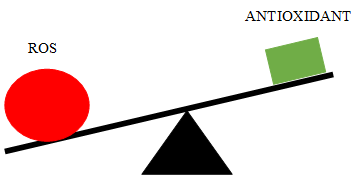
Cancer is a multistage process, mediated by endogenous and exogenous stimuli resulting cellular and molecular changes that transform a normal cell into malignant neoplastic cell. ROS are considered as oncogenic because they have been implicated in initiation, progression, and metastasis of cancers. ROS can cause cellular redox imbalance (Figure 1) which is commonly occur in cancer cells compared to normal cells. It is well known that oxidative DNA damage is responsible for cancer development [12]. In this context, ROS can induce tumor development by direct DNA damage during carcinogenic transformation such as catalyzing the modified DNA base 8-OHdG, resulting in mutation [14].
Oxidative DNA damage occurs due to elevated level of ROS causing single or double strand DNA breaks, replication errors, base modification, base oxidation, DNA cross-linking which leads to cell dysfunction and cell death. Consequently, the mutated DNA which if not repaired will lead to genomic instability and thus, cancer. High level of ROS also can affect various signaling pathways and activates transcription factors such as Nuclear factor erythroid 2-related factor 2 (Nrf2) and Nuclear factor-kappaB (NF-kB), resulting altered gene expression patterns that mediate the cancer progression [15].
Antioxidants Beat The Cancer Cells?
Antioxidants are chemicals that interact with and neutralize free radicals, thus preventing them from causing damage. Antioxidants are also known as “free radical scavengers.” Human body is naturally complemented with antioxidant defense to combat and prevent oxidative stress. The adverse damage caused by oxidants can be reduced by antioxidants by crumbling them before they react with biological targets, thus preventing chain reactions or preventing the activation of oxygen to the highly reactive products [16]. A substance is considered as an ideal antioxidant if it is readily absorbed by body, prevent, or quench free radical formation or chelate metals. It also works in aqueous and membrane domains and affect gene expression [17].
Antioxidants can be classified into enzymatic antioxidant and non-enzymatic antioxidants system. Enzymatic antioxidant consists of superoxide dismutase (SOD), catalase (CAT), gluthatione peroxidase (GSHPx) and gluthatione (GSH) dependent enzymes. Non enzymatic antioxidants further can be divided into metabolic and nutrient antioxidants. Metabolic antioxidants are endogenously produced by metabolism in the body such as GSH, lipoic acid, L-arginine, coenzyme Q10, melatonin, uric acid, and bilirubin. Nutrient antioxidants are obtained exogenously as a part of diet or through dietary supplements including vitamin E, vitamin C, carotenoids and polyphenols [18]. Consumption of these compounds also does not neutralize free radicals but may enhance endogenous activity. Endogenous antioxidants are important for maintaining optimal cellular functions. However, in certain conditions that promote oxidative stress, the endogenous antioxidants are not sufficient. Thus, the exogenous antioxidants need to be supplied to maintain the optimal cellular functions [19].
The antioxidants can also be categorized based on their size. The small-molecule antioxidants such as vitamin C, vitamin E, carotenoids, and GSH neutralize the ROS in a radical scavenging process and carry them away. While the large-molecule antioxidants are enzymes (SOD, CAT, and GSHPx) and sacrificial proteins (albumin) most likely absorb ROS and prevent them from attacking other essential proteins. The other way to categorize the antioxidants is according to their solubility in the water or lipids. They can be categorized as water-soluble and lipid-soluble antioxidants. The water-soluble antioxidants (e.g. vitamin C) are present in the cellular fluids such as cytosol, or cytoplasmic matrix whereas the lipid-soluble antioxidants (e.g. vitamin E, carotenoids, and lipoic acid) are predominantly located in cell membranes [20].
There is increasing evidence from many studies that numerous flavonoids such as daidzein, flavanone hesperetin, and many others, exerted anticancer activity through modulation of ROS-scavenging enzyme activities, cell cycle arrest, induction of apoptosis, autophagy, and suppression of cancer cell proliferation and invasiveness [21, 22]. As an example, daidzein induced apoptosis in the hepatocellular carcinoma cell line, HCCSK-HEP-1 via Bcl-2 homologous antagonist killer (Bak) upregulation and downregulation of anti-apoptotic proteins, resulting in cytochrome c release from mitochondria and activating subsequent apoptotic pathway involving caspases 3 and 9 [23]. However, the mechanisms responsible for this anticancer effect have yet not been fully understood and further studies are required to be done.
Apart from that, phenolic compounds such as gallic acid are also able to introduce potent anticancer effect in cancer cells. These are primarily due to the ability to induce cell cycle arrest, inhibit oncogenic signaling cascades controlling cell proliferation, angiogenesis andapoptosis, modulate ROS levels, promote tumor suppressor proteins such as p53, and enhance the ability to differentiate and transform into normal cells [24]. In this, phenolic compounds, gallic acid was chosen as candidate to be combined with cisplatin.
Generally, polyhydroxy phenolic or polyphenols compound present in nature in the form of conjugated structure with one or over sugar residues linked with a hydroxyl group of phenol. Gallic acid or also known as 3,4,5- trihydroxybenzoic acid is a naturally occurring polyhydroxy phenolic compound that can be found in various natural products, such as green tea, grapes, strawberries, bananas and many other fruits [25]. It was first identified and isolated from plants in 1786 by a famous Swedish chemist named Carl Wilhelm Scheele. His discovery leads to more studies and reports pertaining to the compound together with its derivatives done by other researchers which provided the understanding towards properties, mechanism of actions and benefits of gallic acid [26, 27].
Gallic acid was reported to have anticancer activities in many types of cancer cells including HeLa [26, 28]. Moreover, there are abundant source of gallic acid from plant-based natural product. Thus, it was chosen as candidate to be treated on HeLa cells combined with cisplatin to potentiate chemotherapeutic activity of platinum agent used in cancer treatment [29]. Gallic acid is one of the polyphenolic compounds which can exert anticancer effects through a broad range of mechanisms including inhibition of cancer cells proliferation by modification of signaling pathways, inhibition of cell cycle events, and apoptosis induction [30]. Several studies have demonstrated anticancer activity in various cancer cells, such as gastric, breast, prostate, lung, colon, oesophageal and most importantly in cervical cancer [10, 31]. Previous studies reported that gallic acid single treatment on cancer cells can trigger robust apoptosis, characterized by hallmarks of apoptosis, such as cytochrome c release from mitochondria to cytosol, caspase-3 activation, nuclear condensation, cell shrinkage, and plasma membrane blebbing [32]. Treatment with gallic acid for 24 hours showed significant cell viability reduction in the HeLa cells [28]. The cultured cells treated with varying concentrations (0, 10, 15, 20, 25, 30 and 40 μg/ml) of gallic acid for 24 hours significantly reduced the cell viability in a dose-dependent manner. Gallic acid was capable to reduce the cell viability to 66% at the concentration of 15 μg/ml.
Most of the laboratory and animal studies conducted previously exhibited increased levels of exogenous antioxidants which prevent the types of free radicals damage associated with cancer development. Then further investigations were conducted in human to observe the potentials in lowering risk of developing or dying from cancer upon taking the dietary supplements. There are many observational studies including case–control studies and cohort studies, have been conducted to investigate whether the use of dietary antioxidant supplements is associated with reduced risks of cancer in humans. Overall, these studies have yielded mixed results [33] due to insufficient control for biases that possibly influence study outcomes.
The biases in observational study could be resolved in randomized controlled clinical trials and considered to provide the strongest and most reliable evidence of the benefit and/or harm of a health-related intervention. The dietary antioxidant supplements which were tested on human subjects were beta carotene, tocopherol, alpha tocopherol, vitamin C, selenium and zinc [34, 35, 36, 37]. Overall, the randomized controlled clinical trials did not provide evidence that dietary antioxidant supplements are beneficial in primary cancer prevention [38].
Several randomized controlled trials, some including only small numbers of patients, have investigated whether taking antioxidant supplements during cancer treatment alters the effectiveness or reduces the toxicity of specific therapies [39]. Although these trials had mixed results, some found that people who took antioxidant supplements during cancer therapy had worse outcomes, especially if they were smokers. In some preclinical studies, antioxidants have been found to promote tumor growth and metastasis in tumor- bearing mice and to increase the ability of circulating tumor cells to metastasize [40, 41, 42]. Thus, in cancer patients, taking antioxidant supplements should be with caution until more data can prove the effectiveness of them.
Conclusion
The excessive concentration of ROS can cause oxidative damage to the cell membrane, lipid, protein and DNA that inhibit their normal function. Overproduction of ROS generate oxidative stress when there was deficiency of antioxidants which resulted in the disturbance of equilibrium status of prooxidant/ antioxidant reactions in living organisms. Few in vitro studies utilizing polyphenolic compounds showed good results pertaining response of antioxidant towards reducing the growth of cancer cells. However, in other laboratory and animal studies, mix results were reported. The same observations were recorded in preclinical trials. There were studies recorded the growth of cancer cells upon administration of antioxidant and it depends on few factors such as cancer types and agents used.
- Conflict of Interest: No conflict of interest
- Acknowledgement: The authors would like to express their sincere gratitude to Ministry of Higher Education Malaysia for Fundamental Research Grant Scheme with Project Code: FRGS/1/2020/SKK0/USM/02/38
References
-
Ďuračková Z (2010) Some current insights into oxidative stress. Physiol Res 59(4): 459-469.
-
Jabs T (1999) Reactive oxygen intermediates as mediators of programmed cell death in plants and animals. Biochem Pharmacol 57(3): 231-245.
-
Morry J, Ngamcherdtrakul W, Yantasee W (2017) Oxidative stress in cancer and fibrosis: Opportunity for therapeutic intervention with antioxidant compounds, enzymes, and nanoparticles. Redox Biol 11: 240-253.
-
Poillet Perez L, Despouy G, Delage Mourroux R, Boyer Guittaut M (2015) Interplay between ROS and autophagy in cancer cells, from tumor initiation to cancer therapy. Redox Biol 4: 184-192.
-
Chio IIC, Tuveson DA (2017) ROS in Cancer: The Burning Question. Trends Mol Med 23(5): 411-429.
-
Helfinger V, Henke N, Brandes PR, Schroder K (2017) Hydrogen peroxide formation by Nox4 limits malignant transformation. Free Rad Biol 108 (1): 34.
-
Said M, Al Dalaen, Al Qtaitat AI (2014) Review Article: Oxidative Stress versus antioxidant. Am J of Biosci and Bioeng 2(5): 60-71.
-
Klaunig JE, Wang Z (2018) Oxidative stress in carcinogenesis. Curr Opin in Toxicol 7: 116-121.
-
Pizzino G, Irrera N, Cucinotta M, Pallio G, Mannino F, et al. (2017) Oxidative Stress: Harms and Benefits for Human Health. Oxi Med and Cell Longev.
-
Wang J, Hu S, Nie S, Yu Q, Xie M (2016) Reviews on Mechanisms of In Vitro Antioxidant Activity of Polysaccharides. Oxi Med and Cell Longe.
-
Valko M, Leibfritz D, Moncol J, Cronin MT, Mazur M, et al. (2007) Free radicals and antioxidants in normal physiological functions and human disease. Int J Biochem Cell Biol 39(1): 44-84.
-
Pham Huy LA, He H, Pham Huy C (2008) Free radicals, antioxidants in disease and health. Int J of Biomedical Science 4(2): 89-96.
-
Lobo A, Lopez Anton R, Santabárbara J, de la Cámara C, Ventura T, et al. (2011) Incidence and lifetime risk of dementia and Alzheimer’s disease in a Southern European population. Acta Psychiatr Scand 124(5): 372- 383.
-
Yang X, Sun Z, Wang W, Zhou Q, Shi G, et al. (2018) Developmental toxicity of synthetic phenolic antioxidants to the early life stage of zebrafish. Sci of the Tot Environ 643(1): 559-568.
-
Bisht S, Dada R (2017) Oxidative stress: Major executioner in disease pathology, role in sperm DNA damage and preventive strategies. Front Biosci 9(3): 420-447.
-
Bunaciu, AA, Hassan Y, Aboul Enein Serban F (2012) FTIR Spectrophotometric Methods Used for Antioxidant Activity Assay in Medicinal Plants. Appl Spect Rev 47(4): 245-255.
-
Poljsak B, Šuput D, Milisav I (2013) Achieving the balance between ROS and antioxidants: when to use the synthetic antioxidants. Oxid Med Cell Longev.
-
Singh R, Upadhyaya RAK, Chandra AK, Singh DP (2018) Sodium chloride incites reactive oxygen species in green algae Chlorococcum humicola and Chlorella vulgaris: Implication on lipid synthesis, mineral nutrients and antioxidant system. Biores Tech 270: 489-497.
-
Kurutas EB (2016) The importance of antioxidants which play the role in cellular response against oxidative/ nitrosative stress: current state. Nutr J 15(1): 71.
-
Nimse SB, Pal D (2015) Free radicals, natural antioxidants, and their reaction mechanisms. RSC Adv 27986-28006.
-
Kopustinskiene DM, Jakstas V, Savickas A, Bernatoniene J (2020) Flavonoids as Anticancer Agents. Nutri 12(2): 457.
-
Aggarwal V, Tuli HS, Varol A, Thakral F, Yerer MB, et al. (2019) Role of Reactive Oxygen Species in Cancer Progression. Molecular Mechanisms and Recent Advancements. Biomolecules 9(11): 735.
-
Park HJ, Jeon YK, You DH, Nam MJ (2013) Daidzein causes cytochrome c-mediated apoptosis via the bcl-2 family in human hepatic cancer cells. Food Chem Toxicol 60: 542-549.
-
Anantharaju PG, Gowda PC, Vimalambike MG, et al. (2016) An overview on the role of dietary phenolics for the treatment of cancers. Nutr J 15 (99).
-
Asci H, Ozmen O, Yasar H Bunyamin E, Ercan A, Base NY (2017) The impact of gallic acid on the methotrexate- induced kidney damage in rats. J Food Drug Anal 25(4): 890-897.
-
Sourani Z, Pourgheysari B, Beshkar P, Shirzad H, Shirzad M (2016) Gallic Acid Inhibits Proliferation and Induces Apoptosis in Lymphoblastic Leukemia Cell Line (C121). Iran J Med Sci 41(6): 525-530.
-
Khorsandi K, Kianmehr Z, Hosseinmardi Z, Hossienzadeh R (2020) Anti-cancer effect of gallic acid in presence of low level laser irradiation: ROS production and induction of apoptosis and ferroptosis. Cancer Cell Int 20(18).
-
Zhao B, Hu M (2013) Gallic acid reduces cell viability, proliferation, invasion and angiogenesis in human cervical cancer cells. Oncol lett 6(6): 1749-1755.
-
Aborehab NM, Osama N (2019) Effect of Gallic acid in potentiating chemotherapeutic effect of Paclitaxel in HeLa cervical cancer cells. Can Cell Int 19(154).
-
Bhosale PB, Ha SE, Vetrivel P, Kim HH, Kim SM, et al. (2020) Functions of polyphenols and its anticancer properties in biomedical research: a narrative review. Trans Can Res 9(12): 7619-7631.
-
Abotaleb M, Liskova A, Kubatka P, Büsselberg D (2020) Therapeutic Potential of Plant Phenolic Acids in the Treatment of Cancer. Biomolecules 10(2): 221.
-
Tang HM, Cheung PCK (2021) Gene expression profile analysis of gallic acid-induced cell death process. Sci Rep 11: 16743.
-
Patterson RE, White E, Kristal AR, Neuhouser ML, Potter JD (1997) Vitamin supplements and cancer risk: The epidemiologic evidence. Can Causes Control 8(5): 786- 802.
-
Rautalahti MT, Virtamo JR, Taylor PR, Heinonen OP, Albanes D, et al. (1999) The effects of supplementation with alpha-tocopherol and beta-carotene on the incidence and mortality of carcinoma of the pancreas in a randomized, controlled trial. Cancer 86(1): 37-42.
-
Virtamo J, Edwards BK, Virtanen M, Taylor PR, Malila N, et al. (2000) Effects of supplemental alpha-tocopherol and beta-carotene on urinary tract cancer: incidence and mortality in a controlled trial (Finland). Cancer Causes Control 11(10): 933-939.
-
Albanes D, Malila N, Taylor PR, et al. (2000) Effects of supplemental alpha-tocopherol and beta-carotene on colorectal cancer: results from a controlled trial (Finland). Cancer Causes Control 11(3): 197-205.
-
Wright ME, Virtamo J, Hartman AM, Pietinen P, Edwards BK, et al. (2007) Effects of alpha-tocopherol and beta- carotene supplementation on upper aerodigestive tract cancers in a large, randomized controlled trial. Cancer 109(5): 891-898.
-
Fortmann SP, Burda BU, Senger CA, Lin JS, Whitlock EP (2013) Vitamin and mineral supplements in the primary prevention of cardiovascular disease and cancer: an updated systematic evidence review for the U.S. Preventive Services Task Force. Ann Intern Med 159(12): 824-834.
-
Lawenda BD, Kelly KM, Ladas EJ, Sagar SM, Vickers A, et al. (2008) Should Supplemental Antioxidant Administration Be Avoided During Chemotherapy and Radiation Therapy? J Natl Cancer Inst 100(11): 773-783.
-
Sayin VI, Ibrahim MX, Larsson E, Nilsson JA, Lindahl P, et al. (2014) Antioxidants accelerate lung cancer progression in mice. Sci Trans Medicine 6(221): 221.
-
Le Gal K, Ibrahim MX, Wiel C, Sayin VI, Akula MK, et al. (2015) Antioxidants can increase melanoma metastasis in mice. Sci Trans Med 7(308): 308.
-
Piskounova E, Agathocleous M, Murphy MM, Hu Z, Huddlestun SE, et al. (2015) Oxidative stress inhibits distant metastasis by human melanoma cells. Nature 527(7577): 186-191.
- Origin, Evolution, and Functional Impact of Short Insertion- Deletion Variants in Human Genomes: A Review
- Harnessing Molecular Glues for Next-Generation Vaccine, Cancer and Cardiovascular Disease Drug Development: A Comprehensive Review
- Lateral Cervical Epidermal Inclusion Cyst in a Paediatric Patient: A Rare Case Report
- Malarial Plasmodium Falciparum with Hepatitis B and C Virus Infections among Blood Donors in Ife Central Local Government Area, Ile Ife, Osun State, Nigeria
- Withanolides and Withaferin A- What’s next in Ashwagandha Research
- Designing of Dual Pulse Photoacoustic Tomography for Imaging of Drug-Response and Tumor Growth