Investigating the Diagnostic Utility of Cola acuminata Extract as an Alternative Histological Cytoplasmic Stain
Background: Stains are essential artefacts which aid visualisation and evaluation of histological specimens. However, the use of synthetic dyes for diagnostic purpose has become a major concern a threat to sustainable development owing to the associated toxicity, environmental pollution and potential risk to human health. Several studies have investigated the application of naturally occurring pigments in plants use of natural dyes for clinical and diagnostics purposed. The study investigated the application of naturally occurring pigments of Cola acuminata extract for clinical and diagnostics purpose; specifically, as a histological cytoplasmic stain. Methods: Formalin-fixed, paraffin-embedded tissue sections from cattle heart, liver and spleen were stained by varying concentrations of Cola acuminata nuts extracts, with different extractants including absolute methanol, ethanol and distilled water at different times. Tissue sections stained with dye extracts produced a spectrum of greyish to golden brown cytoplasmic colouration. Results: Cola acuminata dye extracts produced relatively varying staining intensities of golden-brownish cytoplasmic colouration applied on liver, spleen and heart tissue sections cattle tissue with different extractants, at different concentrations and staining times, comparable to eosin, and with blue purple heamatoxylin nuclear stain. Methanolic dye extract (60% w/v) of Cola acuminata at an acidic pH, applied for 15min at room temperature, yielded the best staining result. Conclusion: Cola acuminata nut dye extract is a potential substitute for cytoplasmic synthetic dye utility in histological staining techniques. Further research needed to reinforce and validate its applicability in a wide range of pathological specimens.
Introduction
Dyes have been produced synthetically or naturally from time immemorial and used to enhance the outlook of fabric and other artefacts [1]. They also impart contrast in tissues for histological analysis. After discovering the first synthetic dye, mauve, in 1856 by Henry Perkin [2], many other dyes emerged and are currently used in colouring foods, cosmetics, and medication [3]. However, growing evidence shows these chemical-based dyes have a debilitating effect on human health and the environment in general due to their increased toxicity and non-eco- friendliness in terms of their disposal and the management of their waste products [4, 5, 6].
Natural dyes have shown promising outcomes; their sources are readily available, reproducible, non-toxic, and eco-friendly [7]. These characteristics are indications of their potential to avert the menace associated with the usage of synthetic dyes as suitable alternatives. Increasing advocacy has been made to advance and harness the use of natural dye in various sectors [8, 9]. This is in line with the sustainable development goals (SDGs), which seeks to promote good health and wellbeing by reducing disease burden and environmental problems related to exposure to toxic substances, including synthetic dyes [10].
Cola acuminata plant extract has been identified to possess numerous phytochemicals and medicinal properties and is used as a food colourant [11, 12]. It belongs to the plant family Sterculiaceae, comprising about 125 species of trees found chiefly in the tropical rainforests of Africa. Cultivated mainly in Ghana and Nigeria within the West-African subregion, it is considered a vital commodity for traditional rites [13, 14] and an essential nontraditional agro export in both countries [15, 16]. Extracts from kola nut present with colourful pigments, which is yet to be explored and adopted as a natural dye with prospects for histological assessment. In this view, we aimed to investigate the utility of Cola acuminata nuts dye extract as a potential cytoplasmic stain in diagnostic histopathology.
Materials And Methods
Study Design and Ethics
This study was experimental-exploratory research that sought to assess the staining property of Cola acuminata dye extract on histologically processed heart, liver and spleen of cattle using H&E stain of the same organs as control.
Approval for the study was granted by the Research Ethics Committee of the University of Health and Allied Sciences (UHAS-REC A.10 [32] 20-21) and conducted at the histopathology laboratory of the Ho Teaching Hospital with permission from hospital management.
Tissue Specimen Acquisition
The heart, liver and spleen of cattle were obtained from a veterinary-inspected slaughterhouse and were immediately placed in a 10% neutral buffered formalin-filled container after removal from the animal. Upon arrival in the laboratory, the blood-stained formalin was replaced with a fresh 10% neutral buffered formalin to ensure optimal fixation of the tissue specimens.
Kola Nut Acquisition
Freshly harvested kola nuts were acquired from a farmer in Ho in the province of Volta Region. The nuts were washed with distilled water to eliminate impurities such as dust and soil.
Preparation of the Cola acuminata Extract
The kola nuts were chopped after washing and shade dried. The dried kola nuts were crashed and then pulverised using a new, unused domestic blender to obtain kola nut powder. The extracts were prepared from the kola nut powder using absolute ethanol, absolute methanol and distilled water. Briefly, 20g, 40g and 60g of the powdered kola nuts were dissolved in 100ml of each solvent in amber-coloured airtight bottles for six days with intermittent shaking. To prevent sunlight from directly affecting the extracts, they were stored in amber-coloured airtight bottles after filtering with Whatman No.1 filter paper. Their pH was checked using a pH meter (JENWAY 3510, Staffordshire, UK). The bottles containing the extracts were labelled appropriately and kept safely on the working bench.
Tissue processing
Representative slides of the formalin-fixed tissues (2x3x2mm) were cut into tissue processing cassettes and labelled appropriately (spleen, heart and liver). The tissues in cassettes were taken through the paraffin-embedded tissue processing technique (Appendix A).
Tissue Sectioning
Tissue sections of 3μm thickness were produced from the paraffin-embedded tissue blocks using a rotary microtome (ACCU-CUT SRM 200 CW, Sakura, Japan). The sections were floated out in a thermostatically controlled water bath at 50oC (JP SELECTA, Barcelona, Spain) to facilitate stretching and flattening of the sections. Subsequently, the sections were picked onto clean labelled microscope glass slides and dried at room temperature, followed by heat-fixing in a hot- air oven (GENLAB, Widnes, UK). Twenty-eight sections of each organ were produced and grouped into G1 and G2. G1 contained one section of each of the various organs, and G2 contained twenty-seven sections of each organ serving as a control and test slides, respectively.
Staining Procedure
G1 sections were stained with the routine H&E reagents using the standard staining protocol (Appendix B). G2 sections were further grouped (Appendix D) and stained with the kola nut dye extract based on solvents used in the dye extraction, extract concentration, and staining time using the staining protocol (Appendix C).
Microscopy and Micrograph Analysis
The staining reaction and intensity of the sections were examined microscopically using a binocular light microscope (LEICA DM750, GmbH) and scored (Appendix D and E). Micrographs of the stained sections were taken using a high-resolution smartphone camera (SAMSUNG GALAXY S7, South Korea). The staining intensities were compared among groups (G1:G2) and within groups (G2) depending on the various extracts, the extracts’ concentration, and the staining duration.
Results
Staining Reaction with Kola Nuts Methanolic Extract
Tissue sections stained with haematoxylin and eosin showed bright purple colouration of the nuclei (black arrow) and varied shades of pinkish cytoplasmic staining (green arrow), respectively as shown in Plate 1 (P1) (Figure 1). Methanolic dye-extracts of kola nut used to stain the following tissue sections demonstrated golden-brown cytoplasmic colouration (gold arrows), with haematoxylin, staining the nuclei purple (black arrows) as shown in Figure 1 (P 2 to P 10).
Staining Reaction with Ethanoic Extract
Ethanolic dye-extract of kola nut used to stain the following tissue sections demonstrated grey to golden-brown cytoplasmic colouration (gold arrows), with haematoxylin, staining the nuclei purple (black arrows) as shown in Figure 2 (P11 to P19).
Staining Reaction with Aqeuous Extract
Aqueous (distilled water) dye-extract of kola nut used to stain the following tissue sections demonstrated grey to light-brown cytoplasmic colouration, with haematoxylin, staining the nuclei purple as shown in Figure 3 (P20 – P28).
| 20% | 40% concentration | 60% concentration | |||||||||||
|---|---|---|---|---|---|---|---|---|---|---|---|---|---|
| concentration | |||||||||||||
| Extractant | Tissue | 5 min | 10 min | 15 | Plate No: | 5 min | 10 min | 15 | Plate No: | 5 | 10 | 15 | Plate No: |
| min | min | min | min | min | |||||||||
| Methanol | Spleen | 2+ | 3+ | 3+ | P 2 | 2+ | 2+ | 3+ | P 3 | 3+ | 3+ | 3+ | P 4 |
| Heart | 2+ | 2+ | 3+ | P 5 | 2+ | 2+ | 3+ | P 6 | 3+ | 3+ | 3+ | P 7 | |
| Liver | 2+ | 2+ | 3+ | P 8 | 2+ | 2+ | 3+ | P 9 | 2+ | 3+ | 3+ | P10 | |
| Ethanol | Spleen | 3+ | 3+ | 3+ | P14 | 3+ | 3+ | 3+ | P 15 | 2+ | 2+ | 3+ | P 16 |
| Heart | 2+ | 3+ | 3+ | P11 | 2+ | 2+ | 3+ | P 12 | 2+ | 2+ | 3+ | P 13 | |
| Liver | 2+ | 2+ | 3+ | P17 | 2+ | 2+ | 3+ | P 18 | 2+ | 2+ | 3+ | P 19 | |
| Distilled water | Spleen | 1+ | 1+ | 1+ | P23 | 2+ | 2+ | 3+ | P 24 | 1+ | 1+ | 1+ | P 25 |
| Heart | 1+ | 1+ | 1+ | P20 | 1+ | 1+ | 1+ | P 21 | 1+ | 1+ | 1+ | P 22 | |
| Liver | 1+ | 1+ | 1+ | P26 | 1+ | 1+ | 1+ | P 27 | 1+ | 1+ | 1+ | P 28 |
Table 1: Staining intensities scores of the various dye extracts at differential concentrations and durations.
| Extracts | Concentrations (%) | pH |
|---|---|---|
| Methanol | 20 | 5.84 |
| 40 | 5.78 | |
| 60 | 5.53 | |
| Ethanol | 20 | 5.98 |
| 40 | 5.87 | |
| 60 | 5.76 | |
| Distilled water | 20 | 6.07 |
| 40 | 6.04 | |
| 60 | 6.02 |
Table 2: pH of differential extract concentrations.
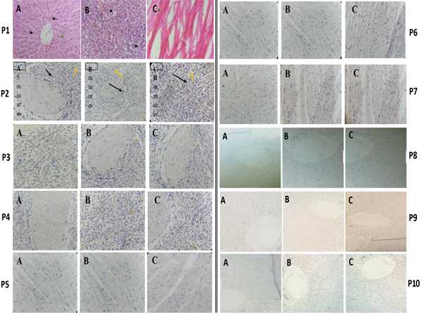
Figure 1: P 1-Photomicrographs of cattle tissue sections of A. Liver; B. Spleen; C. Heart, stained with Haematoxylin and Eosin stain. Magnification: x400. P 2-Photomicrographs of cattle spleen tissue sections stained with haematoxylin and counterstained with 20% methanolic dye extract of kola nut showing relatively indifferent cytoplasmic staining intensity of golden-brown colouration at: A. 5 min; B. 10 min; C. 15 min. Magnification x200. P 3-Photomicrograph of cattle spleen tissue sections stained with haematoxylin and counterstained with 40% methanolic dye extract of kola nut showing relatively indifferent cytoplasmic staining intensity of golden-brown colouration at: A. 5 min; B. 10 min; C. 15 min. Magnification x200. P 4-Photomicrographs of cattle spleen tissue sections stained with haematoxylin and counterstained with 60% methanolic dye extract of kola nut showing relatively intesne cytoplasmic staining reaction of golden-brown colouration at: A. 5 min; B. 10 min; C. 15 min. Magnification x200. P 5-Photomicrographs of cattle heart tissue sections stained with haematoxylin and counterstained with 20% methanolic dye extract of kola nut for A. 5 min; B. 10 min; C. 15 min; (cytoplasmic staining observed as golden-brown colouration without clear distinction at varying staining times). Magnification x200. P 6Photomicrographs of cattle heart tissue sections stained with Haematoxylin and counterstained with 40% methanolic dye extract of kola nut for A. 5 min; B. 10 mi; C. 15 min; (cytoplasmic staining observed as golden- brown colouration showing relatively uniform intensity at varying staining times). Magnification x200. P 7-Photomicrographs of cattle heart tissue sections stained with Haematoxylin and counterstained with 60% methanolic dye extract of kola nut for A. 5 min; B. 10 min; C. 15 min; (cytoplasmic staining observed as golden- brown colour with increasing intensity relative to staining time). Magnification x200. P 8-Photomicrographs of cattle liver tissue sections, stained with Haematoxylin and counterstained with 20% methanolic dye extract of kola nut for A. 5 min; B. 10 min; C. 15 min (cytoplasmic staining observed as golden-brown colouration with staining intensity not relative to staining time). Magnification x100. P 9-Photomicrographs of cattle liver tissue sections stained with Haematoxylin and counterstained with 40% methanolic dye extract of kola nut for A. 5 min; B. 10 min; C. 15 min (cytoplasmic staining observed as golden-brown colouration with relatively uniform staining intensity). Magnification x100. P10-Photomicrographs of cattle liver tissue sections stained with Haematoxylin and counterstained with 40% methanolic dye extract of kola nut for A. 5 min; B. 10 min; C. 15 min (cytoplasmic staining observed as golden-brown colouration with raltively uniform staining intensity). Magnification x100.
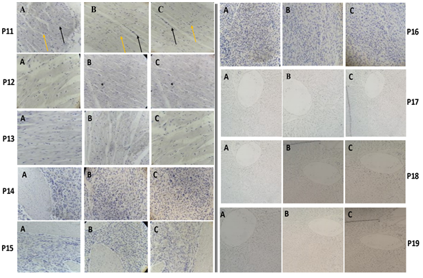
Figure 2: P11-Photomicrograph of cattle heart section stained with haematoxylin and counterstained with 20% ethanolic dye extract of kola nut for A. 5 min; B. 10 min; C. 15 min. Cytoplasmic staining observed as greyish-brown colouration. Nuclei stained blue-black (black arrow) and cytoplasm stained grey to golden brown (yellow arrow) Magnification x200. P12- Photomicrograph of cattle heart section stained with haematoxylin and counterstained with 20% ethanolic dye extract of kola nut for A. 5 min; B. 10 min; C. 15 min. Cytoplasmic staining observed as greyish -brown colouration. Nuclei stained blue-black (black arrow) and cytoplasm stained grey to golden brown (yellow arrow) Magnification x200. P13-Photomicrographs of cattle heart tissue sections stained with haematoxylin and counterstained with 60% ethanolic dye extract of kola nut for A. 5 min; B. 10 min; C. 15 min (cytoplasmic staining observed as grayish-brown colouration with intense and distinct staining pattern). Magnification x200. P14-Photomicrographs of cattle spleen tissue sections stained with haematoxylin and counterstained with 20% ethanolic dye extract of kola nut for A. 5 min; B. 10 min; C. 15 min. (cytoplasmic staining observed as golden-brown colouration with increasing intensity relative to staining time). Magnification x200. P15-Photomicrographs of cattle spleen tissue sections stained with haematoxylin and counterstained with 40% ethanolic dye extract of kola nut for A. 5 min; B. 10 min; C. 15 min. (cytoplasmic staining observed as golden-brown colouration with relatively uniform staining intensity). Magnification x200. P16-Photomicrographs of cattle spleen tissue sections stained with haematoxylin and counterstained with 60% ethanolic dye extract of kola nut for A. 5 min; B. 10 min; C. 15 min. (cytoplasmic staining observed as golden- brown colouration with relatively uniform staining intensity). Magnification x200. P17-Photomicrographs of cattle liver tissue sections stained with haematoxylin and counterstained with 20% ethanol dye extract of kola nut for A. 5 min; B. 10 min; C. 15 min. (cytoplasmic staining observed as grayish-brown colouration with relatively uniform staining intensity). Magnification x100. P18-Photomicrographs of cattle liver tissue sections stained with haematoxylin and counterstained with 20% ethanol dye extract of kola nut for A. 5 min; B. 10 min; C. 15 min. (cytoplasmic staining observed as golden-brown colouration with relatively uniform staining intensity). Magnification x100. P19-Photomicrograph of cattle Liver section. Stain: Haematoxylin and counterstained with 60% ethanol dye extract of kola nut stained for A. 5 minutes; B. 10 min; C. 15 min (cytoplasmic staining observed as golden-brown colouration with relatively uniform staining intensity). Magnification x100.
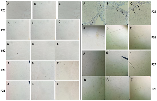
Figure 3: P 20-Photomicrographs of cattle heart tissue sections stained with haematoxylin and counterstained with 20% distilled water dye extract of kola nut for A. 5 min; B. 10 min; C. 15 min. (cytoplasmic staining observed as the grayish -brown colour). Magnification x100. P 21-Photomicrographs of cattle heart tissue sections stained with haematoxylin and counterstained with 40% distilled water dye extract of kola nut for A. 5 min; B. 10 min; C. 15 min. (cytoplasmic staining observed as the grayish brown colour). Magnification x100. P 22-Photomicrographs of cattle heart tissue sections stained with haematoxylin and counterstained with 60% distilled water dye extract of kola nut for A. 5 min; B. 10 min; C. 15 min. (cytoplasmic staining observed as the greyish- -brown colouration). Magnification x100. P23- Photomicrographs of cattle tissue Spleen sections stained with haematoxylin and counterstained with 20% distilled water dye extract of kola nut for A. 5 min; B. 10 min; C. 15 min (cytoplasmic staining observed as golden- brown colouration). Magnification x200. P24- Photomicrographs of cattle tissue spleen sections stained with haematoxylin and counterstained with 40% distilled water dye extract of kola nut for A. 5 min; B. 10 min; C. 15 min (cytoplasm observed as golden-brown colour). Magnification x200. P25- Photomicrographs of cattle tissue spleen sections stained with haematoxylin showing blue nuclear staining (arrows) and counterstained with 60% distilled water dye extract of kola nut for A. 5 min; B. 10 min; C. 15 min (cytoplasm observed as golden-brown colour). Magnification x100. P26- Photomicrographs of cattle liver tissue sections stained with haematoxylin and counterstained with 20% distilled water dye-extract of kola nut stained for A. 5 min. B. 10 min. C. 15 min, (cytoplasm observed as light brownish colouration). Magnification x40. P27- Photomicrographs of cattle liver tissue sections stained with haematoxylin and counterstained with 40% distilled water dye-extract of kola nut stained for A. 5 min. B. 10 min. C. 15 min. (cytoplasm observed as the light brownish colour). Magnification x40. P28- Photomicrographs of cattle liver tissue sections stained with haematoxylin and counterstained with 60% distilled water dye-extract of kola nut stained for A. 5 min. B. 10 min. C. 15 min. (cytoplasm observed as light brownish colouration). Magnification x40
Discussion
The quest for applying natural dyes as valuable sources of stains in diagnostic histopathology is gaining traction. The merit of this cause is aligned with the SDGs, which seek to promote health and well-being by reducing disease burden and environmentally related health problems associated with increasing exposure to toxic substances in the use of synthetic dyes. The present study investigated the use of dye extract from kola nut as a potential source of a histological cytoplasmic stain. The exploratory experiment we carried out resulted in exciting findings of the staining reaction; the dye extracts produced varied cytoplasmic colourations from grey to golden brown depending on the type of extract, as against a blue-black nuclear staining reaction of the haematoxylin stain.
Tissue sections stained with methanolic dye extract of kola nut showed golden-brownish cytoplasmic colouration. Similar staining patterns were observed across all three types of tissue (P2–P10) in Figure 1. The intensity of the staining reaction produced by the methanolic kola nut dye extracts showed increased brightness and clarity with increasing concentration of the extractant. Thus, the 60% concentration of methanolic kola nut extract produced a relatively intense cytoplasmic staining pattern (P4, P7 and P12) compared to 20% and 40% concentration of extract. Additionally, the staining reaction of heart tissue with the 60% concentration of methanolic extract (P12) came out distinctly, a phenomenon which could be explained by the abundance of muscle fibres of the heart tissue. On the contrary, the 40% concentration of extract produced relatively better staining than the 20% concentration in all the tissues. Good staining quality was observed in all tissues with the 60% concentration of extract when the staining time was prolonged to 15min.
Using the ethanolic extract, a greyish-brown cytoplasmic colouration was observed in the following tissue sections in Figure 2; P11, P12, P13 and P17, whilst the others (P14, P15, P16, P18 and P19), stained golden-brown. The staining reaction with the ethanolic extract was not significantly affected by the concentration; however, the intensity across the board showed slight improvement with increasing staining time (notably, P14).
The cytoplasmic staining pattern of the following tissue sections in Figure 3 stained by the aqueous extracts showed a golden-brown colouration (P23 and P24), whilst the following sections: P20, P21, P22, P25, P26, P27 and P28, showed light-brownish staining reaction. This is in keeping with a study conducted by Shehu, et al. [17], which showed slightly similar shades of staining pattern of yellowish-brown colouration. This variation could be due to the tissue type and other staining conditions. Relatively, the intensity of the staining reaction produced by the aqueous-kola nut extracts when applied on spleen tissue sections showed increasing brightness, and clarity as the concentration of the extract was increased (P23-P25). This observation could be due to the high cellularity of the spleen and increased dye-tissue interaction due to the presence of sufficient dye molecules in the extract.
Among the three extracts produced (ethanolic, methanolic and aqueous), it was observed that the methanol- based dye extracts (20%-60% concentration) produced the best staining intensity in all three tissue types followed by the ethanolic extracts (20%-60%), produced in both heart and spleen. Interestingly, distinct staining intensities were observed with aqueous-dye extracts (60% concentration) in the spleen.
The pH of the various concentration of the extracts produced (Table 2) indicated the acidity of the extracted dyes. This means that the natural dye extracts are anionic and have a strong affinity for cationic components of the cell, thus buttressing the relatively strong staining reaction displayed by the acidophilic heart muscle fibers.
The strength of the staining reaction in the present study increased as the duration increased from 5 minutes to 10 minutes and 15 minutes. This supports Ali, et al. [18] findings that the longer the staining time, the higher the colour strength until the dye depletion achieves equilibrium. Ultimately, 60% methanolic extract at a staining time of 15minutes was observed to be the optimal staining reaction in all tissue types. This observation can be attributed to the increased solvent concentration and duration that enhanced the dye-tissue interaction.
Conclusion
Cola acuminata dye extract provided a distinct colouration that enhanced contrast between the nuclear and cytoplasmic staining pattern in formalin-fixed paraffin- embedded tissues from cattle heart, liver and spleen. It may be used as a substitute for synthetic dyes indicative of microscopic histomorphological analysis. However, further studies are needed to broaden the scope of application in other tissue types and should involve some modifications to enhance the staining quality and longevity.
Acknowledgement
The authors are grateful to Ho Teaching Hospital’s management, the Laboratory Department’s staff members, and the technologists at the School of Allied Health Sciences Laboratory Unit, University of Health and Allied Sciences.
Financial Disclosure
This research received no specific grant from any funding agency in public, commercial or not-for-profit sectors.
References
-
Weisburger JH (2002) Comments on the history and importance of aromatic and heterocyclic amines in public health. Mutat Res 506-507: 9-20.
-
Travis AS (1990) Perkin’s Mauve: Ancestor of the Organic Chemical Industry. Technology and Culture 31(1).
-
Lehmkuhler AL, Miller MD, Bradman A, Castroina R, Mitchell AE (2020) Certified food dyes in over the counter medicines and supplements marketed for children and pregnant women. Food and Chemical Toxicology 143: 111499.
-
Benzie IF, Wachtel Galor S (2011) Herbal medicine: biomolecular and clinical aspects. CRC press.
-
Lellis B, Fávaro Polonio CZ, Pamphile JA, Polonio JC (2019) Effects of textile dyes on health and the environment and bioremediation potential of living organisms. Biotechnology Research and Innovation 3(2): 275-290.
-
Velusamy S, Roy A, Sundaram S, Kumar Mallick T (2021) A Review on Heavy Metal Ions and Containing Dyes Removal Through Graphene Oxide-Based Adsorption Strategies for Textile Wastewater Treatment. The Chemical Record 21(7): 1570-1610.
-
Yusuf M, Shabbir M, Mohammad F (2017) Natural Colorants: Historical, Processing and Sustainable Prospects. Natural Products and Bioprospecting 7(1): 123-145.
-
Elsahida K, Fauzi AM, Sailah I, Siregar IZ (2019) Sustainability of the use of natural dyes in the textile industry. IOP Conference Series: Earth and Environmental Science 399(1): 012065.
-
Sigurdson GT, Tang P, Giusti MM (2017) Natural Colorants: Food Colorants from Natural Sources. Annual Review of Food Science and Technology 8(1): 261-280.
-
Racioppi F, Martuzzi M, Matić S, Braubach M, Morris G, et al. (2020) Reaching the sustainable development goals through healthy environments: are we on track? European Journal of Public Health 30: 14-18.
-
Burdock GA, Carabin IG, Crincoli CM (2009) Safety assessment of kola nut extract as a food ingredient. Food Chem Toxicol 47(8): 1725-1732.
-
Lowe HIC, Watson CT, Badal S, Peart P, Toyang NJ, et al. (2014) Promising Efficacy of the Cola acuminate Plant: A Mini Review. Advances in Biological Chemistry 4(4): 240-245.
-
Drucker Brown S (1995) The Court and the Kola Nut: Wooing and Witnessing in Northern Ghana. The Journal of the Royal Anthropological Institute 1: 129-143.
-
Ndagi I, Babalola FD, Mokwunye IU, Anagbogu CF, Aderolu IA, et al. (2012) Potentials and Challenges of Kolanut Production in Niger State, Nigeria. ISRN Agronomy 2012: 492394.
-
Ampadu Agyei O (1995) Non-Traditional Agricultural Exports and Natural Resource Management in Ghana. Practices and Prospects, WRI, Washington, DC, USA.
-
Asogwa EU, Adedeji AR, Oyedokun AVO AH, Mokwunye FC, EA A (2012) Strategies for Improving Production and Storage of Kolanuts in Nigeria. American-Eurasian Journal of Agriculture & Environmental Sciences 12(1): 37-43.
-
Shehu SA, Sonfada ML, Danmaigoro A, Umar AA, Hena SA, et al. (2012) Kola nut (Cola acuminata) extract as a substitute to histological tissue stain eosin. Journal of Veterinary Advances 1(2): 33-37.
-
Ali S, Hussain T, Nawaz R (2009) Optimization of alkaline extraction of natural dye from Henna leaves and its dyeing on cotton by exhaust method. Journal of cleaner production 17(1): 61-66.
- Origin, Evolution, and Functional Impact of Short Insertion- Deletion Variants in Human Genomes: A Review
- Harnessing Molecular Glues for Next-Generation Vaccine, Cancer and Cardiovascular Disease Drug Development: A Comprehensive Review
- Lateral Cervical Epidermal Inclusion Cyst in a Paediatric Patient: A Rare Case Report
- Malarial Plasmodium Falciparum with Hepatitis B and C Virus Infections among Blood Donors in Ife Central Local Government Area, Ile Ife, Osun State, Nigeria
- Withanolides and Withaferin A- What’s next in Ashwagandha Research
- Designing of Dual Pulse Photoacoustic Tomography for Imaging of Drug-Response and Tumor Growth