Spirulina Polypeptides Inhibit the Growth of Human Lung Tumor (H460) Cells
Blue-green algae, or cyanobacteria, are some of the oldest organisms on Earth which grow even in extreme conditions. These algae have biologically active compounds which may contribute to the treatment of cancer due to their inhibitory effect against cell cycle and proliferation. Biologically active polypeptides obtained from the filamentous cyanobacterium called spirulina have considerable amounts of proteins and medicinal properties. Spirulina is shown to have benefits across a range of human health indications from malnutrition to anticancer properties. In this research, polypeptides were hydrolyzed from proteins present in spirulina and their anticancer effects were studied using assays such as MTT, Live/Dead, and Cell proliferation in Human Lung Tumor (H460) Cell model.
Abbreviations
DMEM: Dulbecco’s Modified Eagle Medium; RPMI: Roswell Park Memorial Institute; FBS: Fetal Bovine Serum.
Introduction
Different kinds of microalgae have been found to be the source of number of bioactive compounds having amino acid fragments with the efficiency of various biological applications [1]. Increased levels of protein content in these microalgae have been found to be the reason for their use in various potential applications [2]. Abundantly available in nature, microalgae are photosynthetic organisms used widely as a non-toxic and biocompatible alternative source in potential medical applications such as wound healing, tissue engineering, antitumor activity, and various other therapeutic applications [3]. These organisms are found in environmental sources such as oceans, soils, and various other ecofriendly sources [4]. Spirulina polypeptides are studied for the efficiency depending on their antibacterial, antitumor, antiallergic, and antihypertensive properties [5]. Spirulina, filamentous blue green algae have acquired considerable importance in fields such as pharmacology, food, and agriculture, because of their constitution of proteins, vitamins, minerals, and amino acids [6]. Spirulina has essential effects towards oxidative stress based on the presence of an enzyme called superoxide dismutase [7]. Based on these properties, the extracts from spirulina have been reported to be efficient in inhibiting the growth of various human cancer cells [8]. The evidence of effectiveness of spirulina in cancer is extremely limited as far as the clinical trials are concerned. The spirulina studies conducted on various types of carcinogenesis show a degree of similitude but are in a haphazard state [9]. S. platensis and S. platensis- derived tetrapyrroles were experimentally tested for pancreatic cancer and it was reported that it substantially decreased the proliferation of experimental pancreatic cancer [10]. Arthrospira platensis extract showed a remarkable effect on the lung cancer cell cycle and prevented the proliferation of cancer cells, and it could protect the normal cells by its antioxidant activity [11].
Therefore, there is a need for extensive research to explore novel antioxidant molecules from algae, and their purification strategies. Studies are needed to explore the actual antioxidant compounds present in aqueous extracts of algae that have shown anticancer activities, and to investigate their mechanism of action on the cellular system and their capability to potentiates chemotherapeutic drugs [12].
The current study applied flash hydrolysis process to selectively hydrolyze proteins from Spirulina in the form of water-soluble peptides. Flash hydrolysis is a subcritical water-based continuous process with a residence time in the range of 10-12s [13, 14, 15, 16, 17, 18, 19, 20, 21]. It has been frequently used to hydrolyze proteins from different algae species [13, 19, 21, 22, 23, 24, 25, 26]. The process has been shown to hydrolyze upto 2/3rd of algae proteins in water-soluble peptides form having molecular weight distribution ranging between 200 and 800 mass units (m/z) [15]. The water-soluble peptides produced from the flash hydrolysis of Spirulina slurry were freeze- dried and the recovered powder was applied to study its anticancer effects.
Experimental
Flash Hydrolysis
2 liters of 1% Spirulina (Arthrospira platensis) slurry was prepared using deionized water. The slurry was continuously stirred at 350 rpm for 1 hour using a magnetic stirrer to mix the slurry homogeneously. The final prepared slurry was transferred to a container to perform flash hydrolysis. During the flash hydrolysis process, an electric stirrer was used to continuously mix the slurry to provide a constant input. The slurry was fed into the flash hydrolysis system shown in Figure 1 using a 0.5 HP pump (Lewa pump). The flash hydrolysis system is equipped with a chiller (Pfannenberg), induction heating and a PID controller, (GH Induction Atmospheres), back pressure regulator (Equilibar), thermocouple (Omega), and a stainless-steel tubular reactor (0.79 cm internal diameter, and 40.6 cm long). The flow rate of the Spirulina slurry into the system was adjusted using the pump. A chiller system with 20% glycol mixture as a coolant was used to keep the biomass in a liquid state and keep the system in a set temperature. The system is equipped with an induction heating system of 5 kW with maximum power of 15 kVA at 230V. It is connected to a PID which was used to set the temperature of the reactor, define the heating rate, and monitor the temperature inside the reactor. A thermocouple was inserted below the reactor to measure the real-time temperature of the reactor. Additionally, the pressure within the system was maintained using a back pressure regulator.
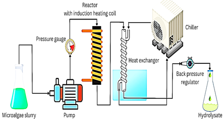
The residence time was calculated using Equation 1 where V is the volume of the reactor (mL), F is the flow rate of the slurry (mL s-1), ρpump is the density of water at pump conditions (g mL-1), ρp,T is the density of water at specific pressure and temperature (g mL-1).
V t
$$ = \frac {V}{F \left(\frac {\rho_ {p u m p}}{\rho_ {p , \mathrm {T}}}\right)} \tag {1} $$ F ρ pump ρ ,T p First, the chiller was operated for 15 minutes to reduce the temperature of the system. The pressure of the system was set to 1550 ± 25 psi using compressed air and back pressure regulator. The system was then heated to 185°C using the induction heating system. The 1 wt% Spirulina slurry was then fed into the system at a flow rate of 92 mL min- 1 with a residence time of 11 ± 2 s. After achieving a constant flow at the outlet, the liquid hydrolysate exiting from the system was collected. The hydrolysate was vacuum filtered, and the remaining liquids were centrifuged. A freeze dryer (Labconco) was used to freeze-dry the liquid hydrolysate. The freeze-drying process was carried out at about -54°C and 0.608 mBar pressure. The freeze-dried sample was placed in a sealed container and used for studying its anticancer effect.
Analyses of Freeze-Dried Hydrolysate
Elemental composition (Table 1) of raw Spirulina and freeze-dried Spirulina hydrolysate was determined by Flash 2000 Elemental Analyzer by Thermo Scientific using helium as a reference gas and oxygen as career gas. Approximately 1 mg of the sample was placed in a tin capsule with dimension of 3.3mm × 5mm for combustion at 950 °C. The elemental analysis was done in duplicate and the reported values are the average of the two values with a standard deviation of less than 3%.
Cell Culture
The human lung epithelial (Beas-2B) cells, and human lung tumor (H460) cells were cultured using Dulbecco’s Modified Eagle Medium (DMEM) and Roswell Park Memorial Institute (RPMI) media supplemented with fetal bovine serum (FBS), and penicillin streptomycin and the cells were stored in the incubator maintained at the temperature of 37°C and 5% CO2. The Beas-2B and H460 cells were sub cultured, and the cells were counted using a hemocytometer to determine the number of cells to be plated for the biocompatibility assays such as MTT, and Live/Dead, and the cell proliferation assay was performed to confirm the biocompatibility assay results of H460 cells.
MTT Assay
A 96-well microtiter plate was used in this analysis and 5000 cells were seeded in each well of the plate. The plate was then placed in the incubator maintained at 37°C and 5% CO2 for 48 hours. After this incubation, the cells were washed with DPBS and treated with lower to higher concentrations of spirulina polypeptides and incubated for another 48 hours. The stock solution of spirulina polypeptides was prepared by adding spirulina polypeptides to RPMI medium in the ratio of 1:1. After the second incubation, the cells were washed with DPBS and treated with the MTT dye and incubated for 3 hours. The MTT dye was prepared by adding them to DPBS in the ratio of 5:1. Live/Dead Assay A 6-well microtiter plate was used for this analysis and 250,000 cells were plated in all the wells. After plating the cells, the plate was incubated for 48 hours at 37°C and 5% CO2. After 48 hours, the cells were washed with DPBS and treated with different concentrations of spirulina polypeptides and incubated again for 48 hours. After this incubation, the cells were treated with the dye mixture prepared by mixing the live and dead components of the dye. This step was carried out in the absence of light and the plate was covered with aluminum foil and placed at room temperature for 30 minutes.
Cell Proliferation Assay
On the first day, 250,000 cells were plated in a 6-well microtiter plate and incubated for 48 hours at 37°C and 5% CO2. After the first incubation, the cells were treated with various concentrations of spirulina polypeptides after washing with DPBS. The plate was then incubated for 48 hours. After this incubation, the cells were treated with the detection reagent containing three components such as nucleic acid stain, background suppressor, and RPMI media. The plate was then incubated for 30 minutes.
Results and Discussion
Elemental Analysis
In Table 1, it is shown that the nitrogen, carbon and hydrogen percentage in the freeze-dried spirulina hydrolysate has decreased in comparison to raw Spirulina which is because of the distribution of it in the solid fraction that is composed of 1.99 % nitrogen, 10% carbon and 1.41% hydrogen. However, the presence of significant amount of these elements in the hydrolysate shows that the algae protein has been hydrolyzed to water-soluble peptides as reported in Kumar S, et al [15].
| Name | Nitrogen % | SD | Carbon % | SD | Hydrogen % | SD |
|---|---|---|---|---|---|---|
| Raw Spirulina | 9.85 | 0.6 | 43.39 | 2.7 | 6.14 | 0.3 |
| Spirulina hydrolysate | 9.43 | 0.4 | 37.48 | 1.3 | 5.58 | 0.2 |
Table 1: Elemental analysis of Spirulina.
MTT Assay
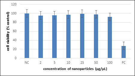
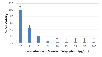
During the 3-hour incubation, the metabolically active Beas-2B and H460 cells absorbed the MTT dye and got converted into insoluble formazan crystals. These formazan crystals were dissolved by adding DMSO after removing the media. The microtiter plate was then placed on a rocker for 20 minutes for properly dissolving the formazan crystals. The absorption of MTT dye by the cells was measured by analyzing the plate using a spectrophotometer at the wavelength of 570 nm. The absorption values were recorded, and a plot was created with cell viability against the concentration of spirulina polypeptides and the results are indicated in the Figures 2 & 3. The results indicated that the viability of Beas-2B cells did not vary much with the spirulina polypeptide concentration whereas the H460 cell viability decreased with dosage-based concentration of spirulina polypeptides, when compared to the control (cells treated with no polypeptides). Significant decrease in the viability of H460 cells was identified from 5μg/μL to 100 μg/ μL concentrations of spirulina polypeptides.
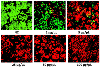
Live/Dead Assay
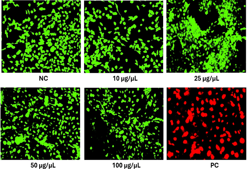
The MTT assay results were further confirmed by the live/dead assay. In the 30-minutes incubation, the metabolically active cells take up the green component of the dye and the dead cells take up the red component of the dye. After the incubation, the media was removed and DPBS was added to the cells and the fluorescence was analyzed using the fluorescence microscope with the FITC filters for live cells and TRITC filters for dead cells. The Live/Dead assay results are shown in Figures 4 & 5. The results of this assay supported the results of MTT assay thereby indicating the anticancer activity of spirulina polypeptides towards the H460 cells based on their metabolic activity.
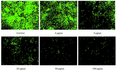
Cell Proliferation Assay
Over the period of the cell proliferation assay, the cells multiply and the population of cells after treatment with spirulina polypeptides was analyzed based on the DNA content of the cells. In this assay, the reagent stains only the healthy cells after cell division. The background suppressor in the reagent suppresses the background fluorescence to obtain better image quality. The results in Figure 6 indicate that the population of cells decreased with the concentration of spirulina polypeptides and significant decrease was identified for the cells treated with 5μg/μL of spirulina polypeptides and further concentrations. This assay determined the anticancer activity of spirulina polypeptides towards the H460 cells based on their DNA content and assured the results of MTT and Live/Dead assays.
Conclusion
The polypeptides hydrolyzed from spirulina plantesis were analyzed using biocompatibility assays towards the human lung epithelial (Beas-2B) cells and human lung cancer (H460) cells. The MTT and Live/Dead assays determined that H460 cells showed anticancer activity towards the spirulina polypeptides based on the metabolic activity of the cells that was further assured by the cell proliferation assay based on the DNA content of the cells.
References
-
Abed RM, Dobretsov S, Sudesh K (2009) Applications of cyanobacteria in biotechnology. Journal of applied microbiology 106(1): 1-12.
-
Lupatini AL, Colla LM, Canan C, Colla E (2017) Potential application of microalga Spirulina platensis as a protein source. Journal of the Science of Food and Agriculture 97(3): 724-732.
-
Zhong D, Du Z, Zhou M (2021) Algae: a natural active material for biomedical applications. View 2(4): 20200189.
-
Fais G, Manca A, Bolognesi F, Borselli M, Concas A, et al. (2022) Wide range applications of spirulina: from earth to space missions. Marine Drugs 20(5): 299.
-
Ovando CA, Carvalho JCD, de Melo Pereira GV, Jacques P, Soccol VT, et al. (2018) Functional properties and health benefits of bioactive peptides derived from Spirulina: A review. Food reviews international 34(1): 34-51.
-
Saranraj P, Sivasakthi S (2014) Spirulina platensis–food for future: a review. Asian J Pharm Sci Technol 4(1): 26- 33.
-
Hassanen MR, Mahfouz MK, Farid AS, Fadlullah AH (2015) Biochemical effects of spirulina platensis against oxidative stress caused by doxorubicin. Benha Veterinary Medical Journal 28(2): 147-154.
-
Ramakrishnan R (2013) Anticancer properties of blue- green algae Spirulina platensis–A review. International Journal of Medicine and Pharmaceutical Science 3(4): 159-169.
-
Kaur K, Kaur S (2021) Spirulina--A Wonder Nutraceutical Against Cancer: A Review. Plant Archives (09725210) 21(1): 1333-1338.
-
Konícková R, Vanková K, Vaníková J, Vánová K, Muchová L, et al. (2014) Anti-cancer effects of blue-green alga Spirulina platensis, a natural source of bilirubin-like tetrapyrrolic compounds. Annals of hepatology 13(2): 273-283.
-
Tajvidi E, Nahavandizadeh N, Pournaderi M, Pourrashid AZ, Bossaghzadeh F, et al. (2021) Study the antioxidant effects of blue-green algae Spirulina extract on ROS and MDA production in human lung cancer cells. Biochemistry and Biophysics Reports 28: 101139.
-
Ferdous UT, Yusof ZNB (2021) Medicinal prospects of antioxidants from algal sources in cancer therapy. Frontiers in Pharmacology 12: 593116.
-
Garcia-Moscoso JL, Obeid W, Kumar S, Hatcher PG (2013) Flash hydrolysis of microalgae (Scenedesmus sp.) for protein extraction and production of biofuels intermediates. The Journal of Supercritical Fluids 82: 183-190.
-
Khandual S, Pokharel U, Rathore S, Bonilla-Ahumada F, Kumar S (2024) A novel method of flash‐hydrolysis assisted pigment extraction (carotenoids) from microalgae biomass. Biofuels, Bioproducts and Biorefining 18(5): 1526-1540.
-
Kumar S, Hablot E, Moscoso JLG, Obeid W, Hatcher PG, et al. (2014) Polyurethanes preparation using proteins obtained from microalgae. Journal of Materials Science 49: 7824-7833.
-
Teymouri A, Kumar S, Barbera E, Sforza E, Bertucco A, et al. (2017) Integration of biofuels intermediates production and nutrients recycling in the processing of a marine algae. AIChE Journal 63(5): 1494-1502.
-
Teymouri A, Adams KJ, Dong T, Kumar S (2018) Evaluation of lipid extractability after flash hydrolysis of algae. Fuel 224: 23-31.
-
Asiedu A, Ben S, Resurreccion E, Kumar S (2018) Techno‐ economic analysis of protein concentrate produced by flash hydrolysis of microalgae. Environmental Progress & Sustainable Energy 37(2): 881-890.
-
Barbera E, Teymouri A, Bertucco A, Stuart BJ, Kumar S (2017) Recycling minerals in microalgae cultivation through a combined flash hydrolysis–precipitation process. ACS Sustainable Chemistry & Engineering 5(1): 929-935.
-
Thakkar A, Pienkos PT, Nagle N, Dong T, Kruger J, et al. (2024) Comparative study of flash and acid hydrolysis of microalgae (Scenedesmus sp.) for the recovery of biochemicals and production of porous biocarbon nanosheets. Biomass Conversion and Biorefinery 14(2): 2253-2262.
-
Barbera E, Sforza E, Kumar S, Morosinotto T, Bertucco A (2016) Cultivation of Scenedesmus obliquus in liquid hydrolysate from flash hydrolysis for nutrient recycling. Bioresource technology 207: 59-66.
-
Talbot C, Garcia-Moscoso J, Drake H, Stuart BJ, Kumar S (2016) Cultivation of microalgae using flash hydrolysis nutrient recycle. Algal research 18: 191-197.
-
Bessette AP, Teymouri A, Martin MJ, Stuart BJ, Resurreccionet EP, et al. (2018) Life cycle impacts and techno-economic implications of flash hydrolysis in algae processing. ACS sustainable chemistry & engineering 6(3): 3580-3588.
-
Thakkar A, Barbera E, Sforza E, Bertucco A, Davis R, et al. (2021) Flash hydrolysis of yeast (Saccharomyces cerevisiae) for protein recovery. The Journal of Supercritical Fluids 173: 105240.
-
Garcia-Moscoso JL, Teymouri A, Kumar S (2015) Kinetics of peptides and arginine production from microalgae (Scenedesmus sp.) by flash hydrolysis. Industrial & Engineering Chemistry Research 54(7): 2048-2058.
- Pattern of Gonadal Hormones in Oral Testosterone-Supplimented Male Wistar Rats with Diabetes-Induced Hypogonadism
- Re-Evaluation of the Genotoxicity of Currently Used Food Dyes in Mouse Multiple Organs Via Continuous Administration by Drinking Using the Comet Assay
- Pharmacogenetics of Type 2 Diabetes Mellitus: Linking Genetic Variability to Drug Efficacy and its Cardiovascular Outcomes
- Exploratory Proteomic Profiling of SARS-CoV-2 Infected THP-1 Macrophages Reveals Alterations in Inflammatory Response and Cellular Metabolism
- Study of Genotoxicity of Hepatocarcinogens in Multiple Organs in Mice by Feeding and Drinking Using the Comet Assay
- The Role of Faculty in Assessment: Traditional vs. Competency- Based Medical Education in Toxicology Learning