Sub-Acute Study on the Cardiotoxic Effects of Monosodium Glutamate Ingestion in Albino Rat
Objective: Many studies has shown that monosodium glutamate (MSG) causes Chinese restaurant syndrome which is associated with significant numbness in the neck, arms, and back, as well as headaches, dizziness, and palpitations especially in high doses. This present study evaluated the sub-acute study on the effect of MSG on the heart. Method: Eighteen (18) albino rats were randomly grouped according to their body weight into 3 groups A, B and C with 6 animals in group A and 7 rats in groups B and C respectively. Group A rats were used as controls. Group B received high dose (1ml/kg) of the monosodium glutamate concentration while group C received a low dose (0.5ml/kg) of the monosodium glutamate concentration for twenty one (21) days. The functionality of the heart was established by estimating the serum level of CK-MB (U/L), LDH (U/L) and AST (U/L), data analysis was done using IBM SPSS version 21 and results from the biochemical assay were reported as mean +/- SEM. The level of significance was tested using one-way analysis of variance (ANOVA) followed by the Tukey post hoc analysis and the probability levels less than 0.05 (p0.05) decreased levels of CK-MB, statistically significant (
Introduction
The white, crystalline powder known as monosodium glutamate is nearly odourless. This sodium salt is a form of glutamic acid, an amino acid that is present in meat, chicken, and other high-protein foods. In the majority of markets, it is sold as “white maggi” or “ajinomoto” and serves as an addition and food enhancer [1, 2]. It is produced by the action of Micrococcus glutamicus on a carbohydrate source, such as sugar beetroot molasses, or by the partial neutralisation and acid hydrolysis of vegetable proteins [3, 4].
Conversely, it has been widely documented that monosodium glutamate poses a risk to people and, consequently, laboratory animals, particularly when administered in larger quantities. There have been instances of hypersensitivity reactions, such as burning and tingling, caused by Chinese restaurant cooks’ over usage of glutamate. “Monosodium glutamate symptom complex” or “Chinese restaurant syndrome” are terms used to describe this illness [5].
Chinese restaurant syndrome is a collection of symptoms, including headaches, dizziness, palpitations, and numbness in the neck, arms, and back, that are thought to affect vulnerable people who eat food that is highly seasoned with monosodium glutamate, according to the Merriam-Webster dictionary. Large quantities of monosodium glutamate are utilised in South Asian, Chinese, and Japanese cooking. The precise origin of this condition is unknown, despite research demonstrating that monosodium glutamate has neurotoxic and neuro-excitatory effects in the hypothalamus region of the central nervous system [6, 7]. Monosodium glutamate at high concentration was also linked to an increased risk of the aforementioned symptoms. Two people had acute bronchial asthma after consuming monosodium glutamate [8].
Toxin buildup causes damage to the heart muscles, which is known as cardiotoxicity. Heart failure or heart dysfunction are other names for it. Myocardial infarction, angina, and acute arrhythmia are among the several kinds [9–11]. Toxicological level can be used to identify sub-acute cardiac damage. Damage to the heart causes changes in the way the heart pumps [12]. Dyspnea, heart palpitations, blood pressure fluctuations, chest pain, and malaise are the most typical signs of cardiotoxicity. Among its complications are congenital heart failure, cardiomyopathy, myocardial infarction, and sudden death [13].
A disruption in the heart tissue’s blood flow causes a heart attack, often referred to as an acute myocardial infarction [14]. When evaluating heart problems, biochemical indicators of the cardiac system might lower the morbidity or fatality rate linked to acute myocardial infarction. The most often utilised diagnostic markers are Lactate Dehydrogenase (LDH), its isoenzyme Creatine Kinase (CK), and Aspartate Amino Transferase (AST). These enzymes are found in kidney, liver, bones, skeletal muscle, brain erythrocytes, gastrointestinal system, uterus, prostate, tongue, and other tissues in addition to heart tissue [15].
Serum levels of cardiac enzymes are typically relatively low. Consequently, when the membrane disrupts these enzymes, they leak out into the plasma and multiply greatly, providing information on the degree of cardiac damage brought on by oxidative stress and lipid peroxidase [16]. AST and LDH are nonspecific and rise significantly in the later stages (24–72 hours) following the onset of symptoms.
The most effective cardiac indicators should be sensitive and specific, able to identify long-term storage problems and early infarctions. Analysis of a single biomarker is not advised because there is no perfect and particular biomarker; instead, enzymes with key characteristics of the ideal biomarker are used. However, it has been noted that the most optimal marker, CK-MB (CK-2), and the LDH1 and LDH2 ratio are more sensitive and specific to acute cardiac damage. Acute myocardial infarction causes an elevation in these enzymes. After myocardial damage, CK-MB peaks within 24 hours, rises 4–9 hours later, and then returns to normal within 48– 72 hours [15, 17]. Along with a number of dietary sweeteners, salt, and hydrogenated fats, monosodium glutamate is an excitatory neurotransmitter that can generate excite-toxicity, which increases safety concerns when used as a flavour enhancer. The purpose of the study is to examine how albino rats’ hearts respond to two oral low doses of monosodium glutamate, 0.5 ml.kg and 1 ml/kg.
Materials and Methods
Chemicals and Reagents
Monosodium glutamate (3g/sachet containing 99+% of MSG-Ajinomoto®) was purchased from Ogbete Main Market, a local market in Enugu metropolis, Enugu State, Nigeria. Distilled water was used for constituting the monosodium glutamate (MSG).
Preparation of Monosodium Glutamate Solution
One sachet of MSG-Ajinomoto® (i.e. 3g of MSG) was dissolved in 1000ml distilled water to give a stock solution of 3mg/ml.
Animals
Eighteen (18) adult albino rats, weighing 120-180g, were obtained from the animal house of the College of Veterinary Medicine, University of Nigeria. The animals were housed in metallic cages under standard conditions of temperature (22 ± 3oC) and a 12 h light, 12 h dark cycle. The animals were kept under observation for about 14 days before the onset of the experiment for acclimatization. Experimental protocol and handling was according to Institutional guidelines describing the use of rats and in accordance with the American Physiological Society guiding principles for research involving animals and human beings [18].
Experimental Design
The Eighteen (18) albino rats were grouped into grouped into (A-C) of 6 rats in each group. They received the following treatments on a daily basis for three weeks:
- Group A: (Normal control): No treatment was administered to this group.
- Group B: received oral administration of high dose of MSG solution (1ml/kg) only.
- Group C: received oral administration of low dose of MSG solution (0.5ml/kg body weight) only.
Sacrificing of Animals and Sample Collection
Blood samples for the determination of serum analyses of CK-MB, Lactate dehydrogenase and Aspartate Transaminase were taken by cardiac puncture of the left ventricle of the heart under chloroform anesthesia and the heart was harvested for histopathological analyses.
Biochemical Analysis
The serum activities of the creatinine kinase isoenzyme, CK-MB and LDH were determined using spectrophotometric method as described by Tietz, [19]. AST was determined using colometric method [20].
Histopathological Analysis
The excised heart was processed using the paraffin wax embedding technique, sectioned at 5 microns and stained using the Haematoxylin and Eosin [H and E] staining procedure [21]. The histological sections were examined using an Olympus TM light microscope.
Statistical Analysis
Data analysis was done using GraphPad prism version 7.0 (GraphPad, San Diego, CA, USA). The results of the biochemical assays were reported as mean±SEM (standard error of mean). The level of significance was tested using one-way analysis of variance (ANOVA), followed by the Tukey post hoc analysis. Probability levels less than 0.05 (p<0.05) was considered significant.
Results
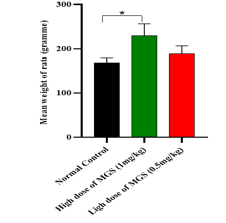
Effects of monosodium glutamate (MSG) on body weight of rats following 21days of administration is represented in Figure 1. It was observed that rats in the High dose of MGS (1ml/kg) group gained significant weight when compared to normal control rats. The mean increase in body weight was highest in the High dose of MGS (1ml/kg) in comparison with other groups.
Biochemical Results
The functionality of the heart was established by estimating the serum level of CK-MB (U/L), LDH (U/L) and AST (U/L). A non-statistically significant (P>0.05) decreased levels of CK-MB (ng/ml (0.5), statistically significant (<0.05) increased levels of LDH (U/L) and statistically significant (<0.05) decreased levels of AST (U/L), were seen in the High dose of MGS (1ml/kg) group B when compared with group A (normal control). We observed the Low dose of MGS (0.5mg/kg) did not show any significant alterations in the biochemical parameters measured Table 1.
| Group | CK-MB (ng/ml (0.5) | LDH (U/L) | AST (U/L) |
|---|---|---|---|
| A: Normal Control | 2.975 ± 1.12 | 775.42± 14.97 | 23.22 ±4.01 |
| B: High dose of MGS (1mg/kg) | 0.867 ± 0.36 | 1278.48 ± 15.12** | 18.11 ± 1.97* |
| C: Low dose of MGS (0.5mg/kg) | 1.022 ± 02.91 | 831.26 ±16.42 | 21.17± 2.23 |
Table 1: Statistical Comparison of cardiac biomarkers of treated groups with negative controls Groups.
Values given as Mean ± SEM. **p˂0.01 or *p˂0.05 is significant when normal control is compared with all other groups. Table 1: Statistical Comparison of cardiac biomarkers of treated groups with negative controls Groups.
Histopathological Results
In Plate 1, myocardial fibres appear normal. The cardiac fibres showed a well conserved morphology. The heart section from High dose of MGS (1mg/kg)-treated group showed normal myocardial fibres with no significant alteration observed (Plate 2). In addition, the heart section low dose of MGS (0.5mg/kg)-treated group showed normal myocardial fibres with no significant alteration observed (Plate 3). The histopathological findings were in tandem with the biochemical results as we observed the monosodium glutamate (MSG) did not pose any serious observable danger to the heart at the doses studied.
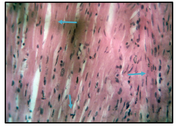
Plate 1: Representative micrograph of the heart of animals in group A. Myocardial fibres (arrows) appear normal. Stain: Haematoxylin and Eosin. Magnification: X400.
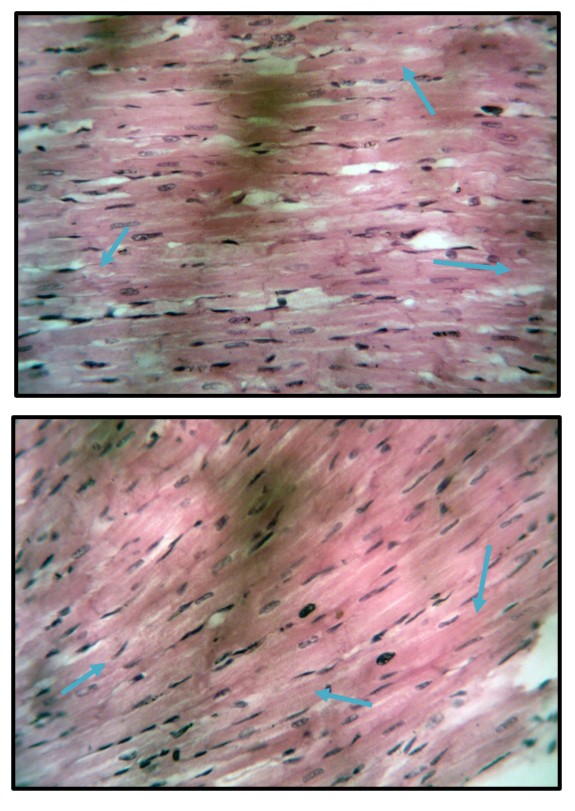
Plate 2: Representative micrograph of the heart of animals in group B. Myocardial fibres (arrows) appear normal. Stain: Haematoxylin and Eosin. Magnification: X400.
Plate 3: Representative micrograph of the heart of animals in group C. Myocardial fibres (arrows) appear normal. Stain: Haematoxylin and Eosin. Magnification: X400.
Discussion
In this study, it was observed that rats in the High dose of MGS (1ml/kg) group gained significant mean increase in weight when compared to normal control rats. This observation agrees with the findings of a study titled “Organ toxicity of monosodium glutamate in adult albino Wister rats.” [22], in their study a significant increase was observed in the Wister rats.
Further findings in this study showed a non-statistically significant decreased levels of CK-MB, a slightly increased levels of LDH and decreased levels of AST, were seen in the High dose of MGS (1ml/kg) group B when compared with group A (normal control). The observation in the low dose (0.5mg/kg) showed no significant alterations in the biochemical parameters measured. It is more common for both levels of LDH and AST to be increased in cardiotoxicity due to cardiac cell damage. This study did not establish cardiotoxicity at the doses used in the study and this is confirmed by the histopathological results. The possible explanation for the slight increase in LDH level may be due to the high presence of LDH in red blood cells as oppose to AST. The breakdown of red blood cells, known as haemolysis, is not directly caused by monosodium glutamate (MSG), but it can have an impact on the lifespan and function of red blood cells. Excessive doses of MSG can raise sodium levels, which may cause red blood cells to shrink and shorten their lifespan and other hematological factors, which could result in anaemia, thus slight increase in LDH level in the plasma [23, 24, 25].
However, the histopathological findings in this study showed normal myocardial fibres in all the study groups A-C. The cardiac fibres also showed a well-conserved morphology. The findings were in tandem with the biochemical results as we observed that monosodium glutamate (MSG) did not pose any serious observable danger to the heart at the doses studied. This finding didn’t agree with a previous study carried out by Okon, et al. [26], titled “Studies of the effect of monosodium glutamate on the cardiac muscle fibres of adult Albino Rats”. Here they found that the histological organisation of the cardiac m`uscles fibres can be significantly altered with continuous or increased use of Monosodium glutamate. However, this disparity can be attributed to the fact that their study was conducted on higher doses (4mg/kg and 8mg/kg) of MSG than this study (0.5ml/kg and 1.ml/kg).
Conclusion
From the investigation, it was observed that rats in the high dose of MGS (1mg/kg) group gained significant weight when compared to control rats. Asides this gain in weight at high dose, it was observed that monosodium glutamate (MSG) did not pose any serious observable danger to the heart at the doses studied. Cardiotoxicity by monosodium glutamate is dose-dependent, therefore it could pose a great risk to the heart if ingested in high amount.
References
-
Utume LN, Ansha PM, Gav TA (2020) The Effects of Orally Administered Monosodium Glutamate (MSG) on the Metabolic Syndrome of Adult Albino Rats. Niger Annals Pure Applied Sci 3(3a): 27-37.
-
Ogbonna CC, Nwafor GO, Kwaor IA, Iwuchi AK, Okpara OM (2024) Effect of Ethanol Extract of Beta Vulgaris on Liver Enzymes of Monosodium Glutamate-Induced Hepatotoxicity in Wistar Rats. Bioequiv & Bioavailab Int J. 8(2): 000240.
-
Thuy LN, Salanta L, Tofana M, Socaci SA, Fărcaș AC (2020) A mini review about monosodium glutamate. Bullet UASVM Food Sci Technol 77(1): 1-12.
-
Liu L, Kong H, Lu B, Wang J, Xie Y (2015) The use of concentrated monosodium glutamate wastewater as a conditioning agent for adjusting acidity and minimizing ammonia volatilization in livestock manure composting. J Environ Manag 161: 131-136.
-
Leussink VI, Hartung HP, Stüve O, Kieseier BC (2016) Vestibular hypofunction after monosodium glutamate ingestion: Broadening the spectrum of ‘Chinese restaurant syndrome. J Neurol 263: 1027-1028.
-
Chang CH, Chen KC, Liaw KC, Peng CC, Peng RY (2020) Astaxanthin protects PC12 cells against homocysteine- and glutamate-induced neurotoxicity. Mol 5 25(1): 214.
-
Okoye CN, Ochiogu IS, Onah CE (2016) The effects of monosodium L-glutamate administration on the reproduction and serum biochemistry of adult male rabbits. Veterinární medicína;61(3): 141-147.
-
Zanfirescu A, Ungurianu A, Tsatsakis AM, Nițulescu GM, Kouretas D (2019) A review of the alleged health hazards of monosodium glutamate. Compr Rev Food Sci food Saf18(4): 1111-1134.
-
Georgiadis N, Tsarouhas K, Dorne JL, Kass GE, Laspa P (2022) Cardiotoxicity of chemical substances: an emerging hazard class. Journal of Cardiovascular Dev Dis 9(7): 226.
-
Hantson P (2019) Mechanisms of toxic cardiomyopathy. Clin Toxicol 57(1): 1-9.
-
Mladěnka P, Applová L, Patočka J, Costa VM, Remiao F (2018) Comprehensive review of cardiovascular toxicity of drugs and related agents. Med Res Rev 38(4):1332- 1403.
-
Raucci A, Di Maggio S, Scavello F, D’Ambrosio A, Bianchi ME (2019) The Janus face of HMGB1 in heart disease: a necessary update. Cell Mol Life Sci 76: 211-229.
-
Layoun ME, Wickramasinghe CD, Peralta MV, Yang EH (2016) Fluoropyrimidine-induced cardiotoxicity: manifestations, mechanisms, and management. Curr Ooncol Rep 18: 1-12
-
Ahmed R, Tanvir EM, Hossen MS, Afroz R, Ahmmed I (2017) Antioxidant properties and cardioprotective mechanism of Malaysian propolis in rats. Evid Based Complementary Altern Med 2017(1): 5370545.
-
Aydin S, Ugur K, Aydin S, Sahin İ, Yardim M (2019) Biomarkers in acute myocardial infarction: current perspectives. Vasc Health Risk Manag 17: 1-10.
-
Hantson P (2019) Mechanisms of toxic cardiomyopathy. Clin Toxicol 57(1): 1-9.
-
Mnafgui K, Hajji R, Derbali F, Khlif I, Kraiem F (2016) Protective effect of hydroxytyrosol against cardiac remodeling after isoproterenol-induced myocardial infarction in rat. Cardiovasc Toxicol 16: 147-155.
-
American Physiological Society (2002) Guiding Principles for research involving Animals and human beings. Am J Physiol-Reg Integrat Comp Physiol 283: R281-R283.
-
Tietz NW (1995) Clinical Guide to laboratory tests. In: Saunders WB (Ed.), 3rd (Edn.), co, Philadelphia.
-
Reitman S, Frankel S (1957) A colorimetric method for the determination of serum glutamic oxaloacetic and glutamic pyruvate transaminases. Am J Clin Pathol 28(1): 56-63.
-
Baker FJ, Silverton RE, Pallister CJ (1998) Baker and Silverton’s Introduction to Laboratory Technology. 7th(Edn.), Butterworth-Heinemann, Wobrun, MA, USA, pp: 448.
-
Ogbuagu OE, Nweke IN, Unekwe PC (2015) Organ toxicity of monosodium glutamate in adult albino Wistar rats. J Med Investig Pract 10(1): 1.
-
Ati UB, Atangwho IJ, Itam EH (2025) Effect of Monosodium Glutamate on Selected Tissue Lipids and Haematology of Neonatal and Adult Wistar Rats. J Biochem Technol 16(1-2025): 1-8.
-
Singh K, Ahluwalia P (2012) Effect of monosodium glutamate on lipid peroxidation and certain antioxidant enzymes in cardiac tissue of alcoholic adult male mice. J Cardiovasc Dis Res 3(1): 12-8.
-
Ashaolu JO, Ukwenya VO, Okonoboh AB, Ghazal OK, Jimoh AA (2011) Effect of monosodium glutamate on hematological parameters in Wistar rats. Int J Med Medical Sci 3(6): 219-222.
-
Okon KA, Ekanem AU, Edem GD, Attah MO (2020) Histological and Morphometric Studies of the Effect of Monosodium Glutamate (MSG) on the Cardiac Muscle Fibres of Adult Albino Rats. License This work is licensed under a Creative Commons Attribution 4.0 International License 56(289): 36-43.
- Effects of 5-HTP and Melatonin on the Sleep Cycle of Medical Students
- Adsorption of Bisphenol A on NH4OH- Modified Rice Husk and Sugar Cane Bagasse Biochar
- Comparative Assessment of the Reinforcement Efficiency of Palm Fruit Fibre and Coconut Fibre in High Density Polyethylene (HDPE) Matrix Composite
- Importance of Bio Compounds Naturally Present in Food with Functionality in Animal Metabolism
- Weight Management and Its Natural Solutions: A Review
- Pharmacokinetics of the Tyrosine Kinase Inhibitor, Alectinib