Cutis Marmarota Telangiectatic Congenita: A Case Report and Review of Literature
Cutis marmorata telangiectatic congenita (CMTC) is a very rare congenital disorder that is characterized by vascular reticulated and fixed patterns on the skin along with discrepancies in limb length. It was first described by Van Louhizen in 1922 and is also called the Lohuizen syndrome. Approximately only 300 cases have been reported till date. It can mimic several congenital disorders, such as Adams–Oliver syndrome (AOS), Bockenheimer’s syndrome, Divry van Bogeart syndrome, Klippel– Trinaunay syndrome, livedo recemosa, M-CMTC syndrome, and reticular haemangioma syndrome. Furthermore, recognizing it is critical since it can affect several organ systems, although rarely. While histopathological and genetic abnormalities have been reported, CMTC is predominantly a clinical diagnosis due to its unclear pathogenesis. Herein, we present the case of a 6-month-old child who was diagnosed with CMTC.
Introduction
Cutis marmorata telangiectatic congenita (CMTC) is a congenital disorder characterized by vascular reticulated and fixed patterns. It was first described by Van Lohuizen in 1922 and is also called the Lohuizen syndrome [1]. Approximately only 300 cases have been reported till date. It can mimic several congenital disorders and recognizing it is critical since it can affect several organ systems. Herein, we present the case of a 6-month-old child with CMTC.
Case Report
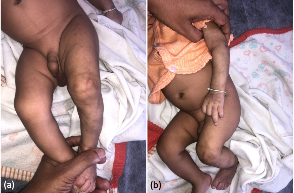
A 6-month-old male infant was brought to the dermatology clinic with complaints of persistent red spider web-like streaks over the left upper and lower limbs since birth. The streaks showed no signs of progression or regression but the limbs had reduced movements. The infant was the first-born of a second-degree consanguineous marriage with an uncomplicated pregnancy and full-term normal vaginal delivery. Reticulate erythema was noted over the left upper and lower limbs (Figure 1). A paediatrician had advised warming the limbs, which did not resolve the lesions. These limbs were leaner than the contralateral limbs with sluggish movements even with painful stimuli. No obvious limb length discrepancy was noted, and the skin over the abdomen was unremarkable. Following a clinical diagnosis of CMTC, the parents were counselled regarding the disorder and its sequelae. Skeletal radiography studies, slit lamp and fundoscopic examinations, and histopathological studies were advised. However, due to severe financial constraints, the parents refused any further evaluation.
Discussion
CMTC is a rare disorder and can present with primary cutaneous manifestations, such as prominent veins, telangiectasia, atrophy, ulceration, and hyperkeratosis. Cutaneous atrophy and ulceration can help in differentiating it from its physiological variant—cutis marmorata—in which the lesions disappear following rewarming [2]. However, lesions of CMTC do not disappear on warming. In addition to the “tram track” sign over the visible lesions, the commonest extracutaneous manifestation that should raise a suspicion of CMTC is a discrepancy in the limb length. The affected limbs usually have signs of atrophy and, rarely, hypertrophy. Other changes include tendinitis stenosans, syndactyly, clubfoot, hip dysplasia, and cleft palate [3]. Vascular malformations are common in CMTC with the commonest lesion being port- wine stain (PWS) [4]. Ocular anomalies in CMTC include glaucoma with or without vascular capillary malformations over the periocular skin [3]. A high concordance has been noted between CMTC and macrocephaly, which has resulted in a new clinical entity called macrocephaly-CMTC (M-CMTC) syndrome [5]. The syndrome may also include neonatal hypotonia, syndactyly, developmental delay, asymmetry, segmental overgrowth, and connective-tissue defects.
CMTC is difficult to diagnosis because it can mimic numerous congenital disorders (Table 1). Anthropometric assessments can help identify discrepancies in limb length. These benign discrepancies may only require an elevation device for the shorter limb. Additional investigations include ophthalmic, cardiac, orthopaedic, and neurological evaluations. There are no standardized criteria to diagnose CMTC. Keinast and Hoeger had suggested that three major and two out of five minor criteria are sufficient to diagnose CMTC (Table 2) [2].
- Adams–Oliver syndrome (AOS)
- Bockenheimer’s syndrome
- Divry Van Bogeart syndrome
- Klippel–Trinaunay syndrome
- Livedo recemosa
- Macrocephaly-CMTC syndrome
- Reticular hemangioma syndrome
- Major criteria
- Congenital reticulate erythema (marmorated)
- Absence of venectasia
- Unresponsiveness to local warming
- Minor criteria
- Fading of erythema within 2 years
- Telangiectasia*
- Port-wine stain outside the area affected by CMTC
- Ulceration*
- Atrophy*
- Three major criteria and at least two minor criteria have been suggested to diagnose CMTC.
Table 1: Differential diagnosis of cutis marmorata
The aetiology of CMTC remains unclear. Interestingly, Rogers and Poyzer had reported four cases that were diagnosed within an 18-month-period within a geographical area, which led to the postulate that a teratogenic agent might be involved in the etiopathogenesis of CMTC [6]. However, there is insufficient evidence to test this theory. Additional theories include failure of development of mesodermal vessels in the early embryonic period or peripheral neural dysfunction [4]. Based on the lack of evidence, Amitai, et al. concluded that the lethal gene theory proposed by Happle could underlie the etiopathogenesis of CMTC [4, 7].
Despite the confusing presentation and several systems affected in CMTC, its prognosis remains good. Amitai, et al. reported an improvement in approximately 46% of their patients within 3 years, which included total disappearance (10%) and marked improvement (36%) of lesions. Various studies have attempted to identify predictors of a favourable outcome or resolution in CMTC with no success. However, annual follow-up for at least 3 years may be useful in screening for associated anomalies and early identification of any systemic manifestations [8]. While the vascular lesions of CMTC fade with time, limb asymmetry persists and may require continued support.
Our report is limited by the lack of histopathological examination and genetic analyses. However, CMTC is a clinical diagnosis and a very rare clinical entity. Its etiopathogenesis and prognosis are unknown due to the paucity of literature on the topic. Reports like this one will aid physicians in suspecting and diagnosing this disorder and advise long- term follow-up to the patients.
References
-
Van Lohuizen CHJ (1922) Uber eine seltene angeborene Hautanomalie (Cutis marmorata telangiectatica congenita). Acta Derm Venereol 3: 202-211.
-
Kienast AK, Hoeger PH (2009) Cutis marmorata telangiectatica congenita: a prospective study of 27 cases and review of the literature with proposal of diagnostic criteria. Clin Exp Dermatol 34(3): 319-323.
-
Garzon MC, Schweiger E (2004) Cutis marmorata telangiectatica congenita. Semin Cutan Med Surg 23(2): 99-106.
-
Amitai D ben, Fichman S, Merlob P, Morad Y, Lapidoth M, et al. (2000) Cutis marmorata telangiectatica congenita: clinical findings in 85 patients. Pediatr Dermatol 17(2): 100-104.
-
Gonzalez ME, Burk CJ, Barbouth DS, Connelly EA (2009) Macrocephaly-capillary malformation: A report of three cases and review of the literature. Pediatr Dermatol 26(3): 342-346.
-
Rogers M, Poyzer KG (1982) Cutis marmorata telangiectatica congenita. Arch Dermatol 118(11): 895- 899.
-
Happle R (1987) Lethal genes surviving by mosaicism: A possible explanation for sporadic birth defects involving the skin. J Am Acad Dermatol 16(4): 899-906.
-
Levy R, Lam JM (2011) Cutis marmorata telangiectatica congenita: a mimicker of a common disorder. Can Med Assoc J 183: E249-251.
- Epithelioid Granuloma; 3cases with Different Clinical Features
- Advancing Representation in Dermatology Clinical Trials: Ethical, Scientific, and Regulatory Imperatives for Inclusion Across all Fitzpatrick Skin Types
- A Case of Atopic Dermatitis with Concurrent Psoriasis Vulgaris: Successful Treatment with Upadacitinib
- Innovation Lifting Eyeshadow: A Synthesis of Makeup and Optical Illusion
- Distinguishing Superficial Actinic Porokeratosis from Actinic Keratosis with UVF Dermoscopy: A Case Report
- High Mobility Group Box 1 (HMGB1) in Cutaneous Inflammation: An Immune Modulator Bridging Cellular Stress, Ferroptosis and Danger Signaling