Dermoscopy in Darier Disease: A Missing Link in an Unusual Hyperkeratotic Disorder
Darier disease is a rare autosomal dominant skin disorder resulting from mutation of the ATP2A2 gene on chromosome 12. Its progressive course makes its precise diagnosis crucial for a good prognosis. A 60-year-old female presented with multiple non-itchy papules over the face, trunk, and extremities since childhood and photosensitivity with similar lesions in the mother and two elder sisters. Examination revealed skin-coloured-to-hyperpigmented, firm and greasy papules with cobblestone appearance. Dirty yellowish-white wart-like lesions were noted over the toes. Histopathology revealed parakeratosis, papillomatosis, suprabasal acantholysis, and cleft with corps rounds in the granular layer. Dermoscopy revealed cobblestone appearance of the toes with tire-tread and cracked river-bed-like appearance and nails showing palmar hemorrhages and V-shaped nicking. The patient was diagnosed with Darier disease and treated accordingly. Uniquely, the cobblestone appearance was noted first over the dorsa of hands and feet. Dermoscopy may be used diagnose the disease while awaiting histopathological evidence.
Introduction
Darier disease is a rare autosomal dominant disorder characterized by greasy hyperkeratotic papules predominantly in the seborrheic areas along with nail and mucous membrane changes [1]. It is caused by a mutation in the gene ATP2A2, which results in a dysfunctional sarcoplasmic/endoplasmic Ca2+ ATPase isoform 2 (SERCA2) proteins [2]. It was first reported by Darier J [3] and White J [4] in 1989 and is also known as Darier–White disease, keratosis follicularis, and dyskeratosis follicularis. Epidemiologically, its reported prevalence is 1:100,000 with a slight male preponderance [1] Due to its rarity, it may be difficult to distinguish it from acne vulgaris, seborrheic dermatitis, acanthosis nigricans, confluent reticulate papillomatosis, prurigo pigmentosa, and reticulate erythematomucinous syndrome [1].
Darier disease has no known cure till date and is characterized by a relapsing and remitting course. It requires early diagnosis and treatment to avoid complications, such as foul-smelling lesions, secondary infections, and poor quality of life [5]. Non-pharmacological interventions include photoprotection, avoidance of synthetic clothing, and frequent use of emollients. Systemic and topical retinoids remain the pharmacological cornerstone of management [6].
Dermoscopy has been found to provide a few clues in diagnosing Darier disease [7, 8, 9, 10]. Herein, we present a case report to highlight the diagnostic utility of dermoscopy in recognizing this rare disorder.
Case Report
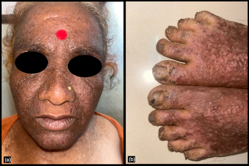
A 60-year-old female presented with multiple non- itchy papules over the face, neck, trunk and extremities since childhood and photosensitivity. Similar lesions were noted in her mother and two elder sisters. Skin-coloured and hyperpigmented, firm, slightly greasy papules with cobblestone appearance were noted over the face, neck, dorsum of hands and feet, and posterior trunk (Figure 1). A dirty-yellow membrane was observed over the posterior aspect of the hard palate with dirty yellowish-white wart- like lesions over the toes.
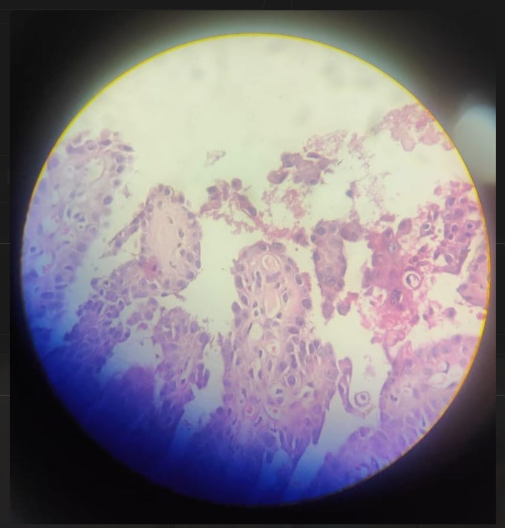
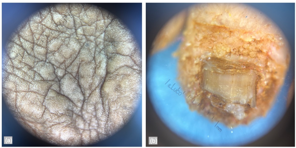
Histopathological examination of the lesions revealed parakeratosis, papillomatosis, and suprabasal acantholysis and cleft with corps ronds in the granular layer (Figure 2). Dermoscopy revealed cobblestone appearance over the toes with tire-tread and cracked riverbed-like appearances along with palmar hemorrhages and V-shaped nicking over the nails (Figure 3). On clinico-pathologico-dermoscopic correlation, the patient was diagnosed with Darier disease and treated with daily oral isotretinoin 10 mg, topical salicylic acid (3%), emollients, and photoprotection.
Discussion
Darier disease generally presents with characteristic cobblestone lesions over seborrheic and intertriginous lesions; similar mucosal lesions are noted in half of patients [1]. Interestingly, in this patient, the lesions began on the extremities; therefore, the patient was misdiagnosed elsewhere with photodermatitis and actinic lichen planus. Dermoscopy provided clues to the disease while awaiting histopathological results. In 2004, Vazquez LF, et al. [11] described the dermoscopic findings of pseudocomedones in five patients with biopsy-proven Darier disease [11]. In contrast, Errichetti E, et al. [12] evaluated 11 patients with biopsy‐proven Darier disease and reported a central yellow‐ brown area (polygonal or star‐like) with a surrounding white halo and pinkish homogeneous structure-less background in all patients. Other dermoscopic features included linear and/ or dotted vessels (63.8%) and whitish scales surrounding polygonal brown‐yellow areas (27.3%) [12]. According to Adya KA, et al. [8] three zones can be distinguished on dermoscopy in Darier disease a central light br own follicular opening, surrounding dark brown structure, and peripheral whitish halo [8].
We, however, noted tire-tread and cracked riverbed-like appearances along with palmar hemorrhages and V-shaped nicking over the nails. The varied findings may be due to the differences in the patient population and the duration of the disease. While Vazquez-Lopez et al. reported from New York, Errichetti E, et al. [12] reported from Italy. Adya KA, et al. [8] reported from India, which is where we are reporting from; however, their patient was a 25-year-old female with a 6-month history of lesions, while our patient was a
60-year-old female with lesions since childhood. Therefore, we believe that the Fitzpatrick skin type and the duration of lesions affect the dermoscopic features in Darier disease.
Clinicopathological correlation is time-consuming and requires dermatopathologists for precise interpretations. In resource-limited and time-limited settings, a more rapid and non-invasive tool of diagnosis will be hugely helpful. Dermoscopy in Darier disease can help distinguishing it from other disorders, especially in atypical or early presentations. The commonest differentials include acne vulgaris, seborrheic dermatitis, acanthosis nigricans, confluent reticulate papillomatosis, prurigo pigmentosa, and reticulate erythematomucinous syndrome. On dermoscopy, acne vulgaris is characterized by papules, pustules, and comedones with varying degrees of erythema [13] while Darier disease demonstrates pseudocomedones and seborrheic dermatitis demonstrates twisted red loops and comma vessels [14]. Pigmentation helps in identifying acanthosis nigricans, while confluent reticulate papillomatosis includes flat lesions predominantly confined to the upper trunk only [1]. Lastly, palpation of rough lesions in Darier disease helps in differentiating it from prurigo pigmentosa and reticulate erythematomucinous syndrome [1].
The limitation our report is the evaluation of a single patient, which limits the generalizability of our findings. Genetic analysis was not performed in this patient due to financial constraints. Evaluation of the dermoscopic features of Darier disease in more patients may enable its utility in diagnosing this disease.
Sources of Support
None
Conflicts of Interest
None
Acknowledgements
None
Informed Consent
Written informed consent was obtained from the patient for publication of anonymized data in the form of a case report. Ethical committee clearance was waived in view of the case report design.
Author Statement
The manuscript has been read and approved by all the authors, that the requirements for authorship as stated earlier in this document have been met and that each author believes that the manuscript represents honest work.
References
-
Suryawanshi H, Dhobley A, Sharma A, Kumar P (2017) Darier disease: A rare genodermatosis. Journal of Oral and Maxillofacial Pathology 21(2): 321.
-
Godi A (2003) Darier disease. A review of pathophysiological mechanisms. Acta Dermatovenerologica Alpina 12(4): 119-126.
-
Darier J (1889) De la psorospermose folliculaire végétante. Ann Dermatol Syphiligr 10: 597-612.
-
White J (1889) A case of keratosis (ichthyosis) follicularis. J Cutan Genitourin Dis 7: 210-219.
-
Ashok KP, Paulraj S, Dutta S (2020) Debilitating Darier’s Disease and Its Impact on the Quality of Life. Cureus 12(5): e8133.
-
Haber RN, Dib NG (2021) Management of Darier disease: A review of the literature and update. Indian J Dermatol Venereol Leprol 87(1): 14-21.
-
Siemianowska D, Wolf B (2021) Importance of dermoscopy in assisting the non-invasive diagnosis of Darier’s disease. Journal of Pakistan Association of Dermatologists 31(1): 103-107.
-
Adya KA, Inamadar AC, Palit A (2020) Dermoscopy of Localized Darier’s Disease in Fitzpatrick Type IV Skin. Indian Dermatol Online J 11(2): 298-300.
-
Ghita S, Jouari El, Gallouj S, Lamouaffaq A, Mernissi FZ (2018) Late onset Darier’s disease: clinical and dermoscopic features. Journal of Dermatology & Cosmetology 2(5): 75-76.
-
Khan S, Farabi B, Zufall A, Safai B, Shulman K, et al. (2023)A case study on the dermatoscopic findings of Darier’s disease in skin type VI. Skin Health and Disease 3(6): e282.
-
Vazquez LF, Lopez EM, Maldonado SC, Perez ON, Marghoob AA (2004) The handheld dermoscope improves the recognition of giant pseudocomedones in Darier’s disease. J Am Acad Dermatol 50(3): 454-455.
-
Errichetti E, Stinco G, Lacarrubba F, Micali G (2016) Dermoscopy of Darier’s disease. Journal of the European Academy of Dermatology and Venereology 30(8): 1392- 1394.
-
Alfaro CP, Mejia SA, Valencia HA, Ramirez S, Mena CC (2012) Dermoscopy Distinction of Eruptive Vellus Hair Cysts with Molluscum Contagiosum and Acne Lesions. Pediatr Dermatol 29(6): 772-773.
-
Kibar M, Aktan S, Bilgin M (2015) Dermoscopic findings in scalp psoriasis and seborrheic dermatitis; Two new signs; Signet ring vessel and hidden hair. Indian J Dermatol 60(1): 41-45.
- Epithelioid Granuloma; 3cases with Different Clinical Features
- Advancing Representation in Dermatology Clinical Trials: Ethical, Scientific, and Regulatory Imperatives for Inclusion Across all Fitzpatrick Skin Types
- A Case of Atopic Dermatitis with Concurrent Psoriasis Vulgaris: Successful Treatment with Upadacitinib
- Innovation Lifting Eyeshadow: A Synthesis of Makeup and Optical Illusion
- Distinguishing Superficial Actinic Porokeratosis from Actinic Keratosis with UVF Dermoscopy: A Case Report
- High Mobility Group Box 1 (HMGB1) in Cutaneous Inflammation: An Immune Modulator Bridging Cellular Stress, Ferroptosis and Danger Signaling