Anatomical Study of Profunda Femoris Artery and it’s Variations-Cadaveric Study
Background and Aims: Profunda femoris artery is the largest branch of femoral artery. It is the principal supply to the muscles of the thigh as well as head and neck of femur. Its branches form anastomosis around the head of the femur. Profunda femoris artery is also used for arteriography. It is frequently used in vascular reconstructive procedures in the proximal thigh. It forms main route of collateral circulation in occlusion of femoral artery. The study of variation of Profunda femoris artery is of great value for radiologists and surgeons during diagnostic and surgical intervention. Aim of the study is to observe the origin of Profunda femoris artery, to measure the distance between midinguinal point and site of origin of Profunda femoris artery and to study the anatomy of Profunda femoris artery and its branches and to find out any variations in its course and its branches. Materials and Methods: The study was conducted as a prospective study from April 2018 to October 2018. All the lower limbs with the intact Femoral artery and Profunda femoris artery and their branches were included. Femoral artery and its branches had got cut during dissection were not included in the study. Thirty two Profunda femoris arteries (20 on the right side and 12 on the left side) were studied in the department of Anatomy. The femoral triangle was exposed by making incisions along the inguinal ligament from pubic symphysis to anterior superior iliac spine. Midpoint was taken as Midinguinal point (MIP). MIP was marked with a coloured pen. The distance between MIP and the site of origin ofProfunda femoris artery was measured. We observed any variation in the site of origin of Profunda femoris artery, medial and lateral circumflex femoral artery and any variations in the branches of each vessel. Results: The Profunda femoris artery was found to be originated from lateral aspect of Femoral artery in 21(65%) of lower limbs. Lateral Circumflex femoral artery was found to be originating from lateral aspect of Profunda femoris artery in 28(87%) of lower limbs. Medial Circumflex femoral artery was found to be originating from medial aspect of Profunda femoris artery in 16(50%) of lower limbs. Lateral Circumflex femoral artery was found to be originating from Femoral artery in 3(10%) of lower limbs. We observed distance of origin of Profunda femoris artery from the midpoint of inguinal ligament as 10 – 20 mm in 10(31%) lower limbs. We observed absence of Lateral Circumflex femoral artery in 1(3%) of lower limbs and absence of medial Circumflex femoral artery in 6(18%) of lower limbs. Conclusions: The Profunda femoris artery is an important branch of the Femoral artery and is of clinical importance to the surgeon. Femoral artery and Profunda femoris artery were used for various imaging procedures including Cathetarization. Such a large & unexpected artery may be damaged while collecting blood in infants from Femoral vein or at the time of exposure of Saphenous vein for ligation at its junction with the femoral vein. During surgery, these vessels may be damaged easily at this region. Knowledge of anatomy and variations of Profunda femoris artery are important for surgeons in reducing intra – operative haemorrhage and postoperative complications.
Introduction
The arteries of lower limb develop from the axis artery, which is derived from fifth lumbar artery. In the developmental process, some of the channels regress and some of them enlarge and form a definitive arterial pattern. The persistence of the channels that are supposed to disappear lead to vascular anomalies [1].
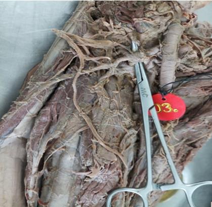
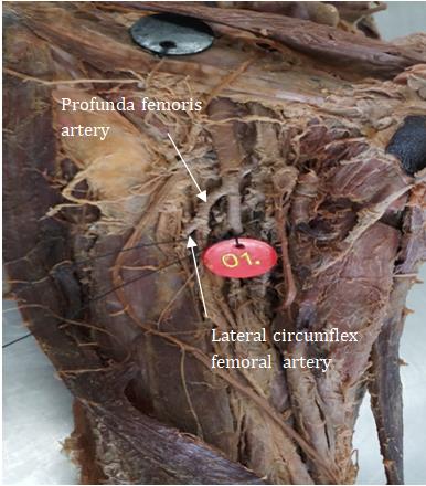
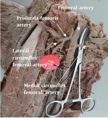
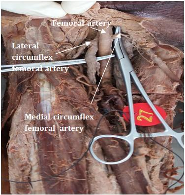
The Profunda femoris artery (PFA) is a major deep branch of Femoral artery given off in the Femoral triangle. This artery is normally located posterolateral to the Femoral artery and given off around 3.5cms distal to the inguinal ligament. The Profunda femoris artery arises sometimes medially and rarely posterior to the Femoral artery [2].
The Profunda femoris artery mainly gives off Lateral circumflex femoral artery and Medial circumflex femoral artery, muscular branches and perforating branches which pierce through Adductor magnus to reach the back of thigh.
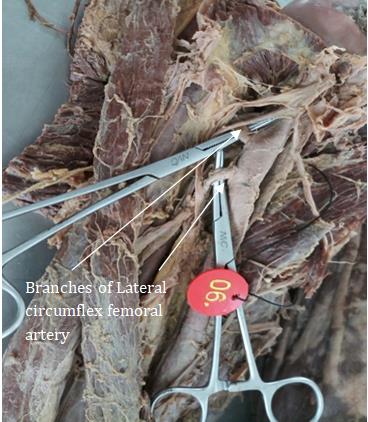
The Lateral circumflex femoral artery arising laterally from Profunda femoris artery is an important landmark to identify the Femoral nerve divisions. The arterial trunk passes between the anterior and posterior divisions of Femoral nerve. Then it branches into ascending, transverse and descending branches. These branches contribute to the anastomoses around the neck of femur and greater trochanter [2].
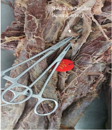
The Medial circumflex femoral artery is given off medially by the Profunda femoris artery. The artery passes between the pectineus and adductor longus to reach close to the neck of femur and contributes blood supply to the head and neck of femur and divides into transverse, ascending and acetabular branches which take part in cruciate and trochanteric anastomoses [2].
The perforating arteries are four in number, the last one being the continuation of Profunda femoris artery. These arteries pierce the adductor magnus and lateral intermuscular septum and supply adductor and Hamstring muscles and end in vasus lateralis muscle where they are connected to one another by a series of anastomoses [3].
Profunda femoris artery is useful for many invasive and noninvasive procedures like Doppler, Ultrasonography and MR angiography. Interventional radiography is a new technique to study the course of Femoral artery and Profunda femoris artery.
Materials and Methods
Objectives of the study: 1. The origin of Profunda femoris artery was identified.
2. The distance of origin of Profunda femoris artery from the bony landmarks were measured.
3. Origin of branches of Profunda femoris artery (Medial
circumflex femoral artery and Lateral circumflex femoral artery) was noted.
4. Any variations in the origin, course of the artery and its branches were identified and noted.
A total of 32 embalmed lower limbs (20 right and 12left lower limbs) were used for the study. The femoral triangle was dissected as per Cunningham’s manual of practical Anatomy [4].
All the lower limbs with the intact Femoral artery and Profunda femoris artery and their branches were included. Femoral artery and its branches had got cut during dissection were not included in the study.
The skin and superficial fascia were reflected from front of the thigh, splitting of femoral sheath on both the side of femoral vein with the exposure of femoral canal and femoral artery. Femoral artery and Profunda femoris artery were identified with their branches after dissection. The distance of origin of femoral artery and Profunda femoris artery from midpoint of inguinal ligament were measured in millimeters with a scale and vernier caliper and recorded in data sheet. The relation of origin of Profunda femoris artery to femoral artery was noted. Branches from Profunda femoris artery and its variations were identified and noted.
Results
Point of Origin of Profunda Femor is Artery
Profunda femoris artery originated from Femoral artery in28 lower limb specimens. Absent Profunda femoris artery was found in 4 lower limbs. Absent Profunda femoris artery and Lateral circumflex femoral artery was found in one lower limb. Absent Medial circumflex femoral artery was found in 4 lower limbs. Medial circumflex femoral artery arose directly from femoral artery in 5 lower limbs. Medial circumflex femoral artery originated from Lateral circumflex femoral artery in one lower limb.
Position of Profunda femoris artery in relation to Femoral artery
The Profunda femoris artery commonly positioned in the posterolateral aspect of Femoral artery in 21(66%) of lower limbs and posterior to Femoral artery in 7(22%) of lower limbs (Table 1).
| Side | Right | Left | Total | ||||||||
|---|---|---|---|---|---|---|---|---|---|---|---|
| 1.Posterolateral | 10 | 11 | 21(66%) | ||||||||
| 2.Posterior | 6 | 1 | 7(22%) |
Table 1: Posterolateral femoral artery in lower limbs and posterior to Femoral artery.
| Serial number | Distance(mms) | Number of cases on the right side | Number of cases on the left side | ||||||||
|---|---|---|---|---|---|---|---|---|---|---|---|
| 1. | 10 - 20 | 6 | 4 | ||||||||
| 2. | 21 – 30 | 5 | 2 | ||||||||
| 3. | 31 – 40 | 3 | 3 | ||||||||
| 4. | 41 – 50 | 1 | 0 | ||||||||
| 5. | 51 – 60 | 1 | 0 | ||||||||
| 6. | 61 – 70 | 1 | 0 | ||||||||
| 7. | >70 | 0 | 0 |
Table 2: Number of cases on
Distance of origin of Profunda femoris artery from the midpoint of inguinal ligament was found most commonly between 10 – 20 mm on both the sides (Table 2).
- Serial number
- Site of origin
- Number of cases on the right side
- 1.
- From Profunda femoris artery – lateral aspect
- 16
- 50
- 12
- 38
- 2.
- From Femoral artery as a common stem with Profunda femoris artery
- 3
- 9
- 0
- 0
- 3.
- From Femoral artery – superior to
- Profunda femoris artery
- 0
- 0
- 0
- 0
- 4.
- From Femoral artery inferior to
- Profunda femoris artery
- 0
- 0
- 0
- 0
- 5.
- From External iliac artery
- 0
- 0
- 0
- 0
- 6.
- Absent Lateral circumflex femoral artery
- 1
- 3
- 0
- 0
Table 3: Site of origin of Lateral circumflex femoral artery.
- Lateral circumflex femoral artery commonly originated from lateral aspect of Profunda femoris artery on both the sides. Lateral circumflex femoral artery originated from Femoral artery as a common stem with
- Absent Profunda femoris artery and Lateral circumflex femoral artery was found in one specimen and Lateral circumflex femoral artery directly from Femoral artery
- Serial number
- Site of origin
- Number of cases on the right side
- Percentage
- (%)
- 1.
- From Profunda femoris artery –medial aspect
- 11
- 34
- 5
- 16
- 2.
- From Femoral artery as a common stem with Profunda femoris artery
- 4
- 12
- 2
- 6
- 3.
- From Femoral artery – superior to
- Profunda femoris artery
- 0
- 0
- 0
- 0
- 4.
- From Femoral artery inferior to
- Profunda femoris artery
- 1
- 3
- 1
- 3
- 5.
- From lateral circumflex femoral artery
- 1
- 3
- 0
- 0
- 6.
- Absent medial circumflex femoral artery
- 3
- 9
- 4
- 12
Table 4: Site of origin of Medial circumflex femoral artery.
| Serial number | Distance (mms) | Number of cases on the right side | Number of cases on the left side | ||||||||
|---|---|---|---|---|---|---|---|---|---|---|---|
| 1. | 0-10 | 11 | 11 | ||||||||
| 2. | 11-20 | 4 | 0 | ||||||||
| 3. | 21-30 | 0 | 1 | ||||||||
| 4. | 31-40 | 0 | 0 | ||||||||
| 5. | 41-50 | 1 | 0 | ||||||||
| 6. | 51-60 | 1 | 0 |
Table 5: Number of cases on
- Lateral circumflex femoral artery commonly originated from lateral aspect of Profunda femoris artery on both the sides. Lateral circumflex femoral artery originated from Femoral artery as a common stem with
- Absent Profunda femoris artery and Lateral circumflex femoral artery was found in one specimen and Lateral circumflex femoral artery directly from Femoral artery
- Serial number
- Site of origin
- Number of cases on the right side
- Percentage
- (%)
- 1.
- From Profunda femoris artery –medial aspect
- 11
- 34
- 5
- 16
- 2.
- From Femoral artery as a common stem with Profunda femoris artery
- 4
- 12
- 2
- 6
- 3.
- From Femoral artery – superior to
- Profunda femoris artery
- 0
- 0
- 0
- 0
- 4.
- From Femoral artery inferior to
- Profunda femoris artery
- 1
- 3
- 1
- 3
- 5.
- From lateral circumflex femoral artery
- 1
- 3
- 0
- 0
- 6.
- Absent medial circumflex femoral artery
- 3
- 9
- 4
- 12
Table 4: Site of origin of Medial circumflex femoral artery.
was found in three specimens were not included in Table 4.
Number of cases on
Percentage
the left side (%)
Site of origin of Medial circumflex femoral artery from Profunda femoris artery was commonly found on the medial side of Profunda femoris artery on both the sides. Origin of Medial circumflex femoral artery as a common stem from Femoral artery was found in six lower limbs and from lateral circumflex femoral artery in one lower limb. Absent Medial circumflex femoral artery was found in seven lower limbs.
| Serial number | Distance (mms) | Number of cases on the right side | Number of cases on the left side | ||||
|---|---|---|---|---|---|---|---|
| 1. | 0-10 | 4 | 5 | ||||
| 2. | 11-20 | 0 | 0 | ||||
| 3. | 21-30 | 0 | 0 | ||||
| 4. | 31-40 | 1 | 0 | ||||
| 5. | 41-50 | 1 | 0 | ||||
| 6. | 51-60 | 0 | 0 |
Table 6: Distance of origin of Medial circumflex femoral artery from the origin of Profunda femoris artery.
femoral artery. Three on both the sides and Absent Profunda femoris artery – Two on the right side were not included in Table 6.
| Serial number | Study | Distance of origin of Profunda femoris artery from Mid | ||
|---|---|---|---|---|
| inguinal ligament in cms | ||||
| 1. | Dixit et al. [5] | 4.75 | ||
| 2. | Aswini C.Appaji et al. [6] | 6.02 | ||
| 3. | Prakash et al. [7] | 4.2 | ||
| 4. | Present study | 3.11 |
Table 7: Comparison of distance of origin of Profunda femoris artery from Mid inguinal ligament.
| Serial number | Study | Postero lateral | Posterior | Postero medial | Lateral/ | Medial | ||
|---|---|---|---|---|---|---|---|---|
| Antero | ||||||||
| lateral | ||||||||
| 1. | Dixit et al [5] | 35.4 | 31.25 | - | - | - | ||
| 2. | Aswini C.Appaji6 et al. [5,6] | 53.3 | 40 | 0.03 | - | - | ||
| 3. | Prakash et al. [7] | 50 | 46.9 | - | - | 3.1 | ||
| 4. | Manjappa et al [8] | 60 | 25 | 5 | 5 | - | ||
| 5. | Present study | - | 22 | - | 66 | - |
Table 8: Showing comparison of various positions of Profunda femoris artery in relation to Femoral artery.
Serial number Study Incidence of Medial circumflex femoral artery (in
Discussion
Anatomical variations reported at the level of division of Femoral artery can be explained as follows. In the lower animals, the Profunda femoris artery is a branch of internal iliac artery. During the course of evolution, the origin shifted distally as a branch of Femoral artery. Ontogeny repeats phylogeny. Hence developmental arrest at different stages may lead to anatomical variations related to the division of femoral artery [7].
The lateral circumflex femoral artery originated from lateral aspect of the Profunda femoris artery in 81.25% and 18.75% originated from femoral artery in Prakash, et al. [7] study. In our study, the lateral circumflex femoral artery originated from lateral aspect of the Profunda femoris artery in 88% and 9% originated from femoral artery and absent lateral circumflex femoral artery in 3% (Table 3).
The medial circumflex femoral artery originated from medial aspect of the Profunda femoris artery in 67.2% and 32.8% originated from femoral artery in Prakash, et al. [7] study. In our study, the medial circumflex femoral artery originated from medial aspect of the Profunda femoris artery in 50% and 18% originated from femoral artery and absent medial circumflex femoral artery in 21% and from lateral circumflex femoral artery in 3% (Table 5).
We observed that origins of Lateral and Medial circumflex femoral arteries were directly from the femoral artery were associated with distal shift of the level of separation of Profunda femoris artery from the Femoral artery.
The Profunda femoris artery acts as a collateral vessel in the occlusion of Femoral artery and for this important function, it has to have a large caliber, which can be based on the aforementioned comparative anatomy [10]. Vazquez, et al. [11] studied 221 embalmed cadavers and classified the pattern of the arteries into Type 1: Both the circumflex arteries arise from Profunda femoris artery Type 1a: The origin of Medial circumflex femoral artery is more proximal than Lateral circumflex femoral artery Type 1b: The origin of Lateral circumflex femoral artery is proximal than Medial circumflex femoral artery Type 1c: Both the arteries arise from a common trunk Type 2: One of the arteries arises from the femoral artery and the other one from the Profunda femoris artery Type 2a: The Medial circumflex femoral artery arises from Femoral artery Type 2b: The Lateral circumflex femoral artery arises from Femoral artery Type 3: Both the arteries arise from Femoral artery In our study, both Medial circumflex femoral artery & Lateral circumflex femoral arteries arise from Profunda femoris artery in 18 lower limb specimens. Type 2a was found in 5 lower limbs and Type 2b was found in 4 lower limbs. Both arteries arise from Femoral artery in 4 lower limbs. In Suthur K, et al. [12] study, the Profunda femoris artery originated from the posterolateral side of Femoral artery in 52% and in Daxsha Dixit, et al. [13] study, it was about 50%. In the present study, the Profunda femoris artery arose from posterolateral aspect of Femoral artery in 66% and from posterior aspect of Femoral artery in 22%. Bergman, et al. [14] said that, if Profunda femoris artery arises from a medial aspect of femoral artery, then Femoral artery may split into three vessels almost of equal caliber that are Profunda femoris artery, medial and lateral circumflex femoral arteries. But in our study, we did not find Profunda femoris artery origin from medial aspect of femoral artery. The mean distance of Profunda femoris artery origin from midinguinal point was 3.1cms in our study which was compared with previous Author’s study (Table 7).
Conclusion
Now -a- days different diagnostic and therapeutic interventions on femoral artery and its branches for various diseases like congenital anomalies of vessels and vascular occlusive diseases may be planned. Care should be given to know the variations in femoral artery, Profunda femoris artery and its branches.
These vessels are useful for catheterization in various diagnostic procedures like angiography and Doppler imaging technique. Knowledge of variation in the origin of artery and variation in its branches are useful to prevent the various serious conditions like Pseudoaneurysm, thrombosis and embolism. Lack of knowledge in variation of these vessels during surgery and procedure can cause severe haemorrhage and complications. The variations may complicate arteriectomies, embolectomies and thromboendarterectomies in cases of atherosclerosis, which are most commonly seen in the lower limb vessels.
Anatomy of Profunda femoris artery and its branches is essential in performing trochanteric and intertrochanteric osteotomies and is also helpful to avoid iatrogenic vascular necrosis of head of femur in reconstructive surgery of the hip and fixation of acetabular fractures through posterior approach. Branches of Lateral and Medial circumflex femoral vessels may be damaged in fracture of neck of femur and acetabulam. These vessels injury may produce intraoperative and postoperative bleeding. Care should be given for those vessels during surgeries. The variations of Profunda femoris artery are vital for the surgeons during femoral hernia repair and surgeries which are planned in the femoral triangle. Because of less chance of flap necrosis, anterolateral thigh flap based on Profunda femoris artery is most preferred choice in most of the reconstructive surgeries. Hence knowledge on Profunda femoris artery and its variations in origin and its branches need great attention for Surgeons and Radiologists before planning and performing any surgeries and interventions in femoral region.
References
-
Lippert H, Pabst R (1985) The arterial variations in Man: The classifications and frequencies. Bermann Munchen, pp: 54-61.
-
Susan Standring (2005) Gray’s Anatomy; The anatomical basis of clinical practice, 40th(Edn.), Elsivier Churchill Livingstone, pp: 1450-1452.
-
Asim Kumar Datta (2009) Essentials of Human Anatomy. 4th(Edn.), Current Books International, pp: 169.
-
Cunningham’s manual of practical Anatomy. Upper and lower limbs. 15th(Edn.), London: Oxford university press, 1: 129-144.
-
Dixit DP, Mehta LA, Kothari ML (2001) Variations in the Origin and Course of Profunda Femoris. J Anat Soc India 50(1): 6-7.
-
Aswini CA, Sanjay CD (2017) Morphometry of Profunda femoris artery and its correlation with Femoral artery: A cadaveric study. Int J Anat Res 5(4.3): 4770-4775.
-
Prakash, Kumari J, Kumar Bhardwaj A, Jose BA, Kumar Yadav S, et al. (2010) Variations in the origins of the Profunda femoris, medial and lateral femoral circumflex arteries: a cadaver study in the Indian population. Rom J Morphol Embryol 51(1): 167-170.
-
Manjappa T, Prasanna LC (2014) Anatomical variations of the Profunda femoris artery and its Branches – A Cadaveric study in South Indian Population. Indian J Surg 76(4): 288-292.
-
Apurva D, Paras S, Hitesh C, Hardik K, Singel TC (2015) A cadaveric study of variations in the origin of medial circumflex femoral artery. IJBAR 6(7): 541-545.
-
Vaas F (1975) Some considerations concerning the deep femoral artery. Arch Chir Neerl 27(1): 25-34.
-
Vazquez MT, Murillo J, Maranilo E, Parkin I, Sanudo J (2007) The patterns of the circumflex femoral artery.revisited. Clin Anat 20(2): 180-185.
-
Suthar K, Patil D, Mehta C, Patel V, Prajapati B, et al. (2013) Cadaveric study: Morphological study of branches of femoral artery in front of thigh. CIB Tech J Surg 2(2): 16-22.
-
Daksha D, Dharati MK, Sureshbhai PR, Mital MP, Tulsibhai CS (2011) A study of variations in the origin of Profunda femoris artery and its circumflex branches. Int J Biol Res 2(4): 1084-1089.
-
Bergman RA, Afifi AK, Mujayichi R (1988) Compendium of human anatomic variations. Urban & Schwarzenberg, Baltimore-Munich, pp: 86-87.
- Pattern of Breast Lesions in Ovu Inland, Delta State, South Southern Nigeria
- Morphometric Analysis of the Human Femur: Exploring Platymetric and Robusticity Indices Among the Nigerian Population
- Anatomical Variation of Arteria Lusoria: Clinical Implications for Dysphagia Lusoria and Surgical Risk
- Morphometric Study of the Vertebral Body and Pedicle of Typical Cervical Vertebrae Using Radiological Image
- Epigenetic Mechanisms Driving Human Evolutionary Changes
- Neuroprotective Effects of Ginkgo Biloba Extract on Bilateral Common Carotid Artery Ischaemic Stroke Induced in Wistar Rat