Study of Morphological Analysis of the Foramen Transversarium and Accessory Foramen Transversarium in Typical Cervical Vertebrae
Foramen Transversarium (FT) is present in the transverse process of cervical vertebrae. One FT is present in each transverse process of cervical vertebrae. The vertebral artery, vein, and sympathetic nerves pass through it. The present study was carried out on 100 typical cervical vertebrae on their foramen transversarium regarding their shape and no of FT on transversarium. The oval shape was mostly observed in 45% and 44% on the right and left sides. The accessory foramen transversarium (AFT) was observed in 22 vertebrae among 100 vertebrae. The AFT was mostly observed on the right side.
Introduction
The typical cervical vertebrae are recognized by a foramen at its transverse process called foramina transversaria (FT), which is pierced by the vertebral artery, vertebral vein, and nerves except for C7 in which only the vertebral vein passes due to its small size. The sympathetic nerves from the inferior cervical ganglion pass through FT. FT protects the vertebral artery on all sides 3. Sometimes, there is the presence of another foramen posterolateral to the foramen transversarium on the transverse process of the cervical vertebrae called the accessory foramen transversarium (AFT). Generally, AFT is located on the lower cervical vertebrae i.e., C6 and C7, and may disturb the course of the vertebral artery due to the irregularities in the shape and size of FT. The twisting course of the vertebral artery with the lushka joint on the medial aspect of FT may lead to various clinical conditions like cervical decompression. The discrepancies in the number, shape, and size of the FT of the cervical vertebrae caused headaches, migraines, and fainting attacks due to compression of the vertebral artery during neck movement. The spasm of the vertebral and basilar artery (supply the inner ear) due to the irritability of sympathetic plexuses may accentuate the labyrinthine disturbance along with neurological symptoms. Therefore, the understanding of AFT and its variations are essential for clinicians while diagnosing a patient presenting with frequent headache, migraine, or fainting attacks. It is equally important for radiologists for clinical interpretation during X-rays and CT scans. The present study is conducted to observe the morphology and variations related to the FT.
Material and Method
The study was carried out on 100 dry adult human cervical vertebrae, which were obtained from the Department of Anatomy at Sri Guru Ram Das Institute of Medical Sciences and Research, Sri Amritsar. Broken and decalcified bones were excluded. The morphology of FT was recognized by analyzing its shape. The morphological observations were made and tabulated [1, 2, 3, 4, 5, 6, 7, 8, 9].
Result
Morphology
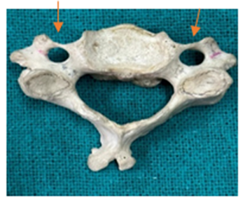
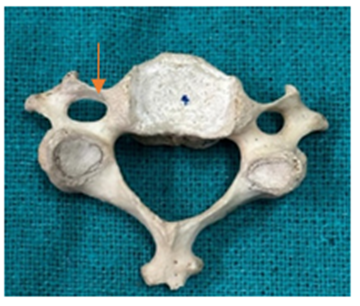
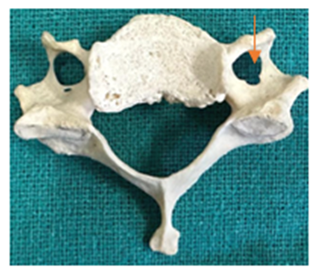
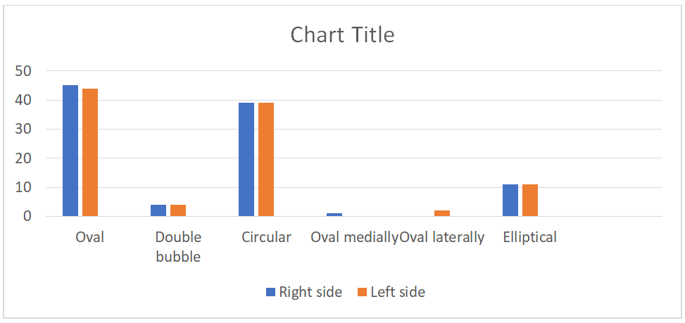
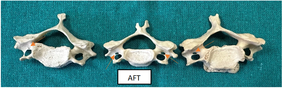
Shape of Main FT: The different shapes of the FT were observed. The six different shapes were observed and categorized into groups Group A, labeled as Oval; Group B as double bubble (incomplete double); Group C as Circular; Group D as Oval medially; Group E as Oval laterally and Group F as Elliptical with a large transverse diameter. Group B shape is unique as it was rarely present. The frequencies of the shape of the FT were noted in Table 1A and Table 1B for the right and left sides, respectively Figures 1-4.
| Shape of Main FT | |||
|---|---|---|---|
| Right Side | Total (%) n=100 | ||
| Oval | 45 | 45 | |
| Double bubble | 4 | 4 | |
| Circular | 39 | 39 | |
| Oval medially | 1 | 1 | |
| Oval laterally | Nil | 0 | |
| Elliptical | 11 | 11 |
Table 1: Accessory Foramen Transversarium in typical cervical vertebrae.
Table 1A: The different shapes of the main Foramen Transversarium of typical cervical vertebrae on the right side.
| Shape of Main FT | |||
|---|---|---|---|
| Left Side | Total (%) n=100 | ||
| Oval | 44 | 44 | |
| Double bubble | 4 | 4 | |
| Circular | 39 | 39 | |
| Oval medially | Nil | 0 | |
| Oval laterally | 2 | 2 | |
| Elliptical | 11 | 11 |
Table 2: Accessory Foramen Transversarium in typical cervical vertebrae.
Table 1B: The different shapes of the main Foramen Transversarium of typical cervical vertebrae on the left side.
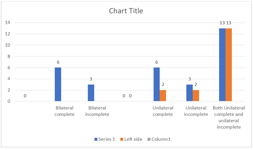
Variations In Number and Shape of AFT: AFT was present in 16% of typical cervical vertebrae. Out of 16%, 6% was present bilateral and 10% was present unilateral. Incomplete AFT was also observed in 9% of the typical cervical vertebrae. AFT was mostly present on the right side of the vertebrae Table 2 and Figures 5 & 6.
| Typical Cervical Vertebrae (n=100) | ||||
|---|---|---|---|---|
| Type of Accessory FT | No of Accessory FT | Accessory FT (%) | ||
| Bilateral complete | 6 | 6 | ||
| Bilateral incomplete | 3 | 3 | ||
| Unilateral complete | 6(R) | 3(L) | 6(R) | 3(L) |
| Unilateral incomplete | 3(R) | 2(L) | 3(R) | 2(L) |
| Total | 22 | 22 |
Table 3: Accessory Foramen Transversarium in typical cervical vertebrae.
*R: Right, L: Left Table 2: Accessory Foramen Transversarium in typical cervical vertebrae.
The present study was carried out on Foramen Transversarium and Accessory Foramen Transversarium morphological observation on 100 typical cervical vertebrae. As depicted in Table 1(A) and (B) the different shapes of the main FT of typical cervical vertebrae, the most common observations were 45% and 44% oval shaped on the right and left side, respectively. The oval shape which is directed laterally was observed in 1% of the typical cervical vertebrae. Compared to the study done by Abdul RS, et al. [6], who observed the most common shape of the main FT was round 34.15% and 32.93% on the right and left side of the vertebrae, respectively. In another study by Rammachandra K, et al. [10], they also observed most common shape of the main FT was round in 63.3% of vertebrae. And Molinet
GM, et al. [11], also observed the round shape was the most common type. In the study of Lalit M, et al. [12], the most common shape of the FT was oval. Taitz C, et al. [13], also observed an oval shape in the main FT. Mostly rounded oval shape was observed. Abdul RS, et al. [6], observed the different shapes of the main FT like round, elliptical, double bubble, irregular, leaf-like, and kite-like shapes. In the present study 2023, various shapes like oval, double bubble, oval directed medially, oval directed laterally elliptical, and most commonly oval shaped were observed. Oval-directed medially and laterally were observed in 1% and 2% on the right and left sides, respectively.
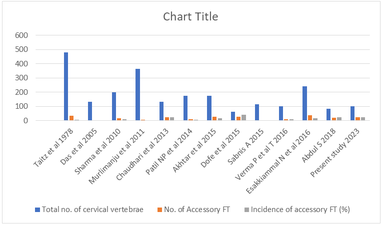
Taitz C, et al. [13], observed 34 AFT among 480 cervical vertebrae. Das S, et al. [14], also conducted a study on 132 cervical vertebrae and observed only 2 AFT. While Sharma A [15], observed 16 AFT among 200 cervical vertebrae, mostly found on the C6 vertebrae. In our study, we observed AFT on a higher side in C6 vertebrae. Murlimanju BV, et al. [16], found 6 AFT from 363 cervical vertebrae, where one cervical vertebra had unilateral AFT and 5 had bilateral AFT. Chaudhari ML, et al. [17], depicted 22 vertebrae with AFT among 133 cervical vertebrae, where 6% bilateral complete and 3% bilateral incomplete whereas unilateral 3% complete AFT on the right side and 2% complete on the left side, respectively. The unilateral incomplete 3% and 2% on the right and left side, respectively. Patel NP, et al. [18], observed 10 AFT among 175 cervical vertebrae from which 6 unilateral and 4 bilateral were observed. Akhtar MJ, et al. [8], carried out a study on 174 cervical vertebrae and 25 vertebrae had AFT (16 typical and 9 atypical), 3 bilateral complete on typical and 2 bilateral complete on atypical cervical vertebrae whereas unilateral AFT was observed on 10 and 3 on the right and left side, respectively. Sabnis A, et al. [3], conducted a study on 114 cervical vertebrae (40 atlas, 50 typical, and 24 seventh cervical vertebrae) and observed 3 unilateral incomplete AFT in atlas. 3 Unilateral incomplete AFT on the right side. Verma P, et al. [7], carried out a study on 200 cervical vertebrae (100 typical and 100 atypical) and observed 16 AFT among 200 cervical vertebrae. Esakkiammal N, et al. [9], studied 241 cervical vertebrae (107 atypical and 134 typical) and observed 37 AFT only in typical cervical vertebrae. In the present study, 22 AFTs were observed among 100 typical cervical vertebrae.
6%bilaterally complete and 3% bilateral incomplete were observed. Unilateral complete was 6% and 2% on the right and left sides, respectively. Unilateral incomplete was 3% and 2% on the right and left side, respectively. The development of vertebral artery is closely related to the development of these variations. The vertebral artery develops from the longitudinal anastomosis fusion that joins the cervical intersegmental arteries, a branch of the primitive dorsal aorta. The intersegmental arteries degenerate, except the seventh artery, which forms the proximal portion of the subclavian artery, including the commencement of the vertebral artery [12]. The failure of controlled deterioration of two intersegmental arteries and a segment of the primitive dorsal aorta causes the duplication of the vertebral artery. The variations in the existence and development of the vertebral vessels would cause variations in FT [2].
| A Study Conducted by Various Authors | Total No. of Cervical Vertebrae | No. of Accessory FT | Incidence of Accessory FT (%) |
|---|---|---|---|
| Taitz C, et al. [13] | 480 | 34 | 7.08 |
| Das S, et al. [14] | 132 | 2 | 1.5 |
| Sharma A, et al. [15] | 200 | 16 | 8 |
| Murlimanju BV, et al. [16] | 363 | 6 | 1.6 |
| Chaudhari ML, et al. [17] | 133 | 22 | 23.15 |
| Patil NP, et al. [18] | 175 | 10 | 5.71 |
| Akhtar MJ, et al. [8] | 174 | 25 | 14.36 |
| Sabnis A [3] | 114 | 2 | 1.7 |
| Verma P, et al. [7] | 100 | 8 | 8 |
| Esakkiammal N, et al. [9] | 241 | 37 | 15.35 |
| Abdul RS, et al. [6] | 82 | 19 | 23.2 |
| Present study 2023 | 100 | 22 | 22 |
Table 4: The comparison of the no. of AFT and incidence of AFT.
If there is an absence of foramen transversarium, then the vertebral artery will be absent. The study of Epstein (1969), depicted that in the double foramen transversarium, one of the foramina may be engaged by the vertebral artery and another by the vertebral vein or by branches of both vessels. The knowledge of morphological variations is clinically significant because the vertebral artery course may get affected in these conditions and can cause vertebral artery compression, which may lead to neurological symptoms like migraine, fainting attack, headache, and hearing disorder. The knowledge of these variations is helpful for neurosurgeons while doing surgery on the posterior cervical region. It is helpful for the Radiologist during MRI and CT scans. Our study will provide further information on the incidence and morphological basis of foramina transversarium Table 3, Figure 7.
Conclusion
In the present study, the various shapes and accessory foramen transversarium in typical cervical vertebrae were observed different factors related to such variations like AFT were analysed. The accessory foramen transversarium is mainly present posterolateral to the main foramen transversarium. In this study, 6% of cases of bilateral complete AFT were observed. Knowing the FT and the connection with the blood vessels is essential to reduce the chance of poor cervical surgery. Clinically, the knowledge of these morphological variations is important because the vertebral artery course gets inaccurate due to the variant state of the foramen transversarium. We concluded from this study that the etiology of variation in foramen transversarium may be related to the development and variation in the course of the vertebral artery.
References
-
Williams PL (1980) Gray’s Anatomy. 36th(Edn.), Churchill Livingstone Edinburgh London Melbourne, New York, pp: 271- 272.
-
Dofe MY, Kasote AP, Meshram MM (2015) The study of cervical vertebrae showing the variational presentation of foramen transversarium. Int J Anat Res 3(2): 1128- 1132.
-
Sabnis A (2015) Anatomical variations in foramen transversarium. Indian Journal of applied research 5(7): 504-506.
-
Yesender M, Devadas P, Saritha S, Vinila BHS (2017) Study on the anatomical variations and morphometry of Foramen Transversaria of the subaxial cervical vertebrae. Int J Anat Res 5(2.1): 3708-3712.
-
Palancar CA, Martínez DG, Monllor DC, Pérez BP, Ferreira MT, et al. (2021) Geometric Morphometrics of the human cervical vertebrae: sexual and population variations. J Anthropol Sci 99: 97-116.
-
Abdul RS, Lazarus L, Rennie C, Satyapal KS (2018) The foramen Transversarium of Typical and atypical cervical vertebrae: Morphology and Morphometry. Int J Morpho 36(4): 1439-1446.
-
Verma P, Seema, Piplani M (2016) Study of incidence of accessory foramina in cervical vertebrae in north Indian population. Int J Anat Res 4(2): 2316-2319.
-
Akhtar MJ, Madhukar PK, Rahman S, Kashyap N (2015) A morphometric study of foramen transversarium of dried cervical vertebrae. Int J Res Med Sci 3(4): 912-916.
-
Esakkiammal N, Chauhan R (2016) Clinical significance of presence of accessory foramen transversarium in typical cervical vertebrae. Int J Res Med Sci 4(12): 5231- 5236.
-
Ramachandran K, Ravikumar PC, Manavalan MS (2014) A study on the foramen transversarium in cervical vertebrae. Int J Health Sci Res 4(12): 178-183.
-
Molinet GM, Robles FP, Roa I (2017) Anatomical variations of the foramen transversarium in cervical vertebrae. Int J Morphol 35(2): 719- 722.
-
Hamilton WJ, Boyd, Mossman (1972) Human Embryology. 4th(Edn.), W. Heffer & Sons Ltd, The Williams & Wilkins Company, USA, pp: 270.
-
Taitz C, Nathan H, arensburg B (1978) Anatomical observations of the foramina transversarium. J Neurology Neurosurgery and Psychiatry 41(2): 170-176.
-
Das S, Suri R, Kapur V (2005) Double foramen transversaria an osteological study with clinical implications. Int Med J 12(4): 311-313.
-
Sharma A, Singh K, Gupta V, Srivastava S (2010) Double foramen transversarium in cervical vertebra an osteological study. J Anat Soc India 59(2): 229-231.
-
Murlimanju BV, Prabhu LV, Shilpa K, Rai R, Dhananjaya KVN, et al. (2011) Accessory transverse foramina in the cervical spine incidence embryological basis morphology and surgical importance. Turk Neurosurg 21(3): 384-387.
-
Chaudhari ML, Maheria PB, Bachuwar SP (2013) Double foramen transversarium in cervical vertebra Morphology and clinical importance. Indian J Basic Appl Med Res 8(2): 1084-1088.
-
Patil NP, Dhapate SS, Porwal S, Bhagwat VB (2014) The study of incidence of accessory foramen transversaria in the cervical vertebra. J Dent Med Sci 13(7): 85-87.
- Pattern of Breast Lesions in Ovu Inland, Delta State, South Southern Nigeria
- Morphometric Analysis of the Human Femur: Exploring Platymetric and Robusticity Indices Among the Nigerian Population
- Anatomical Variation of Arteria Lusoria: Clinical Implications for Dysphagia Lusoria and Surgical Risk
- Morphometric Study of the Vertebral Body and Pedicle of Typical Cervical Vertebrae Using Radiological Image
- Epigenetic Mechanisms Driving Human Evolutionary Changes
- Neuroprotective Effects of Ginkgo Biloba Extract on Bilateral Common Carotid Artery Ischaemic Stroke Induced in Wistar Rat