Study of Morphometric Analysis of Foramen Transversarium and Uncinate Process and Clinical Relevance in Typical Cervical Vertebrae
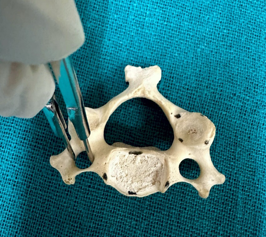
The foramen transversarium differentiates the cervical vertebrae from the other vertebrae of the spinal column. From the first part of the subclavian artery, the vertebral artery arises and passes through the foramen transversarium. FT protects the vertebral artery. The medial boundary of the luschka joint is a safe sight to protect the vertebral artery. The Uncovertebral Joint, also known as the Luschka Joint, is a joint that was formed by the Uncinate Process between the cervical vertebra bodies. These joints form the medial border of an intervertebral foramen in the cervical area below C-2 and are rarely found on the first thoracic vertebra. The present study was carried out on 100 dry human typical cervical vertebrae of unknown age and sex to determine the morphometric dimensions of FT and Uncinate Process in typical cervical vertebrae. The dimensions were taken by the digital vernier caliper on the superior aspect of the FT and Uncinate Process. The length and width of the FT were more on the left side than on the right side which was statistically insignificant (p-value >0.05). The narrow FTs were also observed in the present study. The mean difference between the Right and Left sides of the Uncinate Process was statistically not significant (p-value>0.05).
Introduction
The presence of Foramen Transversarium (FT) in the transverse process in cervical vertebrae is distinguished from other vertebrae in the vertebral column [1]. FT develops from the vestigial costal part along with the body and the true transverse process of the vertebrae [2, 3]. The Vertebral artery, vertebral vein, and sympathetic nerves [4] pass through the FT except for the C7 through which only the vertebral vein passes [5]. The vertebral artery that passes through FT is protected on all the sides [6, 7] and develops from the 32nd to 40th day of intrauterine life. The longitudinal anastomosis of the cervical intersegmental arteries and branches of primitive dorsal aorta form the vertebral artery [8]. The vertebral artery provides posterior circulation to the brain [2]. The vertebral artery is related to the medial aspect of the Luschka joint. The Luschka joint is formed by the bevelled lips projecting upwards on the dorsolateral aspect of the cervical vertebral bodies i.e. Uncinate Process [9, 10]. This articulation creates the medial margin of the Intervertebral Foramen for the emergence of Spinal nerves which later pass through the FT [11]. If there is a narrowing of the FT, the compression on the vertebral and basilar arteries occurs due to the annoyance of sympathetic nerves and it may cause labyrinthine disorders with neurological symptoms [12]. Due to this, the size of the FT and the Uncinate Process are important aspects. The variation in its size may lead to migraine, headache, and fainting attacks due to compression on the vertebral artery [13, 14].
Osteophytes projecting from the Uncinate Process constrict the intervertebral foramina, which is the primary cause of cervical spondylotic radiculopathy and myelopathy [15]. Comparing the cervical Uncinate Process morphometric measures during anterior cervical spine surgery lowers the possibility of damage to neurovascular structures and aids in achieving more effective neural element decompression. As a result, it will assist neurosurgeons in defining the limits of the Uncinate Process and permitting sufficient decompression of neural elements during anterior spinal decompression with a lower risk of neurovascular structures [12].
Therefore, the information about the Formen Transversarium and Uncinate Process is helpful for orthopedics and neurosurgeons during the approaching of cervical spines and for the radiologist while interpreting X-rays, MRI, and CT scans.
Material and Method
The study was conducted on 100 dry human Typical Cervical Vertebrae of unknown age and sex. Decalcified and damaged bones were excluded. The morphometric measurements were taken by using a digital vernier caliper. Dimensions were measured directly from the typical cervical vertebrae. Anteroposterior and transverse diameters of FT were measured. Length and Height of the UP were measured. The mean difference between the Right and Left sides of the FT and UP was statistically insignificant because its P-value was not less than 0.05. Pearson’s chi-square test was also used.
Result
Foramen Transversarium
The width and length of the right and left FT of typical cervical vertebrae were observed with their standard deviations. The mean and standard deviation of the length on the right and left sides were 5.36± 0.81mm and 5.57± 0.67mm, respectively. The mean and standard deviation of width on the right and left sides were 6.37± 1.06mm and 6.44± 0.85mm, respectively. The mean value was lowered on the right side of the FT.
Uncinate Process
The mean and standard deviation of the length of the Uncinate Process was 11.15±1.83mm and 11.10±1.74mm on the right and left sides. The mean on the left side was slightly lower than on the right side. The mean height of the with their standard deviation was 4.86±1.27mm and 4.87±1.21mm on the right and left side of the uncinate process. The mean on the left side was slightly more than the right side.
| Vertebrae | Dimensions on the Right Side | |
|---|---|---|
| Length Mean (mm)& S.D | Width Mean (mm)& S.D | |
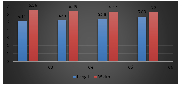
| C3 | 5.11±.501 | 6.56±.635 |
| C4 | 5.25±.438 | 6.39±.775 |
| C5 | 5.38±.806 | 6.32±1.02 |
| C6 | 5.69±1.20 | 6.20±1.60 |
Table 1: Shows the Length and Width on the Right side of the Foramen Transversarium in Typical Cervical Vertebrae.
| Vertebrae | Dimensions on the Left side | |
|---|---|---|
| Length Mean (mm) & S.D | Width Mean (mm) & S.D | |
| C3 | 5.35±.443 | 6.71±.515 |
| C4 | 5.38±.465 | 6.52±.647 |
| C5 | 5.75±.566 | 6.53±.797 |
| C6 | 5.78±.996 | 6.00±1.18 |
| Vertebrae | Uncinate Process | |
| Length Mean (mm)±S.D | Height Mean (mm)± S.D | |
| C3 | 10.64±1.64 | 4.27±1.17 |
| C4 | 11.24±1.53 | 4.88±1.13 |
| C5 | 11.15±1.57 | 4.90±1.17 |
| C6 | 11.56±2.42 | 5.39±1.41 |
Table 2: Shows the Length and Height on the Right side of the Uncinate Process in Typical Cervical Vertebrae.
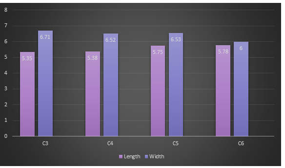
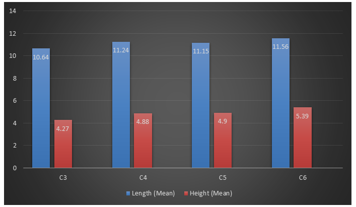
Figure 3a: Length of the FT in Typical Cervical Vertebrae.
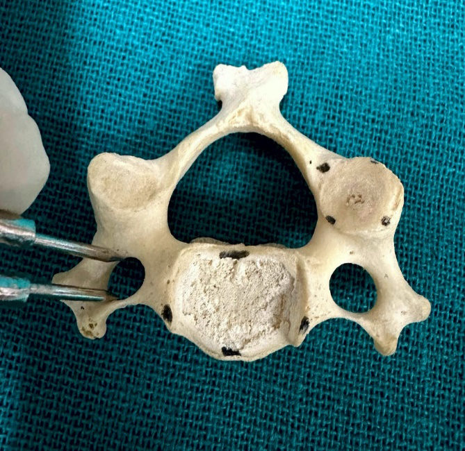
Figure 3b: Width of the FT in Typical Cervical Vertebrae.
| Vertebrae | Uncinate Process | |
|---|---|---|
| Length Mean (mm)±S.D | Height Mean (mm)± S.D | |
| C3 | 10.50±1.36 | 4.33±1.20 |
| C4 | 11.31±1.34 | 4.97±1.16 |
| C5 | 11.10±1.70 | 4.85±1.20 |
| C6 | 11.49±2.33 | 5.32±1.14 |
Table 3: Shows the Length and Height on the Left side of the Uncinate Process in Typical Cervical Vertebrae.
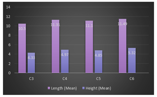
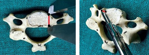
Figure 6a: Shows the Length of the uncinate process of Typical Cervical Vertebrae.
Figure 6b: Shows the Height of the Uncinate Process of the Typical Cervical Vertebrae.
| C3 R(FTL) | C3 L(FTL) | C4 R(FTL) | C4 L(FTL) | C5 R(FTL) | C5 L(FTL) | C6 R(FTL) | C6 L(FTL) | ||
|---|---|---|---|---|---|---|---|---|---|
| C3 R(FTL) | PC | 1 | 0.362 | -0.229 | -0.182 | .582** | .428* | .506** | 0.271 |
| Sig 2 tailed | 0.75 | 0.271 | 0.384 | 0.002 | 0.033 | 0.01 | 0.191 | ||
| N | 25 | 25 | 25 | 25 | 25 | 25 | 25 | 25 | |
| C3 L(FTL) | PC | 0.362 | 1 | 0.101 | .411* | 0.121 | .438* | .555* | 0.361 |
| Sig 2 tailed | 0.075 | 0.632 | 0.041 | 0.565 | 0.029 | 0.004 | 0.077 | ||
| N | 25 | 25 | 25 | 25 | 25 | 25 | 25 | 25 | |
| C4 R(FTL) | PC | .509** | 0.101 | 1 | 0.229 | 0.338 | -0.1 | 0.324 | 0.385 |
| Sig.2 tailed | 0.009 | 0.632 | 0.271 | 0.099 | 0.636 | 0.115 | 0.057 | ||
| N | 25 | 25 | 25 | 25 | 25 | 25 | 25 | 25 | |
| C4 L(FTL) | PC | -0.024 | .411* | 0.229 | 1 | 0.15 | 0.311 | 0.282 | .397* |
| Sig 2 tailed | 0.909 | 0.041 | 0.271 | 0.475 | 0.131 | 0.172 | 0.049 | ||
| N | 25 | 25 | 25 | 25 | 25 | 25 | 25 | 25 | |
| C5 R(FTL) | PC | .582** | 0.121 | 0.338 | 0.15 | 1 | .575** | .399* | 0.184 |
| Sig. 2-tailed | 0.002 | 0.565 | 0.099 | 0.475 | 0.003 | 0.048 | 0.378 | ||
| N | 25 | 25 | 25 | 25 | 25 | 25 | 25 | 25 | |
| C5 L(FTL) | PC | .428* | .0438* | -0.1 | 0.311 | .575** | 1 | .400* | 0.228 |
| Sig. 2-tailed | 0.033 | 0.029 | 0.636 | 0.131 | 0.003 | 0.047 | 0.273 | ||
| N | 25 | 25 | 25 | 25 | 25 | 25 | 25 | 25 | |
| C6R(FTL) | PC | .506** | .555** | 0.324 | 0.282 | .399* | .400* | 1 | 0.372 |
| Sig 2 tailed | 0.01 | 0.004 | 0.115 | 0.172 | 0.048 | 0.047 | 0.067 | ||
| N | 25 | 25 | 25 | 25 | 25 | 25 | 25 | 25 | |
| C6 L(FTL) | PC | 0.271 | 0.361 | 0.385 | .397* | 0.184 | 0.228 | 0.372 | 1 |
| Sig 2 tailed | 0.191 | 0.077 | 0.057 | 0.049 | 0.378 | 0.273 | 0.067 | ||
| N | 25 | 25 | 25 | 25 | 25 | 25 | 25 | 25 | |
| C3 R(FTW) | C3 L(FTW) | C4 R(FTW) | C4 L(FTW) | C5 R(FTW) | C5 L(FTW) | C6 R(FTW) | C6 L(FTW) | ||
| C3 R(FTW) | PC | 1 | 0.078 | .461* | 0.217 | .502* | 0.075 | .468* | 0.351 |
| Sig 2 tailed | 0.71 | 0.02 | 0.299 | 0.011 | 0.722 | 0.018 | 0.086 | ||
| N | 25 | 25 | 25 | 25 | 25 | 25 | 25 | 25 | |
| C3 L(FTW) | PC | 0.078 | 1 | 0.346 | .464* | -0.137 | 0.289 | 0.164 | .398* |
| Sig 2 tailed | 0.71 | 0.09 | 0.02 | 0.514 | 0.212 | 0.433 | 0.049 | ||
| N | 25 | 25 | 25 | 25 | 25 | 25 | 25 | 25 | |
| C4 R(FTW) | PC | .461* | 0.346 | 1 | 0.247 | .437* | -0.256 | .402* | 0.251 |
| Sig.2 tailed | 0.02 | 0.09 | 0.235 | 0.029 | 0.217 | 0.047 | 0.225 | ||
| N | 25 | 25 | 25 | 25 | 25 | 25 | 25 | 25 | |
| C4 L(FTW) | PC | 0.217 | .464* | 0.247 | 1 | -0.06 | 0.241 | 0.085 | 0.299 |
| Sig 2 tailed | 0.299 | 0.02 | 0.235 | 0.776 | 0.246 | 0.687 | 0.147 | ||
| N | 25 | 25 | 25 | 25 | 25 | 25 | 25 | 25 | |
| C5 R(FTW) | PC | .502* | -0.137 | .437* | -0.06 | 1 | 0.065 | .504* | 0.205 |
| Sig. 2-tailed | 0.011 | 0.514 | 0.029 | 0.776 | 0.758 | 0.01 | 0.326 | ||
| N | 25 | 25 | 25 | 25 | 25 | 25 | 25 | 25 | |
| C5 L(FTW) | PC | 0.075 | 0.259 | -0.256 | 0.241 | 0.065 | 1 | 0.248 | 0.123 |
| Sig. 2-tailed | 0.722 | 0.212 | 0.217 | 0.246 | 0.758 | 0.232 | 0.531 | ||
| N | 25 | 25 | 25 | 25 | 25 | 25 | 25 | 25 | |
| C6R(FTW) | PC | .468* | 0.164 | .402* | 0.085 | .504* | 0.248 | 1 | .451* |
| Sig 2 tailed | 0.018 | 0.433 | 0.047 | 0.687 | 0.01 | 0.232 | 0.024 | ||
| N | 25 | 25 | 25 | 25 | 25 | 25 | 25 | 25 | |
| C6 L(FTW) | PC | 0.351 | .398* | 0.251 | 0.299 | 0.205 | 0.132 | .451* | 1 |
| Sig 2 tailed | 0.086 | 0.049 | 0.225 | 0.147 | 0.326 | 0.531 | 0.024 | ||
| N | 25 | 25 | 25 | 25 | 25 | 25 | 25 | 25 |
Table 4: Correlation of the Length of Foramen Transversarium of Typical Cervical Vertebrae. ** Correlation is significant at the
Discussion
Taitz, et al. [16] carried out a study of the FT of 35 cervical vertebrae on its length, and width. The mean and standard deviation of FT Length on the right and the left side were 6.22±0.66mm and 0.68±0.77mm and ranged from 4.6-7.7mm and 5.5-8.8mm, respectively. Yesender, et al. [2] conducted a study on the length and width of the 50 cervical vertebrae. The mean and standard deviation of the length on the right side was 4.88±0.70mm and on the left side was 5.36±0.89mm. The mean & and standard deviation of the width of the right side was 4.80±0.68 and on the left side was 5.53±0.80mm. Sangari, et al. [17], carried out a study on 71 dried cervical vertebrae and observed that the mean width on the right and left sides were 5.60±1.04mm & 5.87±0.89mm and the length was 5.17±0.89mm and 5.13±0.79mm on the right and left sides, and the mean difference was statistically insignificant. Abdul, et al. [18] carried out a study on 82 typical cervical vertebrae. They noted the mean and standard deviation of length on the right side was 4.34±1.63mm and on the left side was 4.60±1.67mm. The mean and standard deviation of the width on the right side was 5.03±1.97mm and on the left side was 5.43±2.00mm.In the present study, the length is the same as compared with other studies, and the width was more than in the study conducted by the other workers. In the study of Polat, et al. [19], they observed 96 cervical vertebrae and the mean AP diameter of FT on the right and left sides was 4.23 and 4.28 mm respectively and the transverse diameter of FT was 4.78mm and 4.95 mm on the right, and left side, respectively. Pramella, et al. [20]
conducted a study on 100 dry cervical vertebrae, the Length of FT on the right side was 4.93 ± 0.82mm, and on the left side was 4.89 ± 0.89mm, and the width of FT on the right and the left side was 5.67 ± 1.03 mm and 5.64 ± 0.89mm respectively.
| Worker | N | Side | FT Length | FT Width | ||
|---|---|---|---|---|---|---|
| Mean (mm) & S.D | Range | Mean (mm) & S.D | Range | |||
| Taitz, et al. [16] | 35 | R | 6.22±0.66 | 4.6-7.7 | 4.89±0.49 | 3.7-5.9 |
| 36 | L | 0.68±0.77 | 5.5-8.8 | 5.15±0.51 | 4.1-6.0 | |
| Yesender, et al. [2] | 50 | R | 4.88 ± 0.70 | - | 4.80 ± 0.68 | - |
| L | 5.36 ± 0.89 | - | 5.53 ± 0.80 | - | ||
| Sangari, et al. [17] | 71 | R | 5.17 ± 0.89 | 2.19-7.21 | 5.69 ± 1.04 | 2.00–8.65 |
| L | 5.13 ± 0.79 | 2.51–6.81 | 5.87 ± 0.89 | 2.62–7.89 | ||
| Abdul, et al. [18] | 82 Typical | R | 4.34 ±1.63 | - | 5.03 ±1.97 | - |
| L | 4.60 ±1.67 | - | 5.43 ±2.00 | - | ||
| Polat, et al. [19] | 96 | R | 4.23 | 1.00-7.00 | 4.78 | 1.90-9.00 |
| L | 4.28 | 1.50-8.00 | 4.95 | 2.00-9.00 | ||
| Pramella, et al. [20] | 200 | R | 4.93 ± 0.82 | - | 5.67 ± 1.03 | - |
| L | 4.89 ± 0.89 | - | 5.64 ± 0.89 | - | ||
| Present, study | 100 | R | 5.36±0.81 | 2.97-7.86 | 6.37± 1.06 | 2.02-10.15 |
| L | 5.57±0.67 | 2.75-7.46 | 6.44±0.85 | 3.16-9.00 |
Table 5: Shows the comparison of the mean, Standard Deviation, and Range of Foramen Transversaium Length and width in typical cer
Table 7: Shows the comparison of the mean, Standard Deviation, and Range of Foramen Transversaium Length and width in typical cervical vertebrae. *N: Number, R: Right, L: Left Two significantly constricted FTs were detected in the current study.[Figures 7(a) and (b)], its Length on the right side was 2.97mm, & on the left side was 2.75mm and the width was 2.02mm on the right side and the left side was 3.16mm.
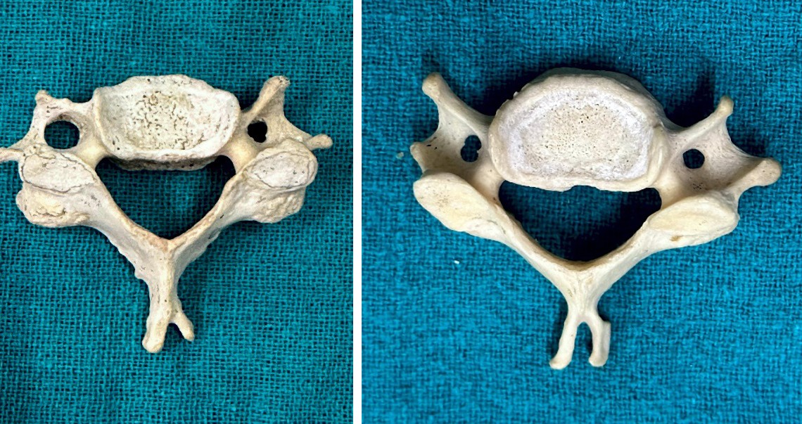
Figures 7(a) and (b): Showing the narrow FT in the Typical Cervical Vertebrae.
The narrowing of FT may cause vertebrobasilar insufficiency during head movement and may lead to thrombus formation [21]. Extreme rotatory movements in the cervical spine may lead to vertebral artery compression and impaired blood flow [22]. Occlusion of the vertebral artery causes Bow hunter’s stroke [17] (compression of the vertebral artery due to hypertrophic osteophytes, mainly at Luschka joints) [23, 24] and it carries more chances of severe transient ischemic attacks due to more risk of formation of thrombus. During anterior cervical disc surgery, the safe sight for the vertebral artery is the medial margin of the Luschka joint [25, 26].
| Worker | N | Side | Uncinate Process Length (Mean±S.D) | |||
|---|---|---|---|---|---|---|
| C3(mm) | C4(mm) | C5(mm) | C6(mm) | |||
| Tubbs, et al. [10] | 40 | R/L | 8.1 | 7.5 | 7.1 | 7.2 |
| Raveendranath, et al. [8] | 80 | R | 9.93±1.41 | 10.39±1.54 | 10.99±2.25 | 12.00±2.84 |
| L | 10.21±1.25 | 10.95±1.66 | 11.32±2.63 | 12.50±2.86 | ||
| Present Study | 100 | R | 10.64±1.64 | 11.24±1.53 | 11.15±1.57 | 11.56±2.42 |
| L | 10.50±1.36 | 11.31±1.34 | 11.10±1.70 | 11.49±2.33 |
Table 6: Shows the comparison of the Uncinate Process Length in the Typical Cervical Vertebrae. *N: Number, R: Right, L: Left
| Worker | N | Side | Uncinate Process Height (Mean±S.D) | |||
|---|---|---|---|---|---|---|
| C3(mm) | C4(mm) | C5(mm) | C6(mm) | |||
| Tubbs, et al. [10] | 40 | R/L | 4.8 | 5 | 4.9 | 5.1 |
| Raveendranath, et al. [8] | 80 | R | 4.34±1.04 | 4.55±1.28 | 4.79±2.02 | 5.31±1.30 |
| L | 3.82±0.85 | 4.19±1.09 | 4.44±1.38 | 5.38±1.18 | ||
| Present Study | 100 | R | 4.27±1.17 | 4.88±1.13 | 4.90±1.17 | 5.39±1.41 |
| L | 4.33±1.20 | 4.97±1.16 | 4.85±1.20 | 5.32±1.14 |
Table 7: Shows the comparison of the Uncinate Process Height in the Typical Cervical Vertebrae. *N: Number, R: Right, L: Left In
Table 9: Shows the comparison of the Uncinate Process Height in the Typical Cervical Vertebrae. *N: Number, R: Right, L: Left In the present study, the mean of the Uncinate Process Length and Height was almost the same as compared with the study of Raveendranath et al. [9] and Tubbs et al. [11]. The statistical data was found insignificant on the Right and Left sides of the Uncinate Process of the Typical Cervical Vertebrae.
Conclusion
FT gets narrowed due to the presence of osteophytes, leading to vertebral artery compression and damage [27, 28]. The morphometric parameters of FT help neurosurgeons protect the vertebral artery while decompressing the vertebral artery through the anterior cervical approach. The medial boundary of the Luschka joint is a safe sight to protect the vertebral artery through the ventral approach to the cervical spine [29]. Uncinectomy is done to cure the narrowing of the vertebral artery or cervical nerve irritation. During surgery of the nerve root decompression, a window is made depending on the dimensions of the uncinate process [9, 15]. The space between the dorsal part of the uncinate process and the lamina facet junction presence of the Intervertebral Foramen [30]. If the Intervertebral foramen gets narrowed due to osteophytes from the uncinate process, it may lead to cervical spondylotic myelopathy, spondylosis, and radiculopathy [31, 32]. To remove osteophytes or a posterolateral disc herniation during surgery, it is crucial to understand the boundaries of the uncinate processes in the cervical spine. Furthermore, a poor surgical outcome has been linked to incomplete removal of osteophytes from the uncinate process [11].
Therefore, the knowledge regarding the morphometric measurements and the structures i.e., vessels and nerves related to the Foramen Transversarium and Uncinate Process, helps to avoid the risk of poor cervical surgery.
References
-
William PL, Soames RW (1995) Axial skeleton Gray’s Anatomy. 38th(Edn.), Churchill Livingstone, Philadelphia, pp: 516.
-
Yesender M, Devadas P, Saritha S, Vinila BHS (2017) Study on the anatomical variations and morphometry of Foramen Transversaria of the subaxial cervical vertebrae. Int J Anat Res 5(2.1): 3708-3712.
-
Standring S (2008) The Back in Gray’s Anatomy: the anatomical basis of clinical practices. In: Standring S, et al. (Eds.), Gray’s Anatomy. 40th(Edn.), Elsevier, Churchill Livingstone, New York, USA, pp: 718-719.
-
William M, Newell RLM, Collin P (2005) The back: cervical vertebrae. In: Standring S, et al. (Eds.), Gray’s Anatomy. 39th(Edn.), Elsevier Churchill Livingstone, Edinburg, London, pp:742-746.
-
Das S, Suri R, Kapur V (2005) Double foramen transversaria: an osteological study with clinical implications. Int Med J 12(4): 311-313.
-
Sabnis A (2015) Anatomical variations in foramen transversarium. Indian Journal of applied research 5(12): 504-506.
-
Sultana Q, Avadhani R, Varalakshmi KL, Sharif MH, Blessina (2015) Variations in foramen transversarium in atlas vertebrae: a morphological study with clinical significance. NUJHS 2(2): 80-83.
-
Hamilton WJ, Boyd JD, Mossman HW (1972) Human Embryology. 4th(Edn.), W. Heffer & Sons Ltd, The Williams & Wilkins Company, Baltimore, Cambridge, USA, pp: 270.
-
Raveendranath V, Kavitha T, Umamageswari A (2019) Morphometry of the Uncinate Process, Vertebral Body, and Lamina of the C3-7 Vertebrae Relevant to Cervical Spine Surgery.Neurospine 16(4): 748-755.
-
Durga P, Dakshayani KR (2020) Morphometric Study of Uncinate Process of Cervical Vertebra and Its Surgical Importance. National Journal of Clinical Anatomy 9(2): 59-62.
-
Tubbs RS, Rompala OJ, Verma K, Mortazavi MM, Benninger B, et al. (2012) Analysis of the uncinate processes of the cervical spine: an anatomical study.J Neurosurg Spine 16(4): 402-407.
-
Akhtar MJ, Madhukar PK, Rahman S, Kashyap N (2015) A morphometric study of foramen transversarium of dried cervical vertebrae. Int J Res Med Sci 3(4): 912-916.
-
Yadav Y, Goswami P, Bharihoke V (2014) An Osteological study of Foramen Trasversarium: Variations and Clinical Aspects. Journal of Evolution of Med and Dent Sci 3(68): 14562-14566.
-
Kaya S, Yilmaz Nd, Pusat S, Kural C, Kirik A, et al.(2011) Double foramen transversarium variation in ancient byzantine cervical vertebrae: preliminary report of an anthropological study. Turk Neurosurg 21(4): 534-538.
-
Lu J, Ebraheim NA, Yang H, Skie M, Yeasting RA(1998) Cervical uncinate process: an anatomic study for anterior decompression of the cervical spine. Surg Radiol Anat 20(4): 249-252.
-
Taitz C, Nathan H, Arensburg B (1978) Anatomical observations of the foramina transversarium. J Neurology, Neurosurgery and Psychiatry 41(2): 170- 176.
-
Sangari SK, Dossous PM, Heineman T, Mtui EP (2015) Dimensions and anatomical variants of the foramen transversarium of typical cervical vertebrae. Anat Res Int: 391823.
-
Abdul RS, Lazarus L, Rennie C, Satyapal KS (2018) The foramen Transversarium of Typical and atypical cervical vertebrae: Morphology and Morphometry. Int J Morpho 36(4): 1439-1446.
-
Polat S, Goker P, Yucel AH, Bozkir MG (2019) Morphometric study of dry cervical vertebrae. Int J Morphol 37(3): 845-851.
-
Pramella MD, Prabhu LV, Murlimanju BV, Pai MM, Rai R, et al. (2020) Anatomical dimensions of the typical cervical vertebrae and their clinical implications. Eur J Anat 24(1).
-
Cagnie B, Barbaix E, Vinck E, D’herde K, Cambier D (2005) Extrinsic risk factors for compromised blood flow in the vertebral artery: anatomical observations of the transverse foramina from C3 to C7. Surg RadiolAnat 27(4): 312-316.
-
Lalit M, Kullar JS, Piplani S, Kullar G, Sharma T (2015) Anatomical observation including morphometric pattern of foramina transversaria of atlas vertebrae in North Indians. Eur J Ant 19(3): 249-255.
-
Citow JS, Macdonald RM (1999) Posterior decompression of the vertebral artery narrowed by cervical stenosis. Surgical Neurology 51(5): 495-499.
-
Seki T, Hida K, Akino M, Iwasaki Y (2001) Anterior decompression of the atlantoaxial vertebral artery to treat bow hunter’s stroke: technical case report. Neurosurgery 49(6): 1474-1476.
-
Ionete C, Omojola MF (2006) MR angiographic demonstration of bilateral duplication of the extracranial vertebral artery: unusual course and review of the literature, American Journal of Neuroradiology 27(6): 1304-1306.
-
Malik SW, Stemper BD, Metkar U, Yoganandan N, Shender BS, et al. (2010)Location of the transverse foramen in the subaxial cervical spine in a young asymptomatic population. Spine 35(12): E514-E519.
-
Burle VS, Panjwani A, Mandalaneni K, Kollu S, Gorantla VR (2022) Vertebral Artery Stenosis: A Narrative Review. Cureus 14(8).
-
Montano M, Alman K, Smith MJ, Boghosian G, Enochs WS (2021)Bow hunter’s syndrome: a rare cause of vertebrobasilar insufficiency. Radiol Case Rep 16: 867- 870.
-
Behera S, Sar M, Mishra SK (2023) Morphological and Morphometric Variations in Foramen Transversarium: A Cross-sectional Study in Dried Cervical Vertebrae of Western Odisha Origin. J Clin of Diagn Res 17(6): AC10- AC14
-
Lee BH, Park JH, Lee JY, Jeon HJ, Park SW (2021) Efficiency of minimal oblique resection of the uncinate process during an anterior cervical discectomy and fusion. Medicine (Baltimore) 100(31).
-
Durga P, Dakshayani R (2020) Morphometric Study of Uncinate Process of Cervical Vertebra and Its Surgical Importance. National Journal of Clinical Anatomy 9(2): 59-62.
-
Hartman J (2014) Anatomy and clinical significance of the uncinate process and uncovertebral joint: a comprehensive review.Clin Anat 27: 431-440.
- Pattern of Breast Lesions in Ovu Inland, Delta State, South Southern Nigeria
- Morphometric Analysis of the Human Femur: Exploring Platymetric and Robusticity Indices Among the Nigerian Population
- Anatomical Variation of Arteria Lusoria: Clinical Implications for Dysphagia Lusoria and Surgical Risk
- Morphometric Study of the Vertebral Body and Pedicle of Typical Cervical Vertebrae Using Radiological Image
- Epigenetic Mechanisms Driving Human Evolutionary Changes
- Neuroprotective Effects of Ginkgo Biloba Extract on Bilateral Common Carotid Artery Ischaemic Stroke Induced in Wistar Rat