Uncommon Association: Primary Malignant Melanoma of the Eyelid and Palpebral Conjunctiva
Ocular melanoma of the eyelid and conjunctival is extremely rare primary ocular malignant tumor in adults. The simultaneous occurrence of both lesions in one eye are uncommonly seen in practice. We report a case of ocular melanoma of the eyelid and conjunctival developed on one eye of a 71-year-old male patient that the reported had grown in size over the last four years.
Introduction
Malignant melanoma of the palpebral conjunctiva and eyelid is a rare disease that is unilateral, can be multicentric, and is associated with a poor prognosis, but its incidence is increasing. It mostly occurs among white adults. In majority of cases it originates from preceding primary acquired melanosis. They remains a therapeutic challenge for ophthalmologists and oncologists. The objectif is To describe the most salient clinical features, and treatment modalities of intraocular melanoma, as well as the novel therapies currently being tested.
Case Report
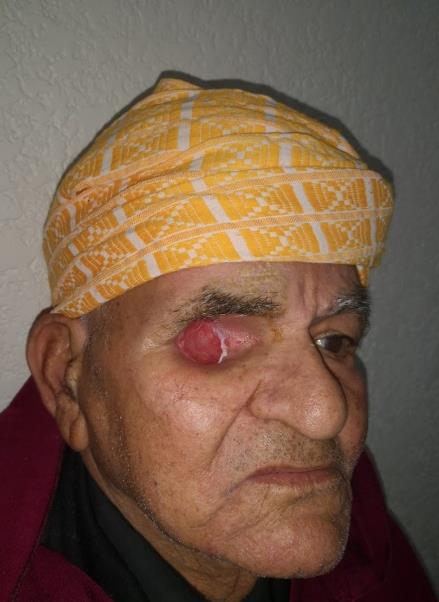
A 70-year-old man developed a slowly enlarging pigmented lesion along the right lower palpebral Uncommon Association: Primary Malignant Melanoma of the Eyelid and Palpebral Conjunctiva conjunctiva and eyelid. Examination showed a pigmented tumor extended to the orbit (Figure 1), the dermoscopic evaluation of the lesion revealed the presence of dermoscopic features typical of melanoma. Biopsy revealed a malignant melanoma in palpebral and conjunctiva. The computed tomography findings were a right eyelid mass with mediastinal lymph nodes. The imaging was completed by an orbital MRI that showed the deep confines of the eyelid mass encompassing the lacrymal gland, the superior and lateral rectus, invading the sclera and the cornea with a retracted anterior chamber (Figure 2). We performed an extended right orbital exenteration, the cavity was left for spontaneous re-epithelization under a “tulle-gras” and gauze dressing (Figure 3). Patient was then referred to oncology for adjuvant chemotherapy.


Discussion
The malignant melanomas of the palpebral conjunctiva although it is the second most common type of melanoma, ocular melanoma is still really rare, rarely occurs in African-American patients [1, 2]. Incidence of conjunctival melanoma is increasing with age [3]. Conjunctival melanomas arise from melanocytes located in the basal layer of the epithelium of the conjunctival membrane, unlike the other mucous membranes is directly exposed to sun radiation. It may arise apparently spontaneously or developing from a benign naevus or from primary acquired melanosis (10-74%) [3, 4]. Malignant melanoma of the eyelid is uncommon, it may arise de novo, from a preexisting naevus. The simultaneous occurrence of both lesions in one eye is really rare. The significance of the relationship between primary conjunctival melanoma and primary eyelid melanoma is not completely clear. Clinically malignant melanomas of the palpebral conjunctiva is manifested by a pigmented tumor raised, lobulated or nodular. Sometimes it has a freely mobile base with the conjunctiva, before it becomes fixed. Occasionally, it is pedunculated as was in this case. However, there are cases of amelanotic tumors that may confuse the clinical picture and delay diagnosis [5, 6]. Although it can appear on any part of conjunctiva, it is most common on bulbar conjunctiva but other locations including palpebral are less common but associated with less favorable prognosis [7]. The Histopathology confirms the diagnosis; reveals epithelioid Cells: The most common type, are large polyhedral cells with round or oval nuclei, prominent nucleoli, and an abundant cytoplasm usually containing fine melanin granules, oc casionally they become multinu clear to form giant cells, Spindle Cells: Large or small fusiform cells with oval nuclei and prolonged at the ends to form fibers, or naevoid Cells: Intermediate in size between benign naevus cells, with round, hyperchromatic nuclei. Current standard treatment for conjunctival melanoma is wide local excision with adjuvant therapy, including brachytherapy, cryotherapy and topical application of chemotherapeutic agent. When he fails to realize a complete and radical exeresis of the mass, or in case of recurrences, he may use a more destructive therapy (enucleation, orbital exenteration).
Conclusion
Compared with cutaneous melanomas, mucosal melanomas are relatively rare. The prognosis is poor; 5- year survival rates are 26.8%. Limited number of therapeutic options for its metastatic power and our context, hence the benefit of early diagnosis, preventive measures and improvement in local treatment and new treatment options in patients with metastatic disease.
References
-
McLaughlin CC, Wu XC, Jemal A, Martin HJ, Roche LM, et al. (2005) Incidence of noncutaneous melanomas in the USA. Cancer 103: 1000-1007.
-
Shields CL, Demirci H, Karatza E, Shields JA (2004) Clinical survey of 1643 melanocytic and nonmelanocytic conjunctival tumors. Ophthalmology 111: 1747-1754.
-
Barrie Jay (1964) Current Developments in Ophthalmology: A Follow-up Study of Limbal Melanomata. Proc Roy Soc Med 57(6): 497-500.
-
Barrie Jay (1963) Naevi & melanomata of the Conjunctiva Brit J. Ophthal 49: 169-204.
-
Jay V, Font RL (1998) Conjunctival amelanotic malignant melanoma arising in primary acquired melanosis sine pigmento. Ophthalmology 105(1): 191-194.
-
Paridaens AD, McCartney AC, Hungerford JL (1992) Multifocal amelanotic conjunctival melanoma and acquired melanosis sine pigmento. Br J Ophthalmol 76(3): 163-165.
-
Shields CL, Shields JA (2009) Ocular melanoma: relatively rare but requiring respect. Clin Dermatol 27(1): 122-33.
- Cancer Diagnosis from RNA Sequence of Blood Cells by Using AI
- Field Cancerization in Oral Cavity, Case Report and Review of Literature. Oncologic Program Salud Integral Hospital, Managua, Nicaragua
- Identification of B Lymphocytes in Cancer Patient’s Blood
- A Case Report of a Breast Cancer Patient Developing Pneumonitis as a Result of Abemaciclib Therapy
- Immune Checkpoint Therapeutics for Today’s Fight and Beyond
- The Amalgamated Sophomore-Gonadoblastoma