Prevalence of Human T-Lymphotropic Virus Type 1 (HTLV-1) Infection among Patients with Hematologic Disorders-Serology and Molecular Based Cross- Sectional Study in Khartoum, Sudan
The human T-cell lymphotropic virus types-1 (HTLV -1) is well-established risk for the developing Hematological malignancies including adult T cell leukemia lymphoma (ATLL). The aim of this study is to evaluate the prevalence of HTLV-1 infection in patients with hematologic malignancies in Khartoum state by using serological and molecular approaches. During the period from April to September 2019 a total of 106 blood samples were obtained from Hematological Malignancy patients including AML, CML, CLL, ALL, MDS and ATLL. The presence of malignancy was confirmed by using IHC, Molecular and cytogenetic studies. Both males and females were included. A volume of 6 ml blood sample was collected from each patient and tested for the presence of anti-HTLV-1 antibodies using enzyme-linked immunosorbent assay (ELISA). Those with positive serology test were confirmed by detection of Proviral gene by using PCR. Data were analyzed by using spss version 19. The study participants were including 72 (67.9%) males and 34 (32.9%) females. The average age of the study population was 41± 12.4. The result of serology test reveals that out of 106 serum samples examined 7 (6.6%) were positive for HTLV-1 antibody test including 5 males and 2 females. All positive cases were confirmed by PCR and identified with the proviral pX-S gene. Of the HTLV-1 positive cases, three were diagnosed with CML, two were diagnosed with AML in addition to one male patient was diagnosed with ATLL as well as another one male patient diagnosed with CLL. In conclusion, our results show a similar prevalence of HTLV-1 infection in patients with hematologic disorders in comparison to other studies conducted on the African general population. Larger case control based studies with inclusion of more sample size are essential to corroborate the study evidence.
Introduction
The human T-lymphotropic viruses’ also known as human T-cell leukemia-lymphoma viruses (HTLV) are RNA group of viruses those belong to retroviruses family. To date, four types of HTLVs (HTLV-1, HTLV-2, HTLV-3, and HTLV-4) identified to cause human disease [1].
Based on the nucleotide diversity in the LTR region, HTLV-1 has seven subtypes (A-F) which has been implicated in several kinds of diseases, including tropical spastic paraparesis, and as a virus cancer link for leukemia (Adult Tcell leukemia lymphoma) [2, 3]. The virus is transmitted through blood contact (blood transfusion, injected drug abuse) in addition they can also transmitted through sexual contact with infected person or through breast feeding [4, 5].
The viral RNA is packed into the icosahedral capsid, which is contained inside the protein inner envelope. The
lipid outer envelope is of host cell origin but contains viral trans membrane and surface proteins. The virion is spherical in shape with a diameter of about 100 nm. This structure encloses the viral capsid) CA), which carries two identical strands of the genomic RNA as well as functional protease integrase, and reverse transcriptase enzymes [5]. The viral genome is dependent on cellular factors for the initial rounds of transcription. The complex retroviral genome codes for the structural proteins Gag (capsid, nucleocapsid, matrix), Protease, polymerase and Envelope from unspliced/singly spliced mRNAs and regulatory and accessory proteins from alternatively spliced mRNA transcripts [5, 6].
HTLV-1 is involved in actively spreading epidemics, affecting 15-20 million people worldwide [6]. It was the most clinically significant over all types of virus with at least 500,000 of the individuals infected with HTLV-1 eventually develop an often rapidly fatal leukemia, while others will develop a debilitative myelopathy, and yet others will experience uveitis, infectious dermatitis, or another inflammatory disorder. However, HTLV-2 is associated with milder neurologic disorders and chronic pulmonary infections. Moreover, there were no specific illnesses have yet been associated with HTLV-3 and HTLV-4 [6].
Up to 2014 the estimated adult seroprevalence of the HTLV-1 was at least 1–2% but it can also reach 20–40% among people aged over 50 years in specific clusters. The main highly endemic areas are the south-western part of Japan, some parts of the Caribbean and its surroundings regions. There are foci in South America, especially in parts of Colombia and French Guyana, some areas of intertropical Africa (such as south Gabon) and in the Middle East (such as the Mashhad region in Iran) and rare isolated clusters in Australia and Melanesia. In Europe, the only country with an endemic HTLV-1 region is Romania. Interestingly and despite different socio-economic and cultural environments, HTLV- 1 seroprevalence increases gradually with age, especially in women, in all the endemic areas. The general increase with age may be related to a cohort effect, as is well demonstrated in Japan, while the increase seen in older women might also be due to an accumulation of sexual exposures with age [7, 8, 9, 10, 11, 12]. While HTLV-2 subtypes are related to highly specific subpopulations and behaviour like injection drug use [13]. For many years studies have established the association of HTLV-1 infection with hematologic malignancies namely Adult T Leukemia lymphoma and lymphosarcoma T-cell leukemia. However, current evidence is quite scarce on the prevalence of HTLV-1 infection in other malignancies such as stomach cancer, colon cancer, breast cancer, and lung cancer in Iran [14, 15].
In Sudan, there was no data regarding the prevalence of the HTLV-1 among Hematological Malignancy patients.
Therefore, this study was the first of its type in our country that seeking to identify the prevalence of the HTLV-1 among Hematological Malignancy patients.
Materials and Methods
This study was cross-sectional–It was conducted in the Radio Isotope center of Khartoum state. During the period from April to September 2019 a total of 106 blood samples were obtained from Hematological Malignancy patients including AML, CML, CLL, ALL, Myelodysplastic syndrome and Adult T Leukemia Lymphoma patients. The presence of malignancy was confirmed by using IHC, Molecular and cytogenetic studies. Both males and females were included.
Specimen’s Collection and Processing
A volume of 6 ml blood was collected from each patient through venepuncture technique then displaced into EDITA anticoagulant containing container (for molecular studies) and plain container (to detect the presence of antibodies). The blood sample in the plain continer was centrifuged at 3000 g for 5 min, and then the serum was gently collected into Eppendorf tube and stored at –20°C until the serological analysis.
Specimens Analysis
Serologyical Detection of HTLV-1 Antibodies
The serum samples were analysed for the presence of anti - HTLV-1 by a commercially available enzyme-linked immune-sorbent assay (Human T-cell lymphotropic Virus antibody, HTLV-1 Ab ELISA Kit - Creative Diagnostics, Shirley, NY, US). The assay was performed following the instructions of the manufacturer. Positive and negative controls were included in each assay. According to the information included in the kit’s insert, the immunoassay used has a sensitivity and specificity of 100% and 99.95% respectively. Procedure of ELISA Test All reagents and specimens were settled to reach room temperature. 50 ul of HRP Conjugate was added to each well except the blank, and then 50 ul of a positive control; negative control and specimen were added to their respective wells. The plate was covered with plate cover and incubated for 60 minutes at 37°C. By the end of the incubation period, each well was washed 5 times with diluted wash buffer. Finally, 50 ul of chromogen A and chromogen B solutions were added to each well including blank, then the plate was incubated at 37°C for 15 minutes. At the end of the incubation 50 ul of stop solution was added to each well and the absorbance was measured at 450nm.
Quality Control and Calculation of the Results
Reagent, standard, and control were checked for storage, stability, and preparation before starting work. Each micro plate was considered separately when the results were calculated and interrelated; the results were calculated by relating each specimen absorbance (A) to the cut off (c.o.) of the plate.
C.O. value was calculated through the equation of (C.O.) =NC+0.18 where NC is mean of the three negative controls = (0.02+0.031+0.01)/3 +0.18=0.20
Interpretation of Results
Positive more than cut-off value. Negative less than cut-off value.
Molecular Analysis
Samples tested positive for HTLV-1 antibodies were confirmed by using molecular Technique.
DNA Extraction
Procedure
DNA extraction from blood sample: About 200 µl of each Whole blood sample was incubated in lysis buffer (20 µl proteinase K (200 mg/ml) and 5µl of RNAase A). DNA extraction was performed by the DNeasy® Blood Kit (QIAGEN Company) according to the manufacturer’s instructions and stored at -80 ºC until further analysis. The quality of DNA was checked using Nano-drop test.
Proviral pX-AS gene amplification: BK viral early gene region (Large T antigen region176bp) was amplified in a master mix reaction volume of 25μl containing 5 μl of DNA sample and specific forward and reverse primer 1µl for pX-S, 5′-CGGATACCCAGTCTACGTGTT-3′; pX-AS, 5′ CAGTAGGGCGTGACGATGTA-3′) [16] and completed to 25µl with 13 µl Distilled water. The amplification was done by using (TC-3000 conventional PCR Thermal Cycler, USA) through 45 cycles of denaturation at 94°C for 1 min proceeded by initial denaturation at 94°C for 5 min, annealing at 55°C for 1 min and extinction at 72°C for 2 min followed by a final extension at the same temperature for 5 min. Finally, the conventional PCR products were subjected to the gel electrophoresis (2% agarose gels) to visualize the band of the amplified target gene region with ethidium bromide (0.5 μg/mL) for 30 min in a UV-gel documentation system.
A method used for data collection: Administrated questionnaire was used to collect patient’s data including the gender and age and marital status.
Data Analysis
SPSS version 15 computerized programs analyzed the data, which were collected from questionnaire and laboratory analysis.
Result
A total of 106 patients were enrolled in the study. Of these, 72 (67.9%) were males and 34 (32.9%) were females. The average age of the study population was 41± 12.4 (ranging from 3 to 76) years (Table 1). The Hematological malignancies were AML 31 (29.2%), CML 37 (34.9%), CLL 9 (8.5%), ALL 17 (16.1%), Myelodysplastic syndrome (MDS) 8 (7.6%) and Adult T Leukemia Lymphoma (ATLL) 4 (3.7%). Most of patients were newly diagnosed 70 (66.0%) while the rest were received Cancer treatment 36 (34.0%). The history of blood transfusion was reported among 27.3% of patients and the rest were not receiving any cancer treatment (Table 1).
| Frequency | Percentage (%) | |
|---|---|---|
| Gender | ||
| Male | 72 | 67.9 |
| Female | 34 | 32.1 |
| The Hematological malignancies | ||
| AML | 31 | 29.2 |
| CML | 37 | 34.9 |
| CLL | 9 | 8.5 |
| ALL | 17 | 16.1 |
| MDS | 8 | 7.6 |
| Adult T LL | 4 | 3.7 |
| Cancer Treatment status | ||
| Newly diagnosed | 70 | 66 |
| Under treatment | 36 | 34 |
| History of Transfusion | ||
| Yes | 29 | 27.3 |
| No | 77 | 72.7 |
Table 1: Demographic and donor baseline characteristics for the study participants.
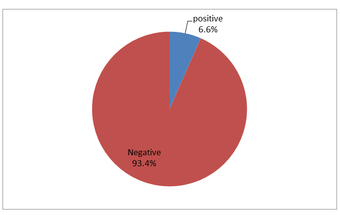
The result of serology test reveals that out of 106 serum samples examined 7 (6.6%) were positive for HTLV-1 antibody test including 5 males and 2 females. All positive cases were confirmed by PCR and identified with the proviral pX-S gene. Of the HTLV-1 positive cases, three were diagnosed with CML, two were diagnosed with AML in addition to one male patient was diagnosed with ATLL, and another one male patient diagnosed with CLL (Table 2) (Figure 1). The
result also showed that none of the HTLV-1 positive patients has history of Blood transfusion. Furthermore, among HTLV- 1 positive cases only one patient was known to be under treatment while the rest were newly diagnosed (Table 2).
| Gender | Age | Diagnosis | Transfusions history | Treatment history | |
|---|---|---|---|---|---|
| 1 | Male | 27 | AML | NO | NO |
| 2 | Male | 6 | CML | NO | YES |
| 3 | Male | 13 | CML | NO | NO |
| 4 | Male | 60 | CLL | NO | NO |
| 5 | Female | 21 | AML | NO | NO |
| 6 | Male | 57 | ATLL | NO | NO |
| 7 | Female | 9 | CML | NO | NO |
Table 2: Characteristics of HTLV-1 positive patients.
Discussion
The HTLV-1 infection through the oncogenic properties of it TAX and HBZ Proteins harbor greet risk for hematological malignancies including ATLL. In the present study, we demonstrated that the prevalence of HTLV-1 infection among patients with hematologic disorders is 7 (6.6%) out of 106 patients. CML was most frequently identified disorder among the study cases followed by AML and ATLL and CLL, none of the MPD patients was identified with virus. The high prevalence of the virus among CML and AML patients may reflect sampling bias since most of our study populations were diagnosed with one othe two-mentioned disorder. This will need further studies with inclusion of more cases affected with the other disorders to verify our result. Although we include only 4 cases with ATLL the prevalence of HTLV-1 among them was 25% and this may confirm the fact that HTLV-1 has an impact on the development of ATLL disorder. Furthermore, the current study demonstrates that the prevalence of HTLV-1 infection was significantly higher in males compared to females and there was only on patient with history of transfusion history. This may suggest that HTLV-1 was transmitted to those cases by route other than blood transfusion such as sexual contact.
Studies from other countries show variable results of HTLV-1 among hematological malignancy patients. The prevelance of the HTLV-1 infection was ranging from 18.2% and 23% in south Chile and japan respectively [17, 18] to 5.12% and 9.01% in Rio de Janeiro, Brazil and Lagos, Nigeria, Respectively [19, 20]. The closest prevalence to our finding is that of Nigeria. This was not surprising because Nigeria located in Africa and near geographically to our country and this may reflect that the geographical distribution of the virus may affect its ability to cause hematological malignancies in certain population. However, our study was differed from that in Nigeria in the sample size and the technique, which were used. We include 106 samples of different hematological malignancies and we confirm the result by detection of proviral gene instead they include only 39 samples of lymphoid malignancies and they did not confirm their result.
Conclusion
This was first study in sudan that high light the prevalence of HTLV-1 among hematological malignancies patients. In conclusion, our results show a similar prevalence of HTLV-1 infection in patients with hematologic disorders in comparison to other studies conducted on the African general population with the most affected group being a patient with CML disorder. We cannot build a clear association of HTLV-1 infection and hematologic disorders due to lacking of the control group. Therefore, larger case control based studies with inclusion of more sample size are essential to corroborate the evidence.
Ethical Approval
The Ethics Committee of Omdurman Islamic University approved the study.
Consent
Informed consent was obtained from all the study participants.
Conflict of Interests
Authors declare no competing financial interests.
Acknowledgments
The authors acknowledge the efforts of Radioisotope center of Khartoum state for availing their facilities and time for this study.
References
-
Poiesz BJ, Ruscetti FW, Gazdar AF, Bunn PA, Minna JD, et al. (1980) Detection and isolation of type C retrovirus particles from fresh and cultured lymphocytes of a patient with cutaneous T-cell lymphoma. Proc Natl Acad Sci USA 77(12): 7415-7419.
-
Mirvish ED, Pomerantz RG, Geskin LJ (2011) Infectious agents in cutaneous T-cell lymphoma. Journal of the American Academy of Dermatology 64(2): 423-431.
-
Roucoux DF, Wang B, Smith D, Nass CC, Smith J, et al. (2005) A Prospective Study of Sexual Transmission of Human T Lymphotropic Virus (HTLV)-I and HTLV-II. The Journal of Infectious Diseases 191(9): 1490-1497.
-
Coovadia HM, Rollins NC, Bland RM, Little K, Coutsoudis A, et al. (2007) Mother-to-child transmission of HIV- 1 infection during exclusive breastfeeding in the first 6 months of life: an intervention cohort study. Lancet 369(9567): 1107-1116.
-
de Thé G, Kazanji M (1996) An HTLV-I/II vaccine: from animal models to clinical trials? J Acquir Immune Defic Syndr Hum Retrovirol 13: S191-198.
-
Desrames A, Cassar O, Gout O, Hermine 0, Taylor GP, et al. (2014) Northern African strains of human T-lymphotropic virus type 1 arose from a recombination event. J Virol 88(17): 9782-9788.
-
Develoux M, Dupont A, Meynard D, Delaporte E (1996) A case of tropical spastic paraparesis associated with HTLV 1 in the Niger Republic. Med Trop (Mars) 56(1): 100-101.
-
Diop S, Calattini S, Abah-Dakou J, Thiam D, Diakhate L, et al. (2006) Seroprevalence and molecular epidemiology of human T-Cell leukemia virus type 1 (HTLV-1) and HTLV-2 in blood donors from Dakar, Senegal.J Clin Microbial 44(4): 1550-1554.
-
Dumas M, Houinato D, Verdier M, Zohoun T, Josse R, et al. (1991) Seroepidemiology of human T-cell lymphotropic virus type 1/II in Benin (West Africa). AIDS Res Hum Retroviruses 7(5): 447-451.
-
El-Farrash MA, Badr MF, Hawas SA, el-Nashar NM, Imai J, et al. (1988) Sporadic carriers of human T-lymphotropic virus type I in northern Egypt. Microbial Immunol 32(9): 981-984.
-
El-ghazzawi E, Hunsmann G, Schneider J (1987) Low prevalence of antibodies to HI V-1 and HTLV-I in Alexandria, Egypt. AIDS Forsch 2(11): 639.
-
Anyanwu NCJ, Ella EE, Ohwofasa A, Aminuet M (2018) Re-emergence of human T-lymphotropic viruses in West Africa. Braz J Infect Dis 22(3): 224-234.
-
Urwijitaroon Y, Barusrux S, Puapairoj C, Romphruk A, Khampeera P (1997)Seroepidemiology of HTLV-1 infection in northeast Thailand: four year surveillance. J Med Assoc Thai 80(1): Sl02-105.
-
Junki K, Ayako M, Keiichiro F, Hisashi T, Takayuki M, et al. (2016) Quantitative PCR for HTLV-1 provirus in adult T-cell leukemia/lymphoma using paraffin tumor sections. Pathology International 66(11): 618-621.
-
Blattner W, Blayney D, Robert-Guroff M, Samgadharan M, Kalyanaraman V, et al. (1983) Epidemiology of human T-cell leukemia/lymphoma virus. J Infect Dis 147(3): 406-416.
-
Yoshida M, Hattori S, Seiki M (1985) Molecular Biology of Human T-cell Leukemia Virus Associated with Adult T-cell Leukemia. Human T-Cell Leukemia Virus. Springer- Verlag, Berlin, pp: 157-75.
-
Barrientos A, Lopez M, Sotomayor C, Pilleux L, Calderón S, et al. (2011) Prevalence of human T-Cell lymphotropic virus type 1 and 2 among patients with malignant hematological diseases in South Chile. J Med Virol 83(4): 745-748.
-
Miyagi J-i, Toda T, Uezato H, Ohshima K, Miyakuni T, et al. (2002) Detection of Epstein-Barr virus and human T-cell lymphotropic virus type 1 in malignant nodal lymphoma, studied in Okinawa, a subtropical area in Japan. Int J Hematol 75(1): 78-84.
-
de Carvalho SMF, de Oliveira MSP, Thuler LCS, Rios M, Coelho RCA, et al. (1997) HTLV-I and HTLV II infections in hematologic disorder patients, cancer patients, and healthy individuals from Rio de Janeiro, Brazil. J Acquir Immune Defic Syndr Hum Retrovirol 15(3): 238-242.
-
Durojaiye I, Akinbami A, Dosunmu A, Ajibola S, Adediran A, et al. (2014) Seroprevalence of human T lymphotropic virus antibodies among healthy blood donors at a tertiary centre in Lagos, Nigeria. Pan Afr Med J 17: 301.
- Cancer Diagnosis from RNA Sequence of Blood Cells by Using AI
- Field Cancerization in Oral Cavity, Case Report and Review of Literature. Oncologic Program Salud Integral Hospital, Managua, Nicaragua
- Identification of B Lymphocytes in Cancer Patient’s Blood
- A Case Report of a Breast Cancer Patient Developing Pneumonitis as a Result of Abemaciclib Therapy
- Immune Checkpoint Therapeutics for Today’s Fight and Beyond
- The Amalgamated Sophomore-Gonadoblastoma