Metastases of the Cranial Vault with Giant Subcutaneous Masses of Breast Cancer
Clinical image
Bone is the second most common site metastasis in breast cancer after lungs, usually occurs in the advanced stages of the disease. Breast cancer metastases to the neurocranium may involve the bone, the dura, or the brain parenchyma. However, metastatic of cranial vault concomitant with subcutaneous mass lesions are exceptionally rare with only small numbers of reported literature. Magnetic resonance imaging is the diagnostic method of choice. The therapeutic option of choice is surgical resection if it’s feasible. Radiation therapy palliative is indicated for patients with painful skull metastases or surgical recused.

Image Article
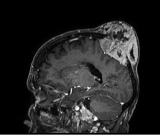
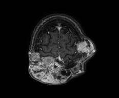
We are reporting a case of 56-year-old women who was operated on 2 years ago for right breast cancer and lost to follow-up. The patient presented 3 years later for diffuse pain. Clinical examination (Figure 1) found a hard cranial swelling painful on palpation. A chest-abdominal-pelvic CT scan showed lytic lesions in the sternum, clavicle, and vertebrae of metastatic allure. The brain MRI showed multiple lesions of the cranial vault, the largest is left fronto-parietal with meningeal extension infiltrating the upper longitudinal sinus that remains permeable and skin extension measuring 90x60x55 mm, of secondary appearance (Figures 2 & 3). The patient received palliative chemotherapy with epiribucin- paclitaxel, with analgesic radiotherapy on the skull at a dose of 30 Gy in 3 Gy/fraction.
- Cancer Diagnosis from RNA Sequence of Blood Cells by Using AI
- Field Cancerization in Oral Cavity, Case Report and Review of Literature. Oncologic Program Salud Integral Hospital, Managua, Nicaragua
- Identification of B Lymphocytes in Cancer Patient’s Blood
- A Case Report of a Breast Cancer Patient Developing Pneumonitis as a Result of Abemaciclib Therapy
- Immune Checkpoint Therapeutics for Today’s Fight and Beyond
- The Amalgamated Sophomore-Gonadoblastoma