Body Irradiation Using Tomotherapy Helical & Direct Technique Using Radixact for Young and Paediatric Patients
Background: We have treated 14 patients of total body irradiation (TBI) in Helical Tomotherapy, out of which 9 are males and 5 are females, age is 5 years to 33 years with acute lymphoblastic leukemia (ALL) remission or acute myeloic leukemia (AML). Total Body Irradiation is an important procedure in the conditioning for Bone Marrow and stem cell transplantation. We present here our treatment technique, treatment planning, delivery and dose verification. Standard Total prescribed dose of target volume 12 Gy in 6 fractions twice daily over 3 days to the whole body with Simultaneous Integrated Boost of 14 Gy/6# to brain. Dose volume constraint for the planning treatment volume (PTV) 95% of prescribed dose is getting by 95% of the volume. Material and Methods: We immobilized the whole body of the patients from Vac-loc and thermoplastic mask for head. We take two computerized tomography (CT) scan for treatment Planning. Result: Average dose D95 to the planning treatment volume was 11.7 Gy corresponding to a mean coverage of the planning treatment volume of 95%. Average dose (D-mean) for the lungs was not exceeding 9 Gy. Conclusions: Total Body Irradiation using helical Tomotherapy is achievable and well tolerated. Each dose distribution is homogeneous and reduce dose to at risk organ.
Introduction
Total Body Irradiation is a treatment in which whole body of patient is treated with radiation. Total body irradiation with helical Tomotherapy purpose to deliver a uniform radiation dose to the entire volume of a patient’s body. Total
body irradiation is used in several types of cancer 1.Leukemia 2.Lymphoma 3.Multiple myeloma. Total body irradiation is different from other types of radiation treatment. Usually total body irradiation treatment is given twice a day for three days. Radiation technologist gives the treatment with team including Radiation Oncologist, Medical Physicist other medical staff (staff nurse) [1]. Helical Tomotherapy (HT) process applies image guided intensity-modulated radiation therapy using megavoltage computed tomography (MVCT), which can deliver radiation slice by slice.
Methods
During the radiation treatment patients and their parents were given information about the treatment, its potential adverse effects, and necessary diagnostic. Written consent by both the patient and the parents, or legal guardians, was gathered in every case [2].
Immobilization Setup
Radiation technologists explain to patient about mould room and simulation procedure. Our Radiation oncologist & medical Physicist review everything. Patient immobilization was achieved by the use of vac-loc and thermoplastic head mask 3 point and also measure the height of patients.
CT-Simulation Planning
Before CT- scan simulation, patient is asked to wear hospital gown or comfortable clothes & asked to remove jewellery or metal objects (such as rings or hairpins).
Radiation technologist help the patient lie down on the CT-Simulation table in supine position the patient is asked not to move or speak during CT simulation because these things may change the position and then CT- simulation procedure start. Planning CT image is acquired in supine position with 5 mm slice thickness [3]. The image sets of a head to midlevel of femur CT1 and a CT2 of the lower extremities from toes to head of the femur. If patient’s height is more than 135 cm. we take two CT- scans CT1 and CT2. When we do HFS CT1 scan fiducial marker is placed in sagittal, coronal and transverse planes on the head mask following. Which we reposition the patient for FFS CT2 scan. Fiducial marker is placed on both legs. We create junction on thigh in CT1 &CT2 with a gap of 5 cm to 10 cm. Patient having height between 110 cm to 120 cm CT scan is done for HFS from vertex to toes in once with at least three fiducial marker placed on head mask.
Treatment Dose and Fractionation
Treatment Dose for total body was 12 Gy in 6 fractions that is 2 Gy in morning and 2 Gy in evening per day, 8 hour gap (interval) in two fractions for three consecutive days with a SIB of 14 Gy is 6# to brain [4]. Paediatric and adult patient are prescribed the same dose. Prescribed dose is received by the target area of 95% in the Helical Tomo therapy.
| Sex/Age | Diagnosis | Dose(Gy) & Fraction | |
|---|---|---|---|
| 1 | 5y/M | B-cell ALL | 12Gy /6 # |
| 2 | 6y/M | ALL | 12Gy /6 # |
| 3 | 13y/F | ALL | 12Gy /6 # |
| 4 | 42y/M | ALL | 12Gy /6 # |
| 5 | 9y/M | ALL | 12Gy /6 # |
| 6 | 24y/M | ALL | 12Gy /6 # |
| 7 | 33y/M | B-cell ALL | 12Gy /6 # |
| 8 | 15y/F | ALL | 12Gy /6 # |
| 9 | 45y/F | ALL | 12Gy /6 # |
| 10 | 31y/F | ALL | 12Gy /6 # |
| 11 | 9y/M | ALL | 12Gy /6 # |
| 12 | 14y/M | ALL | 12Gy /6 # |
| 13 | 10y/M | ALL | 12Gy /6 # |
| 14 | 8y/F | ALL | 12Gy /6 # |
Table 1: Dose & fraction and no. of treated patients since 2020 to 2021.
Treatment Delivery
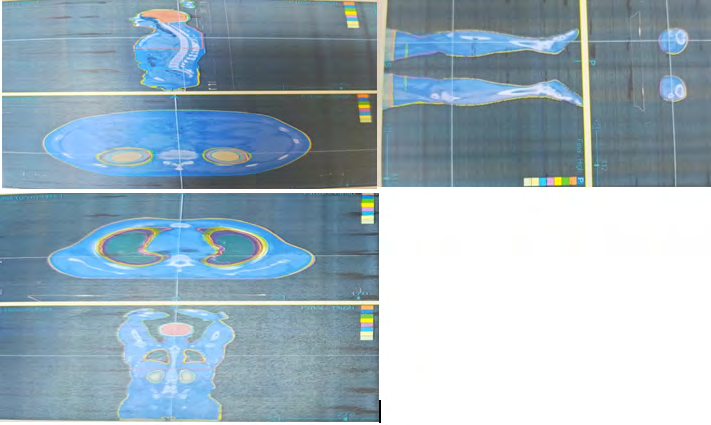
Accuray Precision Treatment planning system is used for contouring and treatment planning. PTV structures were created for body contour outer and inner margin. PTV upper consist of skin, eyes, spinal cord, lungs and central lung, kidneys, liver heart. PTV lower consist right & left leg and Irradiation is split into two parts one is head first supine and other one is feet first supine part [5].
Patients were placed on the treatment couch in the treatment planning position. Each patient underwent a megavoltage computed tomography (MVCT) for treatment alignment before each fraction. Manual fusion was performed on all patients to improve the registration accuracy by verifying axial, coronal and sagittal image from vertex to toes. This divided into 4 parts one includes head & neck second chest & abdomen, third are pelvis region and fourth is knee for each study. We divided the PTV of the patient into 2 parts. When body length exceeding 135 cm because of the machine couch limitation and deliver the TBI in two successive sessions:
- Head first from vertex to cut Plane.
- After the patient reposition, feet first from toes to the cut plane.
Helical Tomotherapy continually delivers a uniform dose to a patient on the treatment couch with 360 degree spiral gantry rotation.
Result
Patient placement setup average time is 5 to 7 minutes for HFS & FFS parts .TBI Patient HFS beam on time is 20 minutes to 25 minutes for treatment and FFS beam on time is 10 minutes to 13 minutes. Distributions are homogeneous and reduce doses to OARs. Lungs average dose (D-mean) does not exceed 9 Gy and minimum dose (D-min) of less 6Gy. We achieved minimum right lung dose is 5.60 Gy and left lung dose is 6.09 Gy [6]. Right kidney dose is 4.63 Gy & left kidney dose is 4.67 Gy. PTV brain coverage is 100%, PTV chest is 89.85%, PTV pelvis is 91.78%, and PTV leg is 90.42%. For adult TBI patient we create junction on thigh for PTV upper and PTV lower with a gap (superior & inferior field) of 5 cm.
Discussion
The Tomotherapy machine, including the next- generation Radixact system, has continuous gantry rotation and couch motion that enables helical delivery of therapy (TomoHelical). This treatment modality produces intensity- modulated radiation from 360 degrees around the patient over very long treatment fields. Helical Tomotherapy is a new concept in radiation therapy IG-IMRT treatment for TBI. Helical Tomotherapy unit 6 MV linear accelerator and a detector array system are mounted opposite each other on a ring gantry that continuously rotates during the imaging and treatment procedures. One of the most important features of the Helical Tomotherapy concept is the ON- BOARD MVCT. We used the same fractionation and dose constraints as in our standard total body irradiation (TBI) protocols, only adding minimum dose for the lungs to reduce the risk of underdosing and relapse [7]. This approach to conditioning prior to stem cell transplantation facilitated the successful engraftment of all 14 patients included in the study. Limitations may occur in patients with high body mass index. For all patients acceptable treatment plan fulfilling the predefined constraints were achievable. An average time of 45-50 minute is required for treatment. Tomo helical mode provides a rotating beam source with a binary multileaf collimator (MLC) moving in a spiral pattern relative to the patients. Treatment plan for all cases satisfied safe dose constraints, including for target volume and risk organs. Moreover, all TBI radiation therapy was completed without any technical problems [8]. Based on the result of this pilot study, our hospital is currently implementing 12 Gy IMRT-TBI as a conditioning regimen.
Conclusion
Patient positioning and immobilization plays a major role for total body irradiation. Total Body Irradiation using Helical Tomotherapy is practical and well tolerated in treatment distribution and homogeneity of dose. Total
body irradiation (TBI) helical Tomotherapy (HT) is two part treatment for taller patient (height> 135).
References
-
Wang H, JunQi, Fan RT, Pi Y, Liu Q, et al. (2020) Technical note: factors affecting dose distribution in the overlap region of two segment total body irradiation by helical Tomotherapy. Radiation Oncology 15(1): 257.
-
Studinski RCN, Fraser DJ, Samant RS, MacPherson MS (2017) Current practice in total body irradiation: results of a Canada- wide survey. Curr. Oncol 24(3): 181-186.
-
Gruen A, Ebell W, Neumann O, Budach V, Marnitz S, et al. (2013) Total body irradiation using helical Tomotherapy in children and young adults undergoing stem cell transplantation. Radiat Oncol 8(1): 92.
-
Buchstab TW, Leitzen C, Schmeel LC, Simon B, Koch D, et al. (2019) Total body irradiation: significant dose sparing of lung tissue achievable by helical Tomotherapy. Journal of Medical Physcis 30(1): 17-23.
-
Sarradin V, Simon L, Huynh A, Gilhodes J, Filleron T, et al. (2018) Total body irradiation using helical Tomotherapy treatment technique, dosimetric results and initial clinical experience. Cancer Radiother 22(1): 17-24.
-
Schultheiss TE, Wong J, Liu A, Olivera G, somlo G (2007) Image- guided total marrow and lymphatic irradiation using helical Tomotherapy. Int J Radiat Oncol Biol Phys 67(4): 1259-1267.
-
Hong CS, Kim MJ, Kim J, chang KH, park K, et al. (2019) Feasibility of hybrid TomoHelical –and Tomodirect- based volumetric gradient matching technique for total body irradiation. Radiat Oncol 14(1): 233.
-
Konishi T, Ogawa H, Najima Y, Hashimoto S, Wada A, et al. (2020) Safety of total body irradiation using intensity- modulated radiation therapy by helical Tomotherapy in allogenic hematopoietic stem cell transplantation: a prospective pilot study. J Radiat Res 61(6): 969-976.
- Cancer Diagnosis from RNA Sequence of Blood Cells by Using AI
- Field Cancerization in Oral Cavity, Case Report and Review of Literature. Oncologic Program Salud Integral Hospital, Managua, Nicaragua
- Identification of B Lymphocytes in Cancer Patient’s Blood
- A Case Report of a Breast Cancer Patient Developing Pneumonitis as a Result of Abemaciclib Therapy
- Immune Checkpoint Therapeutics for Today’s Fight and Beyond
- The Amalgamated Sophomore-Gonadoblastoma