Contribution of MRI in the Diagnosis and Planning of Radiotherapy Treatment in Pituitary Adenomas
Clinical image
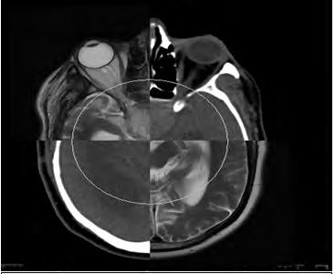
Pituitary adenomas are rare pathologies, in most cases, they are benign tumors, but can be aggressive and pose a problem of local recurrence or even metastases defining pituitary carcinoma. The reference imaging is MRI with the following sequences: 2 to 3 mm coronal slices in T1 sequence with and without injection of gadolinium, and T2 in spin echo. The multi-planar approach and the improved resolution by dynamic injection of gadolinium in a dynamic mode made it easy to diagnosis of microadenomas or even picoadenomas. For macroadenomas, MRI makes it possible Image Article to carry out a local extension assessment of local extension and the search for complications. Classically, adenomas have an iso or hypointense signal in T1, iso or hyperintense in T2, slightly enhanced after injection of gadolinium (Figure 1). The treatment is medico-surgical as a rule, radiotherapy is rarely mentioned as a first-line treatment. In Postoperative, radiotherapy is indicated in the presence of tumor aggressiveness factors. Exclusive radiotherapy is offered in case of a surgical contraindication or inoperable recurrence. MRI allows a precise delineation of the target volume, especially if the tumor is in place by recalling with the dosimetric scanner (Figure 2), and therefore have better coverage of the target volumes while sparing nearby organs at risk (chiasma and optic nerves).
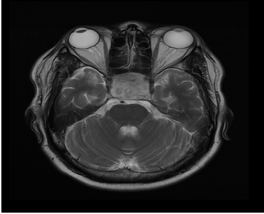
We report the case of a 37-year-old patient, who presents with a recurrence of an operated macroadenoma considered unresectable (Figure 1), treated by exclusive radiotherapy.
MRI has established positive diagnosis, and assist in the delineation of target volumes (Figure 2).
- Cancer Diagnosis from RNA Sequence of Blood Cells by Using AI
- Field Cancerization in Oral Cavity, Case Report and Review of Literature. Oncologic Program Salud Integral Hospital, Managua, Nicaragua
- Identification of B Lymphocytes in Cancer Patient’s Blood
- A Case Report of a Breast Cancer Patient Developing Pneumonitis as a Result of Abemaciclib Therapy
- Immune Checkpoint Therapeutics for Today’s Fight and Beyond
- The Amalgamated Sophomore-Gonadoblastoma