Medicinal plants in combating Breast Carcinogenesis A review on recent investigations
Breast cancer is the most prevalent disease among women and a challenge for the scientific and medical community. Existing treatments for breast cancer are surgery, radiation, chemotherapy, hormone therapy, and targeted therapy. These treatments have the potential to stop the growth and spread of cancer, particularly when the condition is diagnosed at an early stage. As cancer cells reproduce more frequently than the vast majority of normal cells, chemotherapy has a greater chance of successfully killing cancer cells. Certain pharmaceuticals are able to terminate dividing cells by inflicting damage on the section of the cell’s control centre that is responsible for the process of cell division. Other medications are capable of interfering with the chemical processes that are necessary for cell division. On the other hand, as cancer is often diagnosed at an advanced stage, in which, the process of curing the disease is extremely difficult. Therefore, it is prudent to prevent the occurrence of this deadly disease. Several researches have consistently found an inverse relationship between cancer and natural sources, including plant extracts, fractions and active components. The synergistic and cumulative effects of bioactive phytochemicals found in whole plant extracts have been related to these positive effects. In this review, we attempted to scrutinize the antibreast cancer potential of quite a few extracts/ fractions/active components derived from various plant sources such Alpina galaga, Annona muricata, Ficus carica, Murraya koenigii, Nigella sativa, Rosmarinus officinalis, Urtica dioica, According to recent research, above mention plants have showed anticancer properties by inhibiting cell proliferation, induces apoptosis and causing cell-cycle arrest in preclinical in vitro and in vivo breast cancer models. This review will help the scientific and medical community for novel drug discovery against breast carcinogenesis.
Introduction
Breast cancer is caused by unregulated cell proliferation and differentiation of breast cells. It is the commonest in women and a demanding target for the scientific and medical community [1]. From many centuries, naturally occurring medications have been used to cure various different types of diseases. These medications have always relied heavily on plants, plant extracts, and other plant products. The positive effects of plant extracts/products have been linked to the synergistic impact of phytochemicals present in it. Phytochemicals can impact tumor development processes by modifying and detoxifying carcinogens [2]. The majority of medicinal medications for cancer treatment are also derived from plants. At present, four classes of plants derived anti- cancer drugs available in the market such as vinca alkaloids, epipodophyllotoxins, taxanes and camptothecin derivatives [3]. Scientists found in the 1960s that an extract from the bark of the Taxus brevifolia could be used to treat cancer [4]. They found that taxol along with vinca alkaloids were very efficient in arresting cell cycle by blocking microtubules depolarization [5]. According to the latest research, some medicinal plant sources such as Alpina galaga, Annona muricata, Ficus carica, Murraya koenigii, Nigella sativa, Rosmarinus officinalis and Urtica dioica have been effective against breast cancer. In this review, we are principally focused on the most recent research articles of above mention plants. Therefore, the rationale behind this review article is to highlight the protective and beneficial effects of some plant extracts, fractions and active components against breast carcinogenesis which will be helpful for the scientist for novel drug discovery against breast carcinogenesis.
Searching the databases such as PubMed and Google Scholar, yielded information were retrieved by using keywords such as breast cancer+ Alpina galaga, breast cancer+ Annona muricata, breast cancer+ Ficus carica, breast cancer+ Murraya koenigii, breast cancer+ Nigella sativa, breast cancer+ Rosmarinus officinalis, breast cancer+ Urtica dioica, plant extract, anti-cancer, phytochemicals etc. Research articles in English language were considered for this review. First, the abstracts of the research articles were reviewed and if relevant, then the complete articles were analyzed for the anti-cancer properties of various plants against breast carcinogenesis.
Protective Effects of Plants on Breast Carcinoma
Alpinia Galanga
Alpinia galanga (AG) belongs to a Zingiberaceae (ginger) family widely distributed in Asian countries. From many centuries, it is used as a spice in food and as herbal medicine for treating various types diseases. Several pharmacological properties of this plant has been discovered by the researchers such as, antioxidant [6, 7], anti-inflammatory [8, 9] antimicrobial [10], anti-bacterial [11], and anti- osteoarthritic [12]. This plant contains active compounds such as 1’S-1’-acetoxychavicol acetate, 1’-acetoxychavicol acetate, 1′S-1′-acetoxyeugenol acetate, (E)-8, 17-epoxylabd- 12-ene-15, 16-dial, p-hydroxycinnamaldehyde, 1, 7-bis (4-hydroxyphenyl)-1, 4, 6-heptatrien-3-one (BH (BDMC). Tumor volume, a major marker of breast cancer, increases in the untreated group of breast cancer, while the tumors in the treated group typically decrease in size as a result of the AG extract. AG also displayed apoptotic and anti angiogenic potential by activating caspase-3 pathway and inhibiting NF-ƙB, NO and COX-2 in breast cancer mice model [13].
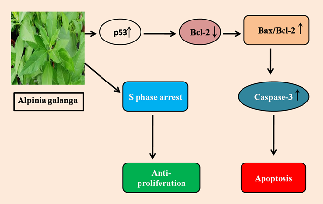
Figure 1: Molecular action of Alpinia galangal: Alpinia galangal stimulates the p53 expression which downregulates the anti- apoptotic Bcl-2 levels followed by upregulation of pro-apoptotic Bax. This upregulation of Bax leads the overexpression of Caspase 3 which finally leading to apoptosis. Additionally, Alpinia galangal also causes S phase arrest in cell cycle and hence acts as an anti- proliferative agent.
Ethanolic extract of AG (96% ethanol) showed cytotoxic effect against MCF7 breast cells lines [14]. According to Song W, et al. [15] galangin, an active component of AG successfully induces apoptosis by TRAIL/Caspase-3/AMPK signalling pathway in human breast cancer cells. AG substantially reduced the proliferation of 4T1 cells at IC50 135 g/mL, and boosted the cytotoxic effect at concentrations of 50 and 100 g/mL. The quantity of senescent cells arrested in the G2/M phase increased in response to AG. Furthermore, AG reduced 4T1 cell migration and lowered MMP-9 production caused by Dox [16]. Awad MG, et al. [17] discovered a synergistic effect of AG leaves extract and cisplatin against various cell lines viz MCF7, HepG2, CaCo2, and PANC1. This action is mediated via cell cycle arrest, and the decrease of several drug resistance genes (MDR1 and MAPK1). AG also stimulates the p53 expression stimulates apoptosis [18] and causes S phase arrest in cell cycle and hence acts as an anti- proliferative agent [19]. AG was found to enhance the cytotoxic effects of cytotoxic T-cells by restricting the proliferation of human triple-negative breast cancer cells hence displayed immunopotentiation effect [20]. AG has the capability to induce cell senescence and intracellular ROS levels, resulting in delayed cell cycle progression, were linked to its antiproliferation action against HER2-overexpressing breast cancer [21]. 1′-acetoxychavicol acetate component of AG down-regulates the human epidermal growth factor receptor 2, pERK1/2, pAKT, estrogen receptor coactivator, cyclin D1, and MYC proto-oncogene by inhibiting the proliferation of human epidermal growth factor receptor 2-overexpressed cell lines in time and concentration dependent manner.. In addition to this, 1′-acetoxychavicol acetate showed significant reduction in tumor mass in in vivo zebrafish- engrafted breast cancer model [22] (Figure 1).
Annona Muricata
Annona muricata (AM) is an everlasting tropical tree plant belongs to the Annonaceae family. It contains a variety of pharmacological activities like anti-inflammatory [23], anticarcinogenic [24], anti-diabetic [25], antioxidant [26], and anti-microbial [27]. Main phytochemicals present in this plant are annonaceousacetogenins, acetogenins and Cyclohexapeptides [28]. In a breast cancer mouse model, the group treated with AM crude extract had a mean tumor volume of 271.7±14.24 mm, which was smaller than the untreated group’s volume of 375 ±25.98 mm. Histological examination revealed the decreased number of mitotic cells per tumor segment upon AM crude extract administration when compared to untreated group. AM crude extract also triggered apoptosis in 4 T1 breast cancer cells, decreased metastasis in vitro and in vivo, regulates the immune system, and reduced cancer-induced inflammation [29]. Ethanolic extract of AM leaves at a dose level of 200mg/kg bw showed significant increase in SOD, MDA, and the histological section of mammary tissue showed lower hyperplasia of mammary epithelial cells [30]. AM leaves extract lowered proliferative indexes of DMBA induced breast cancer, with 300 mg/kg being the most efficacious dose [31]. According to Alshaeri H, et al. [32], Alshaeri HK, et al. [33] AM extract has an anti-proliferative effect via EGFR-mediated signalling pathways such as AKT/MAPK/NF-B pathways and cyclin D1 suppression. Annonacin isolated from Graviola showed marked genotoxicity and inhibitory effect [34] in MCF-7 cells. In breast cancer, ER- functions as a ligand-dependent transcription factor that promotes tumor growth and survival. According to Suhendar U [35] AM extract has the most piperine component, and has cytotoxic action against MCF7 cancer cells which was confirmed by MTT assay. AM extract solid lipid nanoparticles (SLNs) shown a significant apoptotic effect and improved efficacy in killing MCF7 cancer cells [36]. Zeweil MM, et al. [37] reported the downregulation of ER-α gene, increased antioxidants, and reduced lipid peroxidation levels upon Graviola administration. Report by Daddiouaissa D, et al. [38] suggested that the ionic liquid extract of Graviola fruit displayed the anti-proliferative potential on MCF-7 breast cancer cell lines by initiating apoptosis, cell-cycle arrest and by decreasing the cell generation number. AM (ethanol extract) inhibited the proliferation of T47D cell [39], AM (aqueous extract) induces mitochondrial cell death, concealed cell proliferation, and reduced cellular motility in MDA-MB-231 cells [40]. AM leaf (methanol extract) inhibited MCF-7 cells significantly, with an IC50 value of 85.55 g/mL [41]. Another study by Prasad SK, et al. [42] reported that AM seeds extract showed G0/ G1 cell cycle arrest via apoptosis. Ethyl acetate extract of AM leaf resulted in a higher level of cytotoxicity on breast cancer cells, which was responsible for anti-proliferative property of extract. In breast cancer cell lines, mitochondrial membrane integrity were significantly down regulated upon AM treatment resulted in the apoptosis of breast cancer cells [43]. The growths of the MCF-7 and MDA-MB-231 cells were inhibited when incubated with AM leaf extracts- loaded scaffolds [44]. Kariyil J, et al. [45] Chloroform fraction of methanolic extract of AM seeds displayed cytotoxicity via cell membrane lysis, ROS dependent caspase-activated mitochondria-mediated apoptosis, and stopping the S phase of the cell cycle. Flowcytometry with propidium iodide staining indicated that AM extract (13 and 25 g/mL) alone caused cell cycle arrest in the G1 phase and G2/M arrest when combined with dox in 4T1 cells. AM extract at doses of 13g/mL and 25 g/mL reduced intracellular reactive oxygen species (ROS) levels as a single therapy and in conjunction with dox, confirmed by dichloro dihydrofluorescein diacetate staining assay [46]. Rojas A, et al. [47] reported the presence of four sesquiterpenes, viz Z-caryophyllene, α-selinene, β-pinene, and β-elemene in the essential oil of AM leaves which is responsible for reduction in MDA and VEGF and elevation in GSH levels in in vivo breast cancer mouse model (Figure 2).
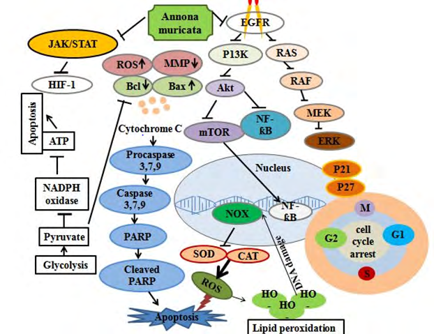
Figure 2: Molecular action of Annona muricata: At a molecular level, AM inhibits the epidermal growth factor receptor (EGFR), which furthers inhibits the other signaling pathways including RAS, phosphatidylinositol-4,5-bisphosphate 3-kinase (PI3K), NF-κB, and ERK. AM also inhibits the JAK/STAT pathway which further inhibits the HIF-1α and it also initiates the intrinsic apoptotic pathways by releasing cytochrome c from mitochondria. In addition to this inhibition of enzymes such as superoxide dismutase (SOD), catalase (CAT), and heme-oxygenase (HO-1) increases the production of reactive oxygen species (ROS). These ROS induces the lipid peroxidation and hence DNA damage takes place. All these signaling pathways finally lead to apoptosis, inflammation and cell cycle arrest.
Ficus Carica
Ficus carica (FC) is a flowering plant belongs to family Moraceae, which is formerly from the West Asia and Middle East, but widely distributed in many other regions in the world [48]. The different parts of this plant are used for its medicinal properties in various disorders such as respiratory, inflammatory, cardiovascular and gastrointestinal disorders [48, 49] Various pharmacological properties are associated with this plant are antibacterial antioxidant [50, 51] anticancer [52, 53], Anti-acne [54] and antipyretic [55]. Bioactive compounds in this plant which is responsible for the medicinal properties are arabinose, βamyrins, β-carotines, glycosides, β-setosterols and xanthotoxol Gilaniet al. 2008. Zubair R, et al. [56] reported the high antiproliferative activity and strong cytotoxic activity of ethyl acetate FC extract towards breast cancer (MCF-7) cells line. Aqueous extract of FC leaves decreases the viability of MDA-MB-231 cell, increases the expression level of proapototic gens (BAX) and tumor suppressor genes (TP53, and TP21) showed the anti-prolifertaion activity. Additionally, decreased breast cancer-marker gene (GATA3) and increased proto-oncogene (ELF5) was observed upon FC extract treatment [57]. Ghandehari F, et al. [58] reported that tumor size and volume were both reduced and growth was halted in rats with breast tumors after they were treated with a latex extract of FC. Histopathological evaluation of breast tissue in the fig latex-treated group demonstrated a reduction in angiogenesis, mitotic characteristics, and an increase in necrosis. Another study by Lightbourn AV, et al. [59] suggests that the fig leaf extract attenuates the single- strand breakage upon Diethylstilbestrol-induction in human breast epithelial cells (MCF10A) which was confirmed by comet assay and phase contrast microscopy. FC leaf extract reduces the growth potential of MDA-MB-231 triple-negative breast cancer cells by reducing the S and G2/M cell cycle stages and causing apoptosis via a p53-independent route [60] A study by Widyaningrum N, et al. [61] reported that the fig extract in combination with olive oil showed cytotoxic activity on T-47D and MCF-7 cells and AlGhalban FM, et al. [62] discovered anticancer activity in MDA-MB-231 cells, demonstrating antiproliferative and antimetastatic effects as well as significant effects on cell shape (Figure 3).

Murraya Koenigii
Murraya koenigii (MK), belongs to the Rutaceae family and widely scattered in Eastern-Asia. Various pharmacological properties this plant are antifungal [63], antioxidant [64, 65], anti-bacterial [66], antidiabetic [67], anti-inflammatory [68, 69] and anticancer [70].
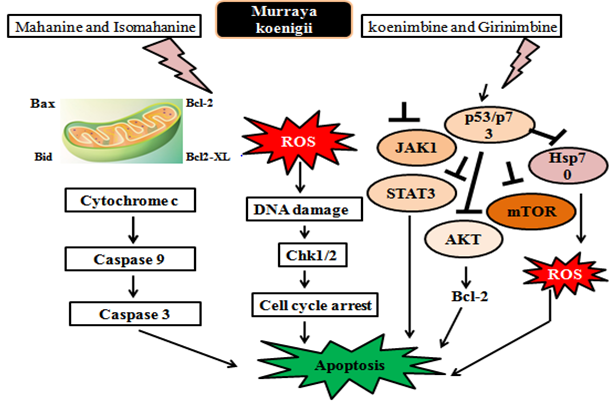
Figure 4: Molecular action on Murraya koenigii: Murraya koenigii and its primary active component regulates multiple signaling pathways, including JAK/STAT, phosphatidylinositol 3 kinase (PI3K)/protein kinase B (AKT) and mammalian target of rapamycin (mTOR). Active constituents viz koenimbine and girinimbine from MK triggered the tumor suppressor gene p53/p73 which in turn inhibits the JAK1, STAT3, mTOR, Hsp90 and AKT. Mahanine and isomahanine from MK responsible for the release of cyctochrome c which furthers initiates cascade of caspases in the intrinsic apoptotic pathway and finally lead to apoptosis. It also triggers the check point of cell cycle and induces the Go/G1 cell cycle arrest.
Various active components of this plants are bismahanine, murrayanine, murrayafoline-A, bi-koeniquinone-A, bismurrayaquinone, mukoenine-A, mukoenine-B, mukoenine-C, murrastifoline, Murrayazolinol, murrayacine, murrayazolidine, murrayazoline, mahanimbine, girinimbine, koenioline, xynthyletin, koenigine-Quinone A and koenigine-Quinone B [71]. Yeap SK, et al. [70] reported that the tumor volume was greatly reduced by MK aqueous extract, and the histological characteristics revealed that MK leaf extract has the ability to manage inflammation, reduction in tumor cells and limit the proliferation of tumor cells. It also reduced nitric oxide levels as well as inflammation-related cytokines and genes such as iNOS, iCAM, NF-kB, and c-MYC, hence stimulates the T cell cytokine production which further helps in reduction of mitotic division and hence delayed the breast cancer formation. MK extract treatment resulted in increased caspase-3 activity and TUNEL-positive cells, indicating accelerated apoptosis [72]. Total alkaloid extract from MK leaves inhibited cell viability (IC50 of 14.4 g/mL), changed growth dynamics, stopped cells in the “S” phase, and promoted cell death of breast cancer cells (MDA-MB-231) [73]. A study by Vijapur LS, et al. [74] revealed the anticancer activity of silver nanoparticles of MK on breast cancer cell lines (MDA- MB-231) by MTT assay. Ethanolic extract of MK showed an anti-cancer activity against DMBA induced breast tumors in rats. Significant decline was observed in tumor mass, quantity of polymorphonuclear leukocytes, multi-layered cuboid epithelium, and proliferated solid collagen fibers upon treatment with MK extract [75]. Additionally Aisyah S, et al. [76] reported the overexpression of caspase-3, linked to apoptosis of cancerous cells and hence showed anti- tumor potency. Apoptotic and anti-angiogenic potential have been displayed by Mahanimbine, an active component of MK against breast cancer cells [77] (Figure 4).
Nigella Sativa
Nigella Sativa (NS), rising as a miracle herb belongs to a Ranunculaceae family. Various pharmacological potential of this herb has been revealed by the researchers such as, anti-inflammatory [78, 79], antihypertensive [80], antioxidant [81, 82], antidiabetic [83, 84, 85], antimicrobial [86, 87] and anticancer [88, 89]. The chemical component present in this plant are Cumin aldehyde, cuminic alcohol, pyrazines, 2-ethoxy-3-isopropylpyrazine, 2-methoxy- 3-sec-butylpyrazine, and 2-methoxy-3-methylpyrazine, terpinene, safranal, p-cymene, pinene, thymoquinone, and monoterpenes. A study by Bhattacharya S, et al. [90] reported that Thymoquinone-Loaded Nanostructured Lipid Carrier displayed the cytotoxicity against Breast Cancer Cell Lines (MDA-MB-231 and MCF-7). Aqueous extract of NS and crude flavonoid extract successfully inhibited the growth of MCF cell lines with the same potency as conventional cisplatin [91, 92]. It is also reported that NS aqueous extract consist of elements such as calcium and magnesium etc., and high amount of these elements inside the cell starts the cell death via apoptosis. Histological investigation of NS seed- supplemented DMBA-treated rats demonstrated mammary gland activation and prevention of breast tumor cell proliferation progression. Tumor volume, MDA, LDH levels, as well as ALP and AST activity, were reduced by NS seed oil and Thymoquinone, an active component of NS. TQ was more efficient in lowering Brca1 and Brca2 gene expression [93] and significantly elevates the P53 gene expression [94].
Another study by Periasamy et al. [95] reported of ultrasonic nanoemulsion formulation of NS essential oil induces apoptosis in MCF-7 cell, hence displayed anti- cancer activity. Another Report by Bumidin MS, et al. [96] demonstrated that NS aqueous extracts can reduce the cell membrane integrity of MCF-7 and thereby can restrain the growth and viability of MCF-7. Thymoquinone synchronize the levels of pro and anti-apoptotic genes hence leading to apoptosis. It can impede the process of metastasis via the JNK and p38 activation and lowered the phosphorylation of NF-κB and IKKα/β [97]. Silver nanoparticles (AgNPs) derived from an aqueous seed extract of NS promote apoptosis in MCF-7 cells through changing the expression of apoptotic proteins Bax and Bcl- 2, as well as COX-2 (inflammatory marker) [98]. Rafati M, et al. [99] observed that using N. sativa gel as a prophylactic measure effectively delayed and reduced the incidence of ARD and moist desquamation in breast cancer patients. Hydroalcoholic extract of NS showed an inhibitory effect against breast cancer cell lines (MCF 7). Reduction in the mRNA expression levels of NFk and IKK demonstrated the anti-inflammatory effects of NS [100]. Khurshid Y, et al. [101] reported that the proteins isolated from NS have apoptotic and anti- proliferative potential against human breast MCF-7 cancer cell line. In breast cancer, regulation of the PIK3CA kinase domain can be diminished by H1047R and p. H1047L mutants resulting in increased PI3K/Akt1 pathway activation. Unal et al. [102] reported the inhibitory effect of Thymoquinone on proliferation and migration of MDA-MB-231 cells by suppressing autophagy. Zhou J, et al. [103] reported that thymoquinone isolated form NS binds to the kinase domain of PI3CA mutants, preventing TQ- mediated PI3K/Akt1 pathway activation. NS seed oil caused a significant cell proliferation reduction and decreased cell viability and act as anti-carcinogenic, anti-proliferative agent [104, 105] (Figure 5).
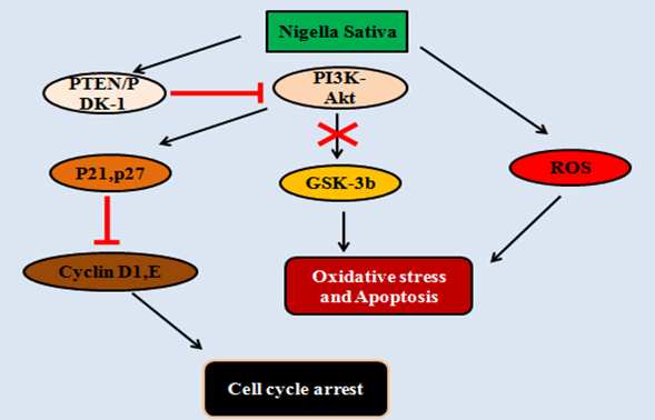
Figure 5: Molecular action on Nigella Sativa: Nigella Sativa suppresses the PI3K-Akt pathways by activating PTEN/PDK-1. After this the inhibition of downstream regulator GSK-3b takes place which further induces the oxidative stress and finally apoptosis. NS also generates the reactive oxygen species which creates oxidative stress condition leading to apoptosis. In addition to this, inhibition of PI3K-Akt activates p21 and p27 which further inhibits the cyclins (D1,E) and leading to cell cycle arrest.
Rosmarinus Officinalis
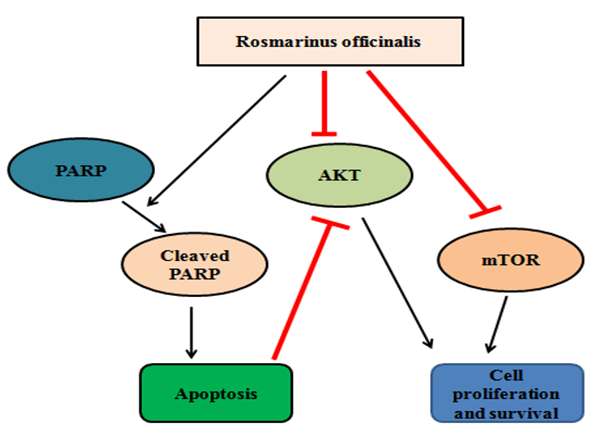
Rosmarinus officinalis (RO) belongs to Lamiaceae family and widely distributed in the Mediterranean region. RO leaves extract has been used as a flavoring agent [106]. Various pharmacological properties are associated with this plant are anti-oxidant, antibacterial [107, 108], anticancer [109] and anti-inflammatory [110, 111] antidiabetic [112, 113]. The main bioactive compounds present in this plant are rosmarinic acid, caffeoylquinic acids, caffeic acid, quinic acid, kaempferol, rutin, quercetin Mena, et al. A study by Moore J, et al. [114] Showed the increased levels of ADP ribose polymerase (PARP) cleavage which is a well-known markers for apoptosis and essential oil of RO repressed the viability of the MCF-7 cell line at a dose-400 µg/ml concentration (IC50 = 48.01 ± 0.94) [115]. Studies have shown the cytotoxic effects of green iron nanoparticles of aqueous extract of RO [116] and anti-prolifeartive activity of RO extract in combination with bleomycin drug [117] on 4T1 and MCF-7 cell lines. RO suppressed MDA-MB-231 and survival at low concentrations (0.5-20 g/mL) and dramatically lowered the phosphorylation/activation levels of Akt and mTOR, two critical actors in cancer cell growth and survival [118, 119]. Shen Y, et al. [120] reported the anti- proliferative property of ethanolic extract of RO against MCF- 7 cancer cell lines. RO suppressed MDA-MB-231 and survival at low concentrations (0.5-20 g/mL) and dramatically lowered the phosphorylation/activation levels of Akt and mTOR, two critical actors in cancer cell growth and survival [121] (Figure 6).
Urtica Dioica
Urtica dioica (UD) is an herbaceous perennial flowering plant, belongs to family Urticaceae and genus Urtica, widely distributed in distinct parts of the world like India, United States, Malaysia, and Iran [122]. Number of pharmacological properties are associated with this plant are anti-inflammatory [123], anticancer [124], antirheumatic [125] and cardiovascular [126], antiaging and antioxidant [127]. Bioactive components present in this plant are mainly flavanoids such as kaempferol, isorhamnetin, quercetin, isoquercitrin, astragalin, rutin, 3-rutinosides and 3-glycosides [128]. A study by Mohammadi A, et al. [129] reported the apoptotic potential of the UD plant extracts against MDA-MB-468 breast cancer cell lines. Later on, another study by Mohammadi A, et al. [130] suggests the cytotoxic activity of UD dichloromethane extract on growth and migration of MDA-MB-468 cells assessed by TUNEL assay and DNA fragmentation analysis. Real-time polymerase chain reaction (PCR) has revealed the increased mRNA expression levels of caspase-3, caspase-9, and decrease in the bcl-2 showed the mechanism of cell death. A study by Telo S, et al. [131] reported the reduced lipid peroxidation and elevation in catalase enzyme activity in rat mammary gland cancer. Histological investigation revealed that animals in the malignant group that were treated with UD had a moderate degree of ductular proliferation along with localised epithelial hyperplasia. A research by UD extract has the ability to regulate miR-21 and its associated gene in breast cancer, as well as limit the development and migration of breast cancer cell lines and in vivo models [132]. Mohammadi A, et al. [133] reported the anti-cancer efficacy of UD extract (dichloromethane solvent) on in vitro 4T1 breast cancer cell line and in vivo mouse 4T1 allograft tumor model. Authors have revealed the cytotoxic potency of UC extract on 4T1 breast cancer cell line and 4T1- induced mouse model by MTT assay. Addtionally, UC treatment induces the apoptosis in 4T1 cells and Balb/c allograft tumor model, confirmed by DNA fragmentation and TUNEL (terminal deoxy transferase (TdT)-mediated dUTP nick- end labeling) assay respectively. Further, growth of breast tumor was also reduced upon treatment with UC extract. Real-Time PCR exhibited the upregualtion of proapoptotic caspase 3 and caspase 9 whereas downregulation of anti-apoptic Bcl- 2. Akbarian et al. [134] reported that cytotoxic effects of zinc oxide nanoparticles on MCF-7 and Fattahi S, et al. [135] demonstrated the anti-cancer effect of UD aqueous extract on MCF-7, MDA-MB-231 cell lines by evaluating adenosine deaminize (ADA) and ornithine decarboxylase (ODC1) gene expression. They observed that the UC induces apoptosis in breast cancer cells by increasing the expression of ODC1 in both cell lines and upregulating the expression of ADA in MCF-7 cell lines. Hydroalcoholic extract of UD at 1200 μg/ml inhibits the proliferation of MCF-7 breast cancer cells [136] (Figure 7) Table 1.
![Figure 7: Molecular action of Urtica dioica: Urtica dioica suppresses metastasis-related genes in breast cancer by regulating the miR-21, MMP1, MMP9, MMP13, vimentin, CXCR4, and E-Cadherin Mansoori B, et al. [133].](/fulltextimages/9547/fig_7.png)
| Plant extract/fraction | Experimental model | Anti-cancer effect | Reference |
|---|---|---|---|
| Alpinia Galanga | |||
| Rhizome extract | MCF-7 cancer cells | Cytotoxic effect | [13] |
| HER-2 over expressing breast cancer cells | Senescence effect, Apoptotic effect | [21] | |
| Root extract | Breast cancer mice model | Anti-proliferative property | [14] |
| 4T1- cancer cells | Cytotoxic effect, Senescence effect, Apoptotic effect, Anti- metastatic | [16,18] | |
| MDA-MB-231 cancer cells | Immunopotentiation effect | [20] | |
| Leaves extract | MCF-7 cancer cells | Cytotoxic effect, Apoptotic effect | [17] |
| Nanoparticles (Rhizome extract) | MDA-MB-231 cancer cells | Anti-proliferative property | [19] |
| Acetoxychavicol acetate (isolated from Alpinia galangal) | MCF-7, HER-2, Zebrafish xenograft model | Anti-tumor effect | [22] |
| Galangin (isolated from Alpinia galangal) | MCF-7 cancer cells, Mice bearing MCF-7 tumor xenografts | Anti-metastatic, Apoptotic effect | [15] |
| Annona Muricata | |||
| Leaves extract | Breast cancer model (in-vivo) | Inhibits carcinogenesisis | [29] |
| Rat breast cancer model | Anti-proliferative property | [31] | |
| MCF-7 cancer cells | Cytotoxic effect, Anti- proliferative, Apoptotic effect, Genotoxic | [30,34,35,40,41,43,44] | |
| MDA-MB-231 cancer cells | Cytotoxic, Suppress cell proliferation, Decreased cell motility | [30,40,44] | |
| 4T1- cancer cells | Cytotoxic effect | [30] | |
| Triple negative breast cancer (TNBC) cells | Anti-proliferative effect | [32,33] | |
| T47D breast cancer cell lines | Cytotoxic effect | [39] | |
| Fruit extract | Rat breast cancer model | Apoptotic effect, Antioxidant effect | [37] |
| MCF-7 cancer cells | Anti-proliferative, Anti- inflammatory, Cytotoxic, Apoptotic effect | [38] | |
| MDA-MB-231 cancer cells | Apoptotic effect | [42] | |
| Pulp extract | MCF-7 cancer cell | Genotoxic effect | [41] |
| Seed extract | MCF-7 cancer cells, MDA-MB-231 cancer cells, Triple negative breast cancer (TNBC) cells, 4T1- cancer cells | Apoptotic effect, Antioxidant effect Cell-cycle arrest | [42,45] |
| Nanoparticles (fruit extract) | MCF-7 cancer cells | Apoptotic effect | [6] |
| Ficus Carica | |||
| Leaves extract | MDA-MB-231 cancer cells | Anti-proliferative, Anti- metastatic, Cell-cycle arrest, Apoptotic effect | [57,59] |
| Leaves latex | Rat breast cancer model | Antioxidant effect, Apoptotic effect, Anti-inflammatory | [58] |
| MDA-MB-231 cancer cells | Anti-proliferative, Anti- metastatic | [62] | |
| Fruit extract | MCF-7 cancer cells, T47D cancer cells, 4T1- cancer cells | Anti-proliferative, Cytotoxic effect | [56,61] |
| Murraya koenigii | |||
| Leaves extract | MDA-MB-231 cancer cells | Cytotoxic effect | [70] |
| In vivo breast cancer model | Immunomodulatory, Anti- inflammatory, Apoptotic effect | [70,72,75,76] | |
| Leaves Alkaloids extract | MDA-MB-231 cancer cells | Apoptotic, Cell cycle arrest | [73] |
| Silver nanoparticles (Leaves extract) | MDA-MB-231 cancer cells | Cytotoxic effect, Apoptotic effect | [74] |
| Nigella Sativa | |||
| Seed extract | MCF-7 cancer cells | Anti-neoplastic, Cytotoxic, Anti- proliferative | [91,100,101] |
| Seed oil | MCF-7 cancer cells | Cytotoxic, Anti-inflammatory, Reduced cell-viability | [95,105] |
| Silver nanoparticles (Seed extract) | MCF-7 cancer cells | Cytotoxic, apoptotic Anti- inflammatory | [98] |
| Thymoquinone (isolated from Nigella sativa) | MCF-7 cancer cells | Cytotoxic | [90] |
| MDA-MB-231 cancer cells | Suppress autophagy, anti- neoplastic | [102,104] | |
| In-vivo breast cancer model | Anti-migratory, Apoptotic, Anti- oxidant effect | [90,93] | |
| Rosmarinus Officinalis | |||
| Leaves extract | MDA-MB-231 cancer cells | Anti-proliferative, Apoptotic, Reduced survival | [119] |
| Leaves oil | MCF-7 cancer cells | Cytotoxic | [115] |
| Aerial part extract (stem, fruit, leaf) | MCF-7 cancer cells | Cytotoxic, Anti-proliferative | [117] |
| Phenolic extract (leaves) | MCF-7 cancer cells | Anti-proliferative, Apoptotic | [120] |
| Iron nanoparticles (Leaves extract) | 4T1- cancer cells | Cytotoxic | [116] |
| Rosmarinic acid (from Rosmarinus officinalis) | In vivo breast cancer model | Anti-inflammatory, Anti- angiogenic, Apoptotic | [121] |
| Urtica Dioica | |||
| Leaves extract | MCF-7 cancer cells, MDA-MB-231 cancer cells 4T1- cancer cells | Anti-metastatic, Growth inhibition, Suppress miR-21 | [130] |
| Urtica dioica extract (stem leaves, roots) | MDA-MB-468 cancer cells | Cytotoxic, Cell-cycle arrest | [128,129] |
| In-vivo breast cancer model | Anti-metastatic, Antioxidant effect | [131] |
Conclusion and Future Perspectives
Prevention can be a good and cost effective strategy for a dreadful disease like cancer. Daily intake of herbs in infusions or in meals could be an advantageous for consumers as it may protects tissue against oxidative stress hence preventing the initiation of cancer. According to recent research, Alpina galaga, Annona muricata, Ficus carica, Murraya Koenigii, Nigella sativa, Rosmarinus officinalis, and Urtica dioica have shown anticancer properties by inhibiting tumor volume, cell proliferation, improving histoarchitecture, inducing apotosis, and causing cell-cycle arrest in preclinical in vitro and in vivo breast cancer models. However, it is necessary to investigate the effects of these plants on humans through clinical trials in order to bring new products to the market either as a chemopreventive medicine or as an anticancer drug.
Declaration of Competing Interest
Authors declare no conflict of interest.
Authors Contribution
Shilpa Sadwal: literature survey, writing the manuscript; Sarvnarinder Kaur: review and editing the manuscript; Aniqa Aniqa: literature survey, editing the manuscript. The final manuscript was read and approved by all of the authors.
References
-
Saha T, Solomon J, Samson AO, Gil Henn H (2021) Invasion and metastasis as a central hallmark of breast cancer. J Clin Medi 10(16): 3498.
-
Abdel BM, Mohamed R, Zaied AE, Gamal A, Smarandache F (2020) Solving the supply chain problem using the best-worst method based on a novel Plithogenic model. In Optimization theory based on neutrosophic and plithogenic sets, pp: 1-19.
-
Upreti S, Pandey SC, Bisht I, Samant M (2021) Evaluation of the target-specific therapeutic potential of herbal compounds for the treatment of cancer. Mol Divers 26: 1823-1835.
-
Wani MC, Taylor HL, Wall ME, Coggon P, McPhail AT (1971) Plant antitumor agents. VI. Isolation and structure of taxol, a novel antileukemic and antitumor agent from Taxus brevifolia. J Am Chem Soc 93(9): 2325- 2327.
-
Zajaczkowska R, Kocot KM, Leppert W, Wrzosek A, Mika J, et al. (2019) Mechanisms of chemotherapy-induced peripheral neuropathy. Int J Mol Sci 20(6): 1451.
-
Tang X, Xu C, Yagiz Y, Simonne A, Marshall MR (2018) Phytochemical profiles, and antimicrobial and antioxidant activities of greater galangal [Alpinia galanga (Linn.) Swartz.] Flowers. Food chem 255: 300-308.
-
Malik T, Pandey DK, Roy P, Okram A (2016) Evaluation of phytochemicals, antioxidant, antibacterial and antidiabetic potential of alpinia galanga and eryngium foetidum plants of Manipur (India). Pharmacog J 8(5): 459-464.
-
Baldo DE, Serrano JE, Diomerl C, Baldo E (2016) Screening for intestinal anti-inflammatory activity of Alpinia galanga against acetic acid-induced colitis in mice (Mus musculus). J Med Plants Stud 4(1): 72-77.
-
Subash KR, Manjunath K, Rao U (1970) Anti- inflammatory activity of ethanolic extract of Alpinia galanga in carrageenan induced pleurisy rats. Natl J Physiol Pharm Pharmacol 6(5): 468-470.
-
Saptiani G, Hardi EH, Pebrianto CA, Agustina A, Ardhani F (2016) Antimicrobial potential of Carica papaya, Ipomoea aquatica, Alpinia galanga and Piper betle against the aquatic microbials. Nusantara Bioscience 8(2): 252-257.
-
Hamad A, Alifah A, Permadi A, Hartanti D (2016) Chemical constituents and antibacterial activities of crude extract and essential oils of Alpinia galanga and Zingiber officinale. Int Food Res J 23(2): 837-841.
-
Chouni A, Paul S (2018) A review on phytochemical and pharmacological potential of Alpinia galanga. Pharmacogn J 10(1): 9-15.
-
Asri A, Winarko S (2016) Antiproliferative Activity by Ethanolic Extract of Red Alpinia galanga (L) Willd in Inoculated Breast Carcinoma Cells of C3H Mice. J Adv Med Pharm Sci 5(4): 1-9.
-
Suhendi A, Wikantyasning ER, Setyadi G, Wahyuni AS, Da’i M (2017) Acetoxy Chavicol Acetate (ACA) concentration and Cytotoxic Activity of Alpinia galanga Extract on HeLa, MCF7 and T47D cancer cell lines. Indonesian Journal of Cancer Chemoprevention 8(2): 81-84.
-
Song W, Yan CY, Zhou QQ, Zhen LL (2017) Galangin potentiates human breast cancer to apoptosis induced by TRAIL through activating AMPK. Biomed Pharmacother 89: 845-856.
-
Nur FNF, Nugraheni N, Salsabila IA, Haryanti S, Da’i M, et al. (2020) Revealing the reversal effect of galangal (Alpinia galanga L.) extract against oxidative stress in metastatic breast cancer cells and normal fibroblast cells intended as a co-chemotherapeutic and anti-ageing agent. Asian Pac J Cancer Prev 21(1): 107-117.
-
Awad MG, Ali RA, El Monem A, Dalia D, El Magd MA (2020) Graviola leaves extract enhances the anticancer effect of cisplatin on various cancer cell lines. Mole Cell Toxicol 16(4): 385-399.
-
Ahlina FN, Nugraheni N, Salsabila IA, Haryanti S, Da’i M, et al. (2020) Revealing the reversal effect of galangal (Alpinia galanga L.) extract against oxidative stress in metastatic breast cancer cells and normal fibroblast cells intended as a co-chemotherapeutic and anti-ageing agent. Asian Pac J Cancer Prev 21(1): 107-117.
-
Raveesha HR, Bharath HL, Vasudha DR, Sushma BK, Pratibha S, et al. (2021) Antibacterial and antiproliferation activity of green synthesized nanoparticles from rhizome extract of Alpinia galangal (L.) Wild. Inorganic Chem Commun 132: 108854.
-
Alif I, Utomo RY, Ahlina FN, Nugraheni N, Hermansyah D, et al. (2021) Immunopotentiation of galangal (Alpinia galanga L.) when combined with T-cells against metastatic triple-negative breast cancer, MDA-MB 231. J App Pharm Sci 11(11): 53-61.
-
Jenie RI, Santoso RA, Salsabila IA, Nugraheni N, Meiyanto E (2021) Alpinia galanga extract induces senescence in human epidermal growth factor receptor 2-overexpressing breast cancer cells. Thai J Pharm Sci 45(1).
-
Pradubyat N, Giannoudis A, Elmetwali T, Mahalapbutr P, Palmieri C, et al. (2022) 1′-Acetoxychavicol Acetate from Alpinia galanga Represses Proliferation and Invasion, and Induces Apoptosis via HER2-signaling in Endocrine- Resistant Breast Cancer Cells. Planta Med 88(02): 163- 178.
-
Oliveira AP, Sa I, Pereira DM, Goncalves RF, Andrade PB, et al. (2017) Exploratory Studies on the in Vitro Anti‐ inflammatory Potential of Two Herbal Teas (Annona muricata L. and Jasminum grandiflorum L.), and Relation with Their Phenolic Composition. Chem Biodivers 14(6): e1700002.
-
Paul J, Gnanam R, Jayadeepa RM, Arul L (2013) Anti- cancer activity on Graviola, an exciting medicinal plant extract vs various cancer cell lines and a detailed computational study on its potent anti-cancerous leads. Curr Top Med Chem 13(14): 1666-1673.
-
Spector PE, Fox S, Penney LM, Bruursema K, Goh A, et al. (2006) The dimensionality of counterproductivity: Are all counterproductive behaviors created equal?. J voc behave 68(3): 446-460.
-
Santhosh SB, Yuvarajan R, Natarajan D (2015) Annona muricata leaf extract-mediated silver nanoparticles synthesis and its larvicidal potential against dengue, malaria and filariasis vector. Parasitol Res 114(8): 3087- 3096.
-
Pai BM, Rajesh G, Shenoy R, Rao A (2016) Anti-microbial efficacy of soursop leaf extract (Annona muricata) on oral pathogens: an in-vitro study. J Clin Diagn Res 10(11): ZC01-ZC04.
-
Gajalakshmi S, Vijayalakshmi S, Devi RV (2012) Phytochemical and pharmacological properties of Annona muricata: a review. Int J Pharm Pharm Sci 4(2): 3-6.
-
Najmuddin SSU, Romli MF, Hamid M, Alitheen NB, Nik Abd Rahman NM (2016) Anti-cancer effect of Annona Muricata Linn Leaves Crude Extract (AMCE) on breast cancer cell line. BMC Complement Altern Med 16(1): 1-20.
-
Muchtaromah B, Romaidi R, Pratiwi KH, Rosyidah I (2015) Effect of Annona muricata leaf extract on antioxidant activity and histology of the mamary tissue in the breast cancer model in Vivo. Aust J Basic Appl Sci 7(9): 92-95.
-
Sulistyoningrum E, Rachmani EP, Baroroh HN, Rujito L (2017) Annona muricata leaves extract reduce proliferative indexes and improve histological changes in rat’s breast cancer. J Appl Pharm Sci 7(1): 149-155.
-
Alshaeri H, Alasmari M, Pino‐Figueroa A (2018) Anticancer Activity of Annona Muricata Extract on Triple Negative Breast Cancer Cells and Isolation/ Charcterization of Active Compounds. The Faseb Journal 32: 565-568.
-
Alshaeri HK, Alasmari MM, Natto ZS, Pino-Figueroa A (2020) Effects of Annona muricata extract on triple- negative breast cancer cells mediated through EGFR signaling. Cancer Manag Res 12: 12519-12526.
-
Mohammed RM, Noshy MM, Mohamed HR (2019) Inhibition of Human Breast cancer cells proliferation by Graviola crude extract. Recent Research in Genetics and Genomics 1(1): 1-7.
-
Suhendar U (2018) Geographycal effect on the cytotoxic activity of Annona muricata L. leaves ethanol extract against MCF-7 cancer cell. Phytopharmaceutical Scientific Journal 8(2): 1-8.
-
Gudykunst WB, Nishida T (1984) Individual and cultural influences on uncertainty reduction. Communications Monographs 51(1): 23-36.
-
Zeweil MM, Sadek KM, Taha NM, El Sayed Y, Menshawy S (2019) Graviola attenuates DMBA-induced breast cancer possibly through augmenting apoptosis and antioxidant pathway and down regulating estrogen receptors. Environ Sci Pollut Res Int 26(15): 15209-15217.
-
Daddiouaissa D, Amid A, Kabbashi NA, Fuad FA, Elnour AM, Epandy MA (2019) Antiproliferative activity of ionic liquid-graviola fruit extract against human breast cancer (MCF-7) cell lines using flow cytometry techniques. J Ethnopharmacol 236: 466-473.
-
Fertilita S, Sandhika W, Suprabawati DG (2020) The Cytotoxic Activity of Annona muricata Linn Leaves Ethanolic Extract (AMEE) on T47D Breast Cancer Cell Line. Med Lab Techno J 6(1).
-
Kim JY, Dao TT, Song K, Park SB, Jang H, et al. (2018) Annona muricata leaf extract triggered intrinsic apoptotic pathway to attenuate cancerous features of triple negative breast Cancer MDA-MB-231 cells. Evid Based Complement Alternat Med 2018: 7972916.
-
Naik AV, Sellappan K (2021) Assessment of Genotoxic potential of Annonacin and Annona muricata L. extracts on human breast cancer (MCF-7) cells. Adv Trad Med 21(4): 779-789.
-
Prasad SK, Veeresh PM, Ramesh PS, Natraj SM, Madhunapantula SV, et al. (2020) Phytochemical fractions from Annona muricata seeds and fruit pulp inhibited the growth of breast cancer cells through cell cycle arrest at G0/G1 phase. J Cancer Res Ther 16(6): 1235-1249.
-
Hadisaputri YE, Habibah U, Abdullah FF, Halimah E, Mutakin M, et al. (2021) Antiproliferation activity and apoptotic mechanism of soursop (Annona muricata l.) leaves extract and fractions on mcf7 breast cancer cells. Breast Cancer 13: 447-457.
-
Akpan UM, Pellegrini M, Salifu AA, Obayemi JD, Ezenwafor T, et al. (2021) In vitro studies of Annona muricata L. extract‐loaded electrospun scaffolds for localized treatment of breast cancer. Journal of Biomedical Materials Research Part B: Appl Biomaterial 109(12): 2041-2056.
-
Kariyil BJ, Ayyappan U, Gopalakrishnan A, George AJ (2021) Chloroform Fraction of Methanolic Extract of Seeds of Annona muricata Induce S Phase Arrest and ROS Dependent Caspase Activated Mitochondria-Mediated Apoptosis in Triple-Negative Breast Cancer. Anticancer Agents Med Chem 21(10): 1250-1265.
-
Salsabila IA, Nugraheni N, Ahlina FN, Haryanti S, Meiyanto E (2021) Synergistic cotreatment potential of soursop (Annona muricata L.) leaves extract with Doxorubicin on 4T1 cells with antisenescence and anti-reactive-oxygen- species properties. Iran J Pharm Res 20(2): 57-67.
-
Armas JPR, Acevedo JLA, Pacheco MP, Sanchez JMO, Calva J, et al. (2022) Phytochemical Constituents and Ameliorative Effect of the Essential Oil from Annona muricata L. Leaves in a Murine Model of Breast Cancer. Molecules 27(6): 1818.
-
Idrus RB, Sainik NQ, Ansari AS, Zulfarina MS, Razali RA, et al. (2018) Ficus carica and bone health: A systematic review. Sains Malaysiana 47(11): 2741-2755.
-
Bouyahya A, Bensaid M, Bakri Y, Dakka N (2016) Phytochemistry and ethnopharmacology of Ficus carica. Int J Biochem Res Rev 14: 1-2.
-
Harzallah A, Bhouri AM, Amri Z, Soltana H, Hammami M (2016) Phytochemical content and antioxidant activity of different fruit parts juices of three figs (Ficus carica L.) varieties grown in Tunisia. Industrial Crops and Products 83: 255-267.
-
Mahmoudi S, Khali M, Benkhaled A, Benamirouche K, Baiti I (2016) Phenolic and flavonoid contents, antioxidant and antimicrobial activities of leaf extracts from ten Algerian Ficus carica L. varieties. Asian Pac J Trop Biomed 6(3): 239-245.
-
Tian J, Zhang Y, Yang X, Rui K, Tang X, et al. (2014) Ficus carica polysaccharides promote the maturation and function of dendritic cells. Int J Mol Sci 15(7): 12469- 12479.
-
Hashemi SA, Abediankenari S (2013) Suppressive effect of fig (Ficus carica) latex on esophageal cancer cell proliferation. Acta Facultatis Medicae Naissensis 30(2): 93-96.
-
Vaghasiya CM, Parth VB, Devang JP (2015) Evaluation of anti-acne activity of Ficus carica as an evidence of current usage in herbal formulations. Int J Pharm Tech Res 8(3): 356-359.
-
Bouyahya A, Bensaid M, Bakri Y, Dakka N (2016) Phytochemistry and ethnopharmacology of Ficus carica. Int J Biochem Res Rev 14: 1-2.
-
Zubair R, Baig A, Aliyu I (2015) Non-toxic antiproliferative effect of Ficus carica fruit extracts on estrogen receptor positive breast cancer cell (MCF-7). J Chem Pharm Res 7(10): 815-821.
-
Zhang Y, Wan Y, Huo B, Li B, Jin Y, et al. (2018) Extracts and components of Ficus carica leaves suppress survival, cell cycle, and migration of triple-negative breast cancer MDA-MB-231 cells. Onco Targets Ther 11:4377.
-
Ghandehari F, Fatemi M (2018) The effect of Ficus carica latex on 7, 12-dimethylbenz (a) anthracene-induced breast cancer in rats. Avicenna J Phytomed 8(4): 286- 295.
-
Lightbourn AV, Thomas RD (2019) Crude edible fig (Ficus carica) Leaf extract prevents diethylstilbestrol (DES)-induced DNA strand breaks in single-cell gel electrophoresis (SCGE)/comet assay: literature review and pilot study. J Bioequivalence Bioavailab 11(2): 19- 28.
-
Valdeolivar CAS, Fitz PA, Gomez AEZ, Quiroz MA, Salazar LC, et al. (2020) Phytochemical profile and antiproliferative effect of Ficus crocata extracts on triple- negative breast cancer cells. BMC Complement Med Ther 20(1):191.
-
Widyaningrum N, Hussana A, Adi RS, Tiastuti M, Utami KM (2020) Cytotoxic activity of combined fig extract and olive oil against breast cancer cells. Jurnal Farmasi Sains dan Praktis 6(1): 1-8.
-
AlGhalban FM, Khan AA, Khattak MN (2021) Comparative anticancer activities of Ficus carica and Ficus salicifolia latex in MDA-MB-231 cells. Saudi J Biol Sci 28(6): 3225- 3234.
-
Tripathi YC, Anjum N, Rana A (2018) Chemical composition and in vitro antifungal and antioxidant activities of essential oil from Murraya koenigii (L.) Spreng. Leaves. Asian J Biomed Pharmaceut Sci 65(8): 1-9.
-
Tomar RS, Banerjee S, Kaushik S (2017) Assessment of antioxidant activity of leaves of Murraya koenigii extracts and it’s comparative efficacy analysis in different solvents. J Pharm Sci Res 9(3): 288-291.
-
Rehana D, Mahendiran D, Kumar RS, Rahiman AK (2017) Evaluation of antioxidant and anticancer activity of copper oxide nanoparticles synthesized using medicinally important plant extracts. Biomed Pharmacother 89: 1067-1077.
-
Erkan N, Tao Z, Rupasinghe HV, Uysal B, Oksal BS (2012) Antibacterial activities of essential oils extracted from leaves of Murraya koenigii by solvent-free microwave extraction and hydro-distillation. Nat prod commun 7(1): 121-124.
-
Husna F, Suyatna FD, Arozal W, Poerwaningsih EH (2018) Anti-diabetic potential of Murraya koenigii (L.) and its antioxidant capacity in nicotinamide-streptozotocin induced diabetic rats. Drug Res 68(11): 631-636.
-
Iman V, Mohan S, Abdelwahab SI, Karimian H, Nordin N, et al. (2017) Anticancer and anti-inflammatory activities of girinimbine isolated from Murraya koenigii. Drug Des Devel Ther 11: 103-121.
-
Mani V, Ramasamy K, Ahmad A, Wahab SN, Jaafar SM, et al. (2013) Effects of the total alkaloidal extract of Murraya koenigii leaf on oxidative stress and cholinergic transmission in aged mice. Phytother Res 27(1): 46-53.
-
Yeap SK, Abu N, Mohamad NE, Beh BK, Ho WY, et al. (2015) Chemopreventive and immunomodulatory effects of Murraya koenigii aqueous extract on 4T1 breast cancer cell-challenged mice. BMC complement Altern Med 15: 306.
-
Aniqa A, Kaur S, Sadwal S (2022) A Review of the Anti- Cancer Potential of Murraya koenigii (Curry Tree) and Its Active Constituents. Nutr Cancer 74(1): 12-26.
-
Noolu B, Gogulothu R, Bhat M, Qadri SS, Reddy SV, et al. (2016) In vivo inhibition of proteasome activity and tumour growth by Murraya koenigii leaf extract in breast cancer xenografts and by its active flavonoids in breast cancer cells. Anticancer Agents Med Chem 16(12): 1605- 1614.
-
Ismail A, Noolu B, Gogulothu R, Perugu S, Rajanna A, et al. (2016) Cytotoxicity and proteasome inhibition by alkaloid extract from Murraya koenigii leaves in breast cancer cells—molecular docking studies. J Med Food 19(12): 1155-1165.
-
Vijapur LS, Hiremath JN, Bonageri NN, Desai AR (2019) Murraya koenigii: biogenic synthesis of silver nanoparticles and their cytotoxic effects against MDA- MB-231, human breast cancer cell lines. W J Pharm Med Res 5: 206-211.
-
Aisyah S, Handharyani E, Bermawie N, Setiyono A (2020) Potency of Murraya koenigii leaves as anti-cancer mammary in 7, 12 dimethylbenz (α) anthracene (DMBA) induced-Sprague dawley rats. InE3S Web of Conferences 151: 01058.
-
Aisyah S, Handharyani E, Bermawie N, Setiyono A (2021) Effects of ethanol extract of curry leaves (Murraya koenigii) on HER2 and caspase-3 expression in rat model mammary carcinoma. Vet World 14(8): 1988-1994.
-
Hobani YH (2022) Cytotoxicity of Mahanimbine from Curry Leaves in Human Breast Cancer Cells (MCF-7) via Mitochondrial Apoptosis and Anti-Angiogenesis. Molecules 27(3): 971.
-
Mokhtari ZA, Norouzi F, Askari VR, Khazdair MR, Roshan NM, et al. (2020) The protective effect of Nigella sativa extract on lung inflammation and oxidative stress induced by lipopolysaccharide in rats. J Ethnopharmacol 253: 112653.
-
Ikhsan M, Hiedayati N, Maeyama K, Nurwidya F (2018) Nigella sativa as an anti-inflammatory agent in asthma. BMC Res Notes 11(1): 744.
-
Lokeswara AW, Afaratu K, Prihastama RA, Farida S (2019) Antihypertensive effects of Nigella sativa: Weighing the evidence. Int J App Pharm 11(6): 135-139.
-
Bordoni L, Fedeli D, Nasuti C, Maggi F, Papa F, et al. (2019) Antioxidant and anti-inflammatory properties of Nigella sativa oil in human pre-adipocytes. Antioxidants 8(2): 51.
-
Figueiredo AK, Rodriguez LM, Fernandez M, Riccobene IC, Nolasco SM (2015) Loss of lipid material during the dehulling of oilseeds with different structural characteristics. J Food Sci Technol 52(12): 7934-7943.
-
Rabey HA, Al-Seeni MN, Bakhashwain AS (2017) The antidiabetic activity of Nigella sativa and propolis on streptozotocin-induced diabetes and diabetic nephropathy in male rats. Evid Based Complement Alternat Med 2017: 5439645.
-
Balbaa M, Abdulmalek SA, Khalil S (2017) Oxidative stress and expression of insulin signaling proteins in the brain of diabetic rats: Role of Nigella sativa oil and antidiabetic drugs. PLoS One 12(5): e0172429.
-
Touati KB, Kacimi G, Haffaf EM, Berdja S, Bouguerra SA (2017) In vivo subacute toxicity and antidiabetic effect of aqueous extract of Nigella sativa. Evid Based Complement Alternat Med 2017: 8427034.
-
Bakal SN, Bereswill S, Heimesaat MM (2017) Finding novel antibiotic substances from medicinal plants— antimicrobial properties of Nigella sativa directed against multidrug resistant bacteria. Eur J Microbiol Immunol 7(1): 92-98.
-
Randhawa MA, Alenazy AK, Alrowaili MG, Basha J (2017) An active principle of Nigella sativa L., thymoquinone, showing significant antimicrobial activity against anaerobic bacteria. J Intercult Ethnopharmacol 6(1): 97- 101.
-
Tabassum H, Ahmad A, Ahmad IZ (2018) Nigella sativa L and its bioactive constituents as Hepatoprotectant: a review. Curr Pharm Biotechnol 19(1): 43-67.
-
Czajkowska A, Gornowicz A, Pawłowska N, Czarnomysy R, Nazaruk J, et al. (2017) Anticancer effect of a novel octahydropyrazino [2, 1-a: 5, 4-a′] diisoquinoline derivative and its synergistic action with Nigella sativa in human gastric cancer cells. Biomed Res Int 2017: 9153403.
-
Bhattacharya S, Ahir M, Patra P, Mukherjee S, Ghosh S, et al. (2015) PEGylated-thymoquinone-nanoparticle mediated retardation of breast cancer cell migration by deregulation of cytoskeletal actin polymerization through miR-34a. Biomaterials 51: 91-107.
-
Reddy YP, Chandrasekhar KB, Sadiq MJ (2015) A study of Nigella sativa induced growth inhibition of MCF and HepG2 cell lines: An anti-neoplastic study along with its mechanism of action. Pharmacognosy Res 7(2): 193- 197.
-
Elkady AI, Hussein RA, El-Assouli SM (2015) Mechanism of action of Nigella sativa on human colon cancer cells: the suppression of AP-1 and NF-κB transcription factors and the induction of cytoprotective genes. Asian Pac J Cancer Prev 16(17): 7943-7957.
-
Linjawi SA, Khalil WK, Hassanane MM, Ahmed ES (2015) Evaluation of the protective effect of Nigella sativa extract and its primary active component thymoquinone against DMBA-induced breast cancer in female rats. Arch Med Sci 11(1): 220-229.
-
Dastjerdi MN, Mehdiabady EM, Iranpour FG, Bahramian H (2016) Effect of thymoquinone on P53 gene expression and consequence apoptosis in breast cancer cell line. Int J Prev Med 7: 66.
-
Ma Y, Ye X, Hao Y, Xu G, Xu G, et al. (2008) Ultrasound- assisted extraction of hesperidin from Penggan (Citrus reticulata) peel. Ultrason Sonochem 15(3): 227-232.
-
Bumidin MS, Johari FA, Risan NF, Nasir MH (2018) The effect of aqueous extracts of nigella sativa on breast cancer cell line Mcf-7: An in vitro study. Sci Heritage J (GWS) 2(1): 13-17.
-
Imran M, Rauf A, Khan IA, Shahbaz M, Qaisrani TB, et al. (2018) Thymoquinone: A novel strategy to combat cancer: A review. Biomed Pharmacother 106: 390-402.
-
Rohini B, Akhter T, Waseem M, Khan J, Kashif M, et al. (2019) AgNPs from Nigella sativa control breast cancer: An in vitro study. J Environ Pathol Toxicol Oncol 38(2): 185-194.
-
Rafati M, Ghasemi A, Saeedi M, Habibi E, Salehifar E, et al. (2019) Nigella sativa L. for prevention of acute radiation dermatitis in breast cancer: A randomized, double-blind, placebo-controlled, clinical trial. Complement Ther Med 47: 102205.
-
Kordestani Z, Farjah MS, Rouholamini SEY, Saberi A (2020) Reduced ikk/nf-kb expression by Nigella sativa extract in breast cancer. Middle East J Cancer 11(2):150-
-
Khurshid Y, Syed B, Simjee SU, Beg O, Ahmed A (2020) Antiproliferative and apoptotic effects of proteins from black seeds (Nigella sativa) on human breast MCF-7 cancer cell line. BMC Complement Med Ther 20(1): 1-5.
-
Khurshid Y, Syed B, Simjee SU, Beg O, Ahmed A (2020) Antiproliferative and apoptotic effects of proteins from black seeds (Nigella sativa) on human breast MCF- 7 cancer cell line. BMC Complement Med Ther 20(1):1-1.
-
Zhou J, Imani S, Shasaltaneh MD, Liu S, Lu T, et al. (2022) PIK3CA hotspot mutations p. H1047R and p. H1047L sensitize breast cancer cells to thymoquinone treatment by regulating the PI3K/Akt1 pathway. Mol Biol Rep 49(3): 1799-1816.
-
Hussain SF, Munireddy D, Pandurangan AK (2022) Effects of Nigella sativa and Its Active Ingredient Thymoquinone on Breast Cancer. In Handbook of Research on Natural Products and Their Bioactive Compounds as Cancer Therapeutics, pp: 249-263.
-
Baig WA, Alwosaibai K, Al-Jubran KM, Chaudhry TM, Al-Dowish N, et al. (2022) Synergistic anti-cancer effects of Nigella sativa seed oil and conventional cytotoxic agent against human breast cancer. Drug Metabol Personal Ther.
-
Hassani FV, Shirani K, Hosseinzadeh H (2016) Rosemary (Rosmarinus officinalis) as a potential therapeutic plant in metabolic syndrome: a review. Naunyn Schmiedebergs Arch Pharmacol 389(9): 931- 949.
-
Takayama C, Faria FMD, Almeida ACAD, Dunder RJ, Manzo LP, et al. (2016) Chemical composition of Rosmarinus officinalis essential oil and antioxidant action against gastric damage induced by absolute ethanol in the rat. Asian Pac J Trop Biomed 6(8): 677- 681.
-
Bajalan I, Rouzbahani R, Pirbalouti AG, Maggi F (2017) Antioxidant and antibacterial activities of the essential oils obtained from seven Iranian populations of Rosmarinus officinalis. Industrial Crops and Products 107: 305-311.
-
Soundararajan G, Babu NGR, Johney J, Ragunathan R (2017) Extraction of bioactive compounds from Rosmarinus officinalis L. and its anticancer activity against HeLa cell line. Int J Sci Res 6(8): 165-168.
-
Harris PA, Taylor R, Minor BL, Elliott V, Fernandez M, et al. (2019) The REDCap consortium: Building an international community of software platform partners. J Biomed Inform 95: 103208.
-
Rocha J, Figueira ME, Barateiro A, Fernandes A, Brites D, et al. (2015) Anti‐inflammatory effect of rosmarinic acid and an extract of Rosmarinus officinalis in rat models of local and systemic inflammation. Basic Clin Pharmacol Toxicol 116(5): 398-413.
-
Belmouhoub M, Chebout I, Ouada MI (2018) Antidiabetic and anti-hypercholesterolemic effects of flavonoid-rich fractions of Rosmarinus officinalis in streptozotocin-induced diabetes in mice. Phytotherapie 16(4): 204-210.
-
Ahamad J, Uthirapathy S, Ameen MSM, Anwer ET (2019) Essential oil composition and antidiabetic, anticancer activity of Rosmarinus officinalis L. leaves from Erbil (Iraq). J Essential Oil Bearing Plants 22(6): 1544-1553.
-
Tsiani E, Moore J, Yousef M (2016) Anticancer Effects of Rosemary (Rosmarinus officinalis L.) Extract and Rosemary Extract Polyphenols. Nutrients 8(11): 731.
-
Tabatabaei SM, Kianinodeh F, Nasiri M, Tightiz N, Asadipour M, et al. (2018) In vitro inhibition of MCF-7 human breast cancer cells by essential oils of Rosmarinus officinalis, Thymus vulgaris L., and Lavender x intermedia. Arch Breast Cancer 5(2).
-
Farshchi HK, Azizi M, Jaafari MR, Nemati SH, Fotovat A (2018) Green synthesis of iron nanoparticles by Rosemary extract and cytotoxicity effect evaluation on cancer cell lines. Biocatalysis and Agricultural Biotechnology 16: 54-62.
-
Mrdjanovic J, Bogdanovic V, Kiprovski B, Malencic D, Petkovsek MM, et al. (2019) Novel insights to the anti- proliferative activity of rosemary (Rosmarinus officinalis L.) co-treatment. Lekovite Sirovine 39: 44-51.
-
Jaglanian A (2019) Investigation of the anti-cancer effects of rosemary (Rosmarinus Officinalis L.) extract in human breast and prostate cancer cells. pp: 1-122.
-
Jaglanian A, Tsiani E (2020) Rosemary extract inhibits proliferation, survival, Akt, and mTOR signaling in triple-negative breast cancer cells. Int J Mol Sci 21(3): 810.
-
Shen Y, Han J, Zheng X, Ai B, Yang Y, et al. (2020) Rosemary leaf extract inhibits glycation, breast cancer proliferation, and diabetes risks. App Sci 10(7): 2249.
-
Mahmoud MA, Okda TM, Omran GA, Abd Alhaseeb MM (2021) Rosmarinic acid suppresses inflammation, angiogenesis, and improves paclitaxel induced apoptosis in a breast cancer model via NF3 κB-p53-caspase-3 pathways modulation. J Appl Biomed 19(4): 202-209.
-
Badirzadeh A, Kharaji MH, Omrani VF, Dabiri H, Araghi A, et al. (2020) Antileishmanial activity of Urtica dioica extract against zoonotic cutaneous leishmaniasis. PLoS Negl Trop Dis 14(1): e0007843.
-
Liao JC, Wei ZX, Ma ZP, Zhao C, Cai DZ (2016) Evaluation of a root extract gel from Urtica dioica (Urticaceae) as analgesic and anti-inflammatory therapy in rheumatoid arthritis in mice. Trop J Pharm Res 15(4): 781-785.
-
Mohammadi A, Mansoori B, Baradaran PC, Khaze V, Aghapour M, et al. (2017) Urtica dioica extract inhibits proliferation and induces apoptosis and related gene expression of breast cancer cells in vitro and in vivo. Clin Breast Cancer 17(6): 463-470.
-
Riehemann K, Behnke B, Osthoff KS (1999) Plant extracts from stinging nettle (Urtica dioica), an antirheumatic remedy, inhibit the proinflammatory transcription factor NF-κB. FEBS letters 442(1): 89-94.
-
Saleem A, Husheem M, Harkonen P, Pihlaja K (2002)Inhibition of cancer cell growth by crude extract and the phenolics of Terminalia chebula retz. fruit. J Ethnopharmacol 81(3): 327-336.
-
Bourgeois C, Leclerc EA, Corbin C, Doussot J, Serrano V, et al. (2016 Nettle (Urtica dioicaL. ) as a source of antioxidant and anti-aging phytochemicals for cosmeticapplications. CR Chim 19(9): 1090-1100.
-
Aledo NM, Carrillo DN, Pinero EO (2020) Medicinal plants: active compounds, properties and antiproliferative effects in colorectal cancer. Phytochem Rev 19(1): 123-137.
-
Mohammadi A, Baradaran B (2015) Apoptotic effect of the Urtica dioica plant extracts on breast cancer cell line (MDA-MB-468). J Ardabil Univ Med Sci 15(3): 283-
-
Mohammadi A, Mansoori B, Aghapour M, Shirjang S, Nami S, et al. (2016) The Urtica dioica extract enhances sensitivity of paclitaxel drug to MDA-MB-468 breast cancer cells. Biomed Pharmacother 83: 835-842.
-
Mansoori B, Mohammadi A, Hashemzadeh S, Shirjang S, Baradaran A, et al. (2017) Urtica dioica extract suppresses miR-21 and metastasis-related genes in breast cancer. Biomed Pharmacother 93: 95-102.
-
Telo S, Halifeoglu I, Ozercan IH (2017) Effects of stinging nettle (Urtica Dioica L.,) on antioxidant enzyme activities in Rat Model of mammary gland cancer. Iran J Pharm Res 16(Suppl): 164-170.
-
Mohammadi A, Mansoori B, Baradaran PC, Khaze V, Aghapour M, et al. (2017) Urtica dioica extract inhibits proliferation and induces apoptosis and related gene expression of breast cancer cells in vitro and in vivo. Clin Breast Cancer 17(6): 463-470.
-
Akbarian M, Mahjoub S, Elahi SM, Zabihi E, Tashakkorian H (2018) Urtica dioica L. extracts as a green catalyst for the biosynthesis of zinc oxide nanoparticles: characterization and cytotoxic effects on fibroblast and MCF-7 cell lines. New J Chem 42(8): 5822-5833.
-
Fattahi S, Ghadami E, Asouri M, Ardekanid AM, Niaki HA (2018) Urtica dioica inhibits cell growth and induces apoptosis by targeting Ornithine decarboxylase and Adenosine deaminase as key regulatory enzymes in adenosine and polyamines homeostasis in human breast cancer cell lines. Cell Mol Biol (Noisy-le-grand) 64(3): 97-102.
-
Soltani L, Darbemamieh M, Mohebi Z, Moarrefzadeh N (2021) Comparison of Anti-Cancer Effects of Hydroalcoholic Extract of Syzygium Aromaticum and Utrica Dioica on Breast Cancer Cells (Mcf-7) and Normal Cells (Huvec). Stud Med Sci 32(3): 175-186.
- Cancer Diagnosis from RNA Sequence of Blood Cells by Using AI
- Field Cancerization in Oral Cavity, Case Report and Review of Literature. Oncologic Program Salud Integral Hospital, Managua, Nicaragua
- Identification of B Lymphocytes in Cancer Patient’s Blood
- A Case Report of a Breast Cancer Patient Developing Pneumonitis as a Result of Abemaciclib Therapy
- Immune Checkpoint Therapeutics for Today’s Fight and Beyond
- The Amalgamated Sophomore-Gonadoblastoma