Carbohydrates Test and In Vitro Inhibitory Activities of Alpha- Glucosidase and Alpha-Amylase of Stem Bark Extracts of Musanga Cecropioides
Elevated postprandial hyperglycemia is one of the risk factors linked to diabetes. Decreasing postprandial hyperglycemia is possible by inhibiting certain carbohydrate hydrolyzing enzymes like α-amylase and α-glucosidase. Our study aimed to assess Oral Maltose and Starch tolerance Test, in the presence of the aqueous extract of Musanga cecropioides in male rats and to measure the α-glucosidase and α-amylase inhibiting activities in vitro in the presence of the fractions obtained from M. cecropioides. The results obtained show that the aqueous extract at the doses used did not significantly reduce the Area Under the Curve in comparison with the groups of animals receiving only water and maltose or those receiving water with starch. In contrast, glibenclamide at a dose of 10 mg/kg induced a significant drop in both the Area Under the Curve induced by maltose and that induced by starch respectively. As regards the inhibition of α-glucosidase activity, the fractions A, B, C, and D showed respective median inhibitory concentrations of 14.84, 83.10, 69.86, and 0.70 μg/mL compared to acarbose (positive control) and fraction D had the best half-maximal inhibitory concentration (IC50) of 0.70 μg/mL. In terms of inhibition of α-amylase activity, the median inhibitory concentrations (IC50) for fraction A, Fraction B, Fraction C, and fraction D were 63.51, 221.5, 691.1 and 604.4 μg/mL respectively. This study shows that fraction A has α-glucosidase and α-amylase inhibiting activities, while fraction D has very pronounced α-glucosidase activity. It would be necessary to further split fractions A and D, to determine the compounds responsible for their activity of inhibiting α-glucosidase and α-amylase.
Introduction
Musanga cecropioides is known in Cameroon and Equatorial Guinea as “Asseng” in Fang, in the Yoruba language as “Aga”, in Linguala (the Democratic Republic of Congo) as “Mombambo”; in English as West African corkwood, corkwood African umbrella tree. Musanga cecropioides is used in Cameroon, Gabon, Southwest of Nigeria and the Democratic Republic of Congo in the treatment of many diseases, amongst which, cough, constipation, schizophrenia, chest infection, rheumatism, leprosy, trypanosomiasis, hypertension, toothache, malaria, wounds, and jaundice [1, 2, 3]. Several parts of the plant are used in traditional medicine. A bark decoction is drunk in case of cough or high blood pressure [4, 5]. The juice of the stems is administered in the event of dysmenorrhea and as a galactagogue, the juice of the roots for stomach spasm, diarrhea, gonorrhea, pulmonary affection, trypanosomiasis, skin disease, otitis, rheumatism, edema and epilepsy, and to facilitate childbirth [4]. In Cameroon, the leaves are used in the treatment of hypertension. A decoction of inflorescences is prescribed to facilitate childbirth. The plant is employed as dehydrant, anthelminthic, antidysenteric and analgesic, for treating frailty in infants, for the restoration of appetency. The barks macerate for treating ache and as boiling for respiratory organ troubles. It’s conjointly helpful as a cough medication, for dressing wounds and bruises. Bark scrapings may be used as an aphrodisiac [6, 7, 8]. The aerial stilt-roots and conjointly the younger branches are known for their capability of yielding an outsized quantity of consumable sap, “a large portion of a bucketful” is affirmed to be available from one tree overnight [7].
Several scientific studies have demonstrated the hypotensive, hypoglycemic, antidiabetic, antidiarrheal and bactericide properties of M. cecropioides extracts [3, 9, 10, 11]. Previous studies established uterotonic effects of leaf in rats [8], and the hypotensive effects of the water extracts of the leaf and stem bark [12,13 ]. The sap is absorbed as blood-purifier, to wash abdomen, for gonorrhea, cough and chest infections, as a galactogogue, and ordinarily as a wash for persons with cephalitis, Hansen’s disease and fevers to alleviate aches and rheumatism; the wood is additionally used for construction works [8, 14]. Between doses of 1-4 g/ kg, the water extract of M. cecropioides was ascertained to be tolerated in mice as no obvious signs of toxicity were found on the animals [15]. Additionally, Medou, et al. [16] found no acute nor subacute toxicity in rats with the water extract of M. cecropioides stem barks.
Lontsi, et al. [17] isolated many compounds from the stem bark of M. cecropioides: a novel triterpenic A-ring seco derivative has been obtained, methyl 16α, 19α-dihydroxy-2, 3-secours-12-ene-2, 3. 28-trioate 3, oleanolic and ursolic acids, 2α-hydroxy oleanolic and ursolic acids, benthamic acid, 3-rhamnosyl benthamic acid, and tormentic acid [18]. Lontsi, et al. [19] identified from the trunk wood of M. cecropioides, cecropic acid methyl ester, euscaphic and tormentic acid methyl esters, methyl cecropiaceate, and methyl pomolate, methyl arjunolate and ß-D-glucosyl tormentate. Ojinnaka and Okogun [20] reported the isolation of tormentic acid, 2-acetyl tormentic acid, 3-acetyl tormentic acid, and euscaphic acid, from the rootwood of M. cecropioides. From the same part, three seco-triacidic triterpenes, cecropiacic acid, musangic acids A (2, R1 = H) and B (3, R1= H) were isolated [21], and two A-ring contracted triterpenes, musancropic acids A and B, from the rootwood of Musanga cecropioides [22]. Methyl musangicate and methyl euscaphate are two pentacyclic triterpenes isolated from the methylated rootwood extract of Musanga cecropioides [23]. Methyl kalaate, methyl ursolate, methyl oleanolate, methyl 2α-hydroxyursolate, methyl 2α-hydroxyoleanolate, and methyl pomolate have been identified from the methylated stem bark polar extracts of Musanga cecropioides [14]. Methyl tormentate, methyl 2-acetyltormentate, methyl 28-glucosyltormentate, methyl pomolate, methyl euscaphate, and methyl cecropioate are other triterpenes separated from rootwood extracts of Musanga cecropioides [24]. In furtherance of studies on Musanga cecropioides, protocatechuic acid (3, 4-dihydroxybenzoic acid) and protocatechualdehyde (3, 4-dihydroxybenzaldehyde were obtained from the stem bark [25]. Protocatechuic acid has been found also in the leaves of M. cecropioides [14], and Hibiscus sabdariffa in which it was reported to exhibit remarkable inhibitory effects against lipopolysaccharide-induced hepatic damage [26]. The plant sap contains female hormones. Isovitexin, vitexin, chlorogenic acid, catechin, and procyanidins are isolated from the leaves of M. cecropioides. Alternative phytochemical studies have additionally also suggested the presence of kalaic acid within the stem bark and a few different triterpenoid acids within the leaves, stem bark and also the root [14]. Additionally, Lacaille Dubois, et al. [27] reported the isolation of isovitexin, vitexin, orientin, isoorientin, chlorogenic acid, flavane-3-ols (+)-catechin and (–)-epicatechin, the flavone isovitexin and the flavonol isoquercitrin, procyanidins B2 and C1 from the leaves of this plant.
Following the previous study done by Nyunaï, et al. [28] which demonstrated the anti-hyperglycemic potential of the water-ethanol extract of the stem bark of Musanga cecropiodes, This work aims to evaluate the inhibitions of α-glucosidase and α-amylase activities by Musanga cecropioides fractions, and to determine the effect of the aqueous extract in the maltose tolerance test, and the starch tolerance test.
Material and Methods
Chemicals
p-Nitrophenyl-ß-D-Glucopyranoside (PNPG), α-glucosidase Type I from baker’s yeast, 3,5-Dinitrosalicylic Acid, α-Amylase Type Vi-B From Porcine Pancreas, Acarbose were obtained from Sigma Chemical Co (Taufkirchen, Germany) and were used as standard. All reagents were of analytical grade or better.
Plant Material Collection and Extraction
Fresh M. cecropioides stem-barks were gathered at Mbankomo, Yaoundé suburbs (Centre Region, Cameroon) in February 2018 and identified by Dr. Onana Jean Michel, a botanist of the National Herbarium of Cameroon. The samples were cleaned with tap water, chopped into small pieces; pieces were dried under room temperature and ground into powder with an electrical blender.
Aqueous Extract Preparation
225 g of the powder of M. cecropioides Aqueous Extract (MCAE) was introduced into 4 L of distilled water and stewed for 20 minutes. The subsequent decoction was separated through Whatman paper N°3 and further lyophilized. A purple concentrate powder (14.625 g) was obtained, giving a yield of 6.5%.
Fractionation and Isolation of Compounds
38 g of crude extract was obtained by percolation with ethyl acetate of Musanga cecropioides stem barks and 36 g of the said extract was fixed on the silica (72 g) and the mixture obtained was subjected to fractionation on a column of silica gel of particle size 70-230 mesh with 350 g of silica and eluted with the mobile phase n-hexane/EtOAc (hexane/ Ethyl Acetate) by increasing polarities and then with another mobile phase EtOAc/MeOH (Ethyl Acetate /Methanol), by increasing polarities. 80 fractions of 500 mL each were collected and grouped into four (04) series A (1-29), B (30- 44), C (45-57), D (58-80). These extracts/fractions were serially diluted to get a required concentration to perform both α-amylase and α-glucosidase enzyme inhibition assays.
Animals
Male Wistar rats produced in the Institute of Medical Research and Medicinal Plants Studies (IMPM) weighing 160-220 g were acclimatized at the temperature of 23 ± 2°C with controlled humidity conditions (50-55%) at 12 h light and dark cycle. The rats were kept in polypropylene cages and were fed ad libitum.
Carbohydrates Tolerance Tests in Normal Rats
Oral Starch Tolerance Test (OSTT)
Thirty rats were divided into six groups of five rats (n = 5) each and treated as follows: Group I (normal control rats, distilled water), Group II (diabetic control, loaded with starch and distilled water), Group III (Glibenclamide (10 mg/kg), reference drug), Group IV (MCAE: 250 mg/kg) and Group V (MCAE: 500 mg/kg). After an overnight fast (14 h), the blood glucose of all groups was taken and considered as time zero (0) before the administration of the aqueous extract.
Thirty minutes after administration of plant extract (or distilled water or glibenclamide), all rats were administered starch (3 g/kg body weight) orally. Postprandial blood glucose levels were then measured at 30, 60, 90, 120 and 180 min after oral administration of starch by using a CERA- CHEK brand glucometer. Postprandial blood glucose (PBG) curves were plotted and the Area Under the Curve (AUC) calculated.
Oral Maltose Tolerance Test (OMTT)
The procedure for the maltose tolerance test was similar to that of starch tolerance test except that maltose (5 g/kg body mass) instead of starch was orally administrated to all groups of rats [29]. Postprandial Blood Glucose (PBG) curves were plotted and the Area Under the Curve (AUC) calculated.
Evaluation of α-Glucosidase Inhibitory Activity
The method used to evaluate the α-glucosidase inhibitory activity of M. cecropioides was based on Sheikh, et al. [30]. Concentrated aqueous and fractions were reconstituted with distilled water and/or DMSO (7%; v/v), respectively, at concentrations of 6.25, 12.5, 25, 50 and 100 µg/mL concentrations. Extracts (50 μL of different concentrations) were incubated with 50 μL α-glucosidase (0.5 Units/mL) and 50 μL of potassium phosphate buffer (0.2 M, pH 6.8) at 37oC in a water bath for 15 min. Then, 100 μL of p-Nitrophenyl-ß- D-Glucopyranoside (PNPG) (3 mM) was added as a substrate. The reaction was incubated again for ten minutes and then stopped by the addition of 750 μL of 0.1 M Na2CO3. The absorption of 4-nitrophenol, a product after the reaction, was measured at 405 nm using a UV-visible spectrophotometer. The reaction mixture without the sample served as negative control and reaction mixture without the substrate served as blank. This experiment was performed in triplicate. Acarbose was used as a positive control.
The percentage inhibition of α-glucosidase was computed as follows:
Inhibition rate (%) = (1-(Abs S-Abs B)/ Abs C) x 100
Where: Abs S = absorbance of the experimental sample, Abs B = absorbance of the blank Abs C = absorbance of the negative control
Evaluation of α-Amylase Inhibitory Activity
The assay was done according to the protocol of Kim, et al. [31]. The α-amylase activity was carried out by t h e starch-iodine method. Briefly 10 µL of α-amylase solution (0.025 mg/mL) was mixed with 390 µL of phosphate buffer (0.02 M containing 0.006 M NaCl, pH 7.0) containing the plant extracts at different concentrations (6.25, 12.5, 25, 50 and 100 µg/mL concentrations). After incubation at 37 °C for 10 min, 100 µL of starch solution (1%) was added, and the mixture was re-incubated for 1 h. Next, 0.1 mL of 1% iodine solution was added, the absorbance was taken at 565 nm using the UV-visible spectrophotometer. Sample, substrate and α-amylase blank determinations were carried out under the same reaction conditions. Inhibition of enzyme activity was calculated as (%) = (A-C) X 100/(B- C), where, A = absorbance of the sample, B= absorbance of blank (without α-amylase), and C= absorbance of the control (without starch).
Statistical Analysis
difference in Postprandial Blood Glucose (PBG) and AUC between control and treatment groups was determined using GraphPad Prism software version 5.03. One-way analysis of variance (ANOVA) followed by Dunnet test for post hoc analysis. Estimated median inhibitory concentration (IC50) values were determined using the nonlinear regression analysis functions within the software. For all the comparisons in the study, P< 0.05 was considered significant.
Results
Oral Starch Tolerance Test (OSTT)
The Area under the Curve from the Starch Tolerance Test shows a significant value only in the group treated with glibenclamide (15075 ± 460.4 to 12941 ± 985.5; p≤ 0.01), when compared with the control group. The extract doses of 250 mg/kg and 500 mg/kg respectively (14948 ± 382.7; 15161 ± 800.7) does not induce a variation in the Area under the Curve induced by the group haven received only distilled water and starch (12941 ± 985.5) (Figure 1).

Figure 1: (a) Effect of M. cecropioides aqueous extract on glycemia during Oral Starch Tolerance Test (OSTT) and (b) Area Under Curve associated with this effect of M. cecropioides aqueous extract. Data are expressed as means ± S.D (n = 5). **p ≤ 0.01 compared with the corresponding value for vehicle control rats. M250- STARCH: a group treated with M. cecropioides (250 mg/kg) plus Starch; M500-STARCH: a group treated with M. cecropioides (500 mg/kg) plus Starch; GLIB-STARCH: a group treated with Glibenclamide (10 mg/kg) plus Starch.
Oral Maltose Tolerance Test (OMTT)
The results show that the Area Under the Curve of rats treated with glibenclamide and maltose is significantly reduced when compared to that of rats having received water and maltose (22009 ± 611.7 to 16815 ± 1826, p≤ 0.001) (Figure 2). The extract at the doses of 250 µg/mL and 500 µg/mL used did not significantly decrease the Area under the Curve induced by maltose; however, there is a reduction of 12.17% of the Area under the Curve at the dose of 250 mg/ kg in comparison to the Water-Maltose group.
Figure 2: (a) Effect of Musanga cecropioides aqueous extract on glycemia during Oral Maltose Tolerance Test (OMTT) and (b) Area Under Curve associated with the effect of Musanga cecropioides aqueous extract. Data are expressed as means ± S.D (n = 5). ***p ≤ 0.001 compared with the corresponding value for vehicle control rats. M250-MALTOSE: a group treated with Musanga cecropioides (250 mg/kg) plus Maltose; M500-MALTOSE: a group treated with Musanga cecropioides (500 mg/kg) plus Maltose; GLIB-MALTOSE: a group treated with Glibenclamide (10 mg/kg) plus Maltose.
Alpha-glucosidase Inhibitory Activity of Fractions from M. cecropioides
The best alpha-glucosidase inhibitory activity was obtained with acarbose (IC50= 0.037 μg/mL), followed by the fraction D of M. cecropioides extract showing the inhibitory activity of IC50 = 0.7 μg/mL; the fraction A had IC50 = 14.84 μg/mL. The fraction B and fraction C had shown the inhibitory concentrations 50 (IC50) of 83.1 and 69.86 μg/mL. The extract fractions of M. cecropioides showed a concentration-dependent inhibition of the enzyme. The in vitro α-glucosidase inhibitory activity of the fractions is summarized in Table 2 and Figure 3. Fraction D showed the highest activity, with an Inhibitory percentage of 87.10 % and IC50 of 0.7 µg/ml. Acarbose, which exhibited 93.01 % inhibition with IC50 of 0.037 µg/mL (Table 1).
| Inhibitory activity (%) | |
|---|---|
| 25 | 93.01 ±0.93 |
| 12.5 | 85.22 ±0.93 |
| 6.25 | 83.06 ±0.81 |
| 3.125 | 80.38 ±0.93 |
| 1.5625 | 79.03 ±0.81 |
| 0.78125 | 73.39 ±0.81 |
| IC50 (Acarbose) | 0.037 |
Table 2: α-glucosidase inhibitory activity of Acarbose, positive control.
b
25000 Water-MALTOSE
***
20000 Area Under Curve
M250-MALTOSE M500-MALTOSE GLIB-MALTOSE
| IC50 (μg/mL) | |
|---|---|
| Fraction A | 14.84 |
| Fraction B | 83.1 |
| Fraction C | 69.86 |
| Fraction D | 0.7 |
Table 1: α-glucosidase inhibitory activity of Fraction A, Fraction B, Fraction C, and Fraction D.
Alpha-amylase Inhibitory Activity of Fractions from M. cecropioides
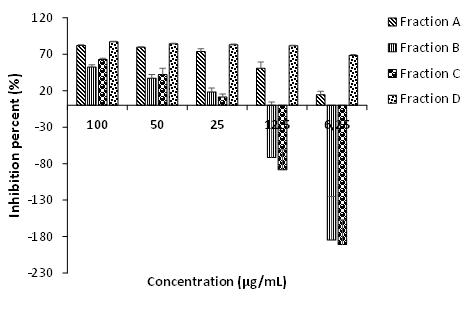
The percentage of inhibition of α-amylase inhibitory activity (percentage of inhibition) is dose-dependent except at 100 µg/mL concentration where it decreases with the fraction D, from 61.95% ± 5.05 at 50 µg/mL to 37.68% ± 4.71 at 100 µg/mL (Figure 4). Fraction C and fraction D had the highest inhibitory concentrations of 691.1 and 604.4 µg/mL.
| Concentration (μg/mL) | Inhibitory activity (%) | |
|---|---|---|
| 500 | 31.43±3.99 | |
| 250 | 28.68±1.77 | |
| 125 | 26.47±2.23 | |
| 62.5 | 23.90±3.35 | |
| 31.25 | 20.77±1.77 | |
| IC50 | ≈ 0 |
Table 3: α-amylase inhibitory activity of Acarbose, positive control. The highest inhibitory concentrations were obtained with fr
| IC50(μg/mL) | |
|---|---|
| Fraction A | 63.51 |
| Fraction B | 221.5 |
| Fraction C | 691.1 |
| Fraction D | 604.4 |
Table 4: α-amylase inhibitory activity of Fraction A, Fraction B, Fraction C, and Fraction D.
Discussion
With the increasing number of diabetics worldwide, and given the limitations of existing drugs, more and more research has intensified to find new molecules that are effective and accessible. Plants are an inexhaustible source of these new molecules. Musanga cecropioides is an herb traditionally used in the treatment of diabetes [9]. Nyunaï, et al. [28] highlighted the anti-hyperglycemic potential of the water-ethanol extract of Musanga cecropioides stem bark effect. It is, therefore, seemed useful to us from the perspective of determining the active compounds of this plant to determine the α-glucosidase and α-amylase activity of the fractions derived from this plant and evaluate the effect of aqueous extract in maltose tolerance test, and starch tolerance test.
The carbohydrate test revealed that the aqueous extract of M. cecropioides used at doses of 250 mg/kg and 500 mg/ kg for the hyperglycemia induced by maltose or starch carried out in normal male rats does not reduce the Area under the Curve, unlike glibenclamide. This would suggest that the aqueous extract of M. cecropioides does not act in the same way as glibenclamide. Previous work has shown that the water-ethanol extract of stem bark of Musanga cecropioides extracts reduced blood glucose levels in diabetic rats and normal rats during the glucose tolerance test [28]. The ineffectiveness of the aqueous extract in this study could be explained by the fact that maltose and starch are polysaccharides and release more glucose molecules.
Results revealed a dose-dependent response because increasing the concentration of fractions extract increased the inhibitory activity. Likewise, in terms of half-maximal inhibitory concentration (IC50) which is the concentration needed for the sample to inhibit α-glucosidase by 50%, Table 2 shows that fraction B has a highest IC50 value of 83.10 µg/ mL compared to fraction D with IC50 value of 0.7 µg/mL and the highest inhibitory activity of 87,097% with IC50 at 0.7 µg/ mL. This suggests the presence of α-glucosidase inhibitors separated from other compounds in the extract. The results of the present study indicate that out of acarbose, fraction A and fraction D showed the maximum α-glucosidase inhibitory activity. Fraction D is the most active extract in this test because it has the lowest IC50 value. This result is related to phytochemical compounds in the fraction extract that can inhibit α-glucosidase, in contrast to acarbose which is a pure compound, besides, to determine the bioactive compounds of these fractions, it is necessary to characterize them. The expected bioactive compounds could be flavonols or phenolic acids as the literature indicates a big link among polyphenols and anti-diabetic action of plant extracts [32]. As friedelan-3-one (triterpenoid) is the major compound in Fraction A (result not shown), suggest that this compound has α-amylase inhibition properties and might be responsible for a major part of that activity of the total extract of M. cecropioides.
Fractions B and C showed a negative value, which indicated that no inhibition occurred at 6.25 µg/mL and 12 µg/mL. Some of the negative values were quite large, e.g. especially fraction C at 6.25 µg/mL gave a negative value of -190.86% and this could indicate that the α-glucosidase is activated rather than inhibited. If this were to occur in vivo, it would aggravate, rather than alleviate, the diabetic condition since the rate of glucose production would be increased and thereby the serum levels rise more rapidly. However, it might be that an increase in the reaction product levels may be due to a conformational change derived from the binding of compounds to the enzyme [33]. This could also indicate that these fractions no longer contained α-glucosidase inhibitors. Since these fractions were collected in the latter part of elution, they may already contain wash residues, pigments and other flow-through components which are not α-glucosidase inhibitors but larger molecules that might mask the activity of the α-glucosidase inhibitor activity.
The treatment goal of diabetes patients is to maintain near-normal levels of glycemic control, in both the fasting and postprandial states. One of the strategies to monitor blood glucose for type II diabetes is to either inhibit or reduce the release of glucose from the small intestine. Many natural resources have been investigated for the suppression of glucose production from carbohydrates in the gut or glucose absorption from the intestine [34]. Thus, natural products of great structural diversity are still a good source for finding such inhibitors, thereby motivating to explore biologically active compounds from the extremely various plants. α-amylase catalyzes the hydrolysis of α-(1,4)-glucosidic linkages of starch, glycogen, and various oligosaccharides and α-glucosidase further breaks down the disaccharides into simpler sugars, readily available for the intestinal absorption. The inhibition of their activity in the digestive tract of humans is considered to be effective to control diabetes by diminishing the absorption of glucose decomposed from starch by these enzymes [35]. Therefore, effective and non- toxic inhibitors of α-amylase and α-glucosidase have long been sought. Several α-glucosidase inhibitors have been remoted from medicinal plants for the development of the latest drugs with increased efficiency and decrease adverse effects than the present medication [36].
Conclusion
The results show for the first time, the potential role of fractions from M. cecropioides to inhibit α-glucosidase and α-amylase inhibition activities. Due to it having the best inhibitory α-glucosidase effect, the fraction D of M.
cecropioides will be selected for further investigation, involving bioassay-guided fractionation, to isolate the constituents responsible for the inhibitory effect of the Fraction. Given the results obtained, the mechanism of action of this plant would mainly be through the inhibition of α-glucosidase, but also secondarily through the presence of the inhibiting α-amylase compounds.
Acknowledgment
The authors are thankful to The World Academy of Sciences (TWAS), for the research grant N° TWAS Research Grant Award_17-432 RG_BIO_AF awarded to Dr. NYUNAÏ NYEMB, Senior Researcher at Medical Research Centre, Institute of Medical Research and Medicinal Plants Studies (IMPM), Yaoundé, Cameroon.
Availability of Data and Materials
The datasets generated during and/or analyzed during the study are available from the corresponding author on request.
Conflict of Interest
The authors declare no conflict of interest.
References
-
Akendengué B (1992) Medicinal plants used by the Fang traditional healers in Equatorial Guinea. J Ethnopharmacol 37(2): 165-173.
-
Akendengué B, Louis AM (1994) Medicinal plants used by the Masango people in Gabon. J Ethnopharmacol 41(3): 193-200.s
-
Fomogne FMCY, Van VS, Ndinteh DT, Krause RWM, Olivier DK (2014) Antibacterial activities of plants from Central Africa used traditionally by the Bakola pygmies for treating respiratory and tuberculosis-related symptoms. J Ethnopharmacol 155(1): 123-131.
-
Burkill HM (1985) The Useful Plants of West Tropical Africa. In: Farinhes AD, (Eds.), 2nd (Edn.), Royal Botanical Gardens, Kew 1: 346-349.
-
Adeneye AA (2005) The hypotensive and toxicity studies on the aqueous crude extract of stem bark of Musanga cecropioides. MSc Pharmacology Dissertation. Postgraduate School, Usmanu Danfodiyo University, Sokoto.
-
Gill LS (1994) Ethnomedical uses of plants in Nigeria. University of Benin Press, Benin City, pp: 170.
-
Irvine FR (1961) Wood plants of Ghana. Oxford University Press, London, pp: 446-447.
-
Kamanyi A, Bopelet M, Tatchum TR (1992) Contractile effect of some extracts from the leaves of Musanga cecropioides (Cecropiaceae) on Uterine Smooth Muscle of the rat. Phytotherapy Research 6(3): 165-167.
-
Adeneye AA, Ajagbonna OP, Ayodele OW (2007) Hypoglycemic and antidiabetic activities of the stem bark aqueous and ethanol extracts of Musanga cecropioides in normal and alloxan-induced diabetic rats. Fitoterapia 78(7-8): 502-505.
-
Owolabi OJ, Ayinde BA, Nworgu ZA, Ogbonna OO (2010) Antidiarrheal evaluation of the ethanol extract of Musanga cecropioides stem bark. Methods Find Exp Clin Pharmacol 32(6): 407-411.
-
Ayinde BA, Omogbai EKI, Onwukaeme DN (2010) Hypotensive effects of 3, 4-dihydroxybenzyaldehyde isolated from the stem bark of Musanga cecropioides. Journal of Pharmacognosy and Phytotherapy 1(1): 4-9.
-
Dongmo AB, Kamanyi A, Bopelet M (1996) Saponins from the leaves of Musanga cecropioides constitute a possible source of potent hypotensive principle. Phytotherapy Research 10(1): 23-27.
-
Ayinde BA, Omogbai EKI, Onwukaeme DN (2003) Pharmacognostic characteristics and hypotensive effects of the stem bark of Musanga cecropioides. West African journal of pharmacology and drug research 19 (1-2): 37- 41.
-
Lontsi D, Sondengam BL, Bodo B, Martin MT (1998a) Kalaic acid, a new Ursane – type Saponin from Musanga cecropioides. Planta Med 64(2): 189-191.
-
Ayinde BA, Onwukaeme DN, Nworgu ZAM (2006) Oxytocic effects of the water extract of Musanga cecropioides R. Brown (Moraceae) stem bark. African Journal of Biotechnology 5 (14): 1350-1354.
-
Medou F, Nyunaï N, Bika Lele E, Oumarou G, Metsadjio AN (2019) Acute and 28 days Toxicity Assessment of Aqueous Extract of Stem Back of Musanga cecropioides (Urticaceae). Advances in Complementary & Alternative Medicine 4(3): 337-345.
-
Lontsi D, Sondengam BL, Ayafor JF, Connolly JD (1987) Cecropiacic acid, A new pentacyclic A-Ring Seco triterpenoid from Musanga cecropioides. Tetrahedron Letters 28(52): 6683-6686.
-
Lontsi D, Sondengam BL, Ayafor JF (1989) Chemical Studies on the Cecropiaceae - A novel A-ring seco triterpene from Musanga cecropioides. Journal of Natural Products 52(1): 52-56.
-
Lontsi D, Sondengam BL, Ayafor JF, Tsoupras MB, Taracchi R (1990) Further Triterpenoids of Musanga cecropioides - the structure of Cecropic acid. Planta Med 56(3): 287-289.
-
Ojinnaka Chukwunonye M, Okogun JI (1985) The Chemical Constituents of Musanga cecropioides. J Nat Prod 48(2): 337-337.
-
Lontsi D, Sondengam BL, Martin MT, Bodo B (1991a) Seco-ring-A triterpenoids from the rootwood of Musanga cecropioides. Phytochemistry 30(5): 1621-1624.
-
Lontsi D, Sondengam BL, Martin MT, Bodo B (1991b) Musancropic acids A and B: A-ring contracted triterpenes from Musanga cecropioides. Phytochemistry 30(7): 2361-2364.
-
Lontsi D, Sondengam BL, Martin MT, Bodo B (1992) Musangicic acid, a triterpenoid constituent of Musanga cecropioides. Phytochemistry 31(12): 4285-4288.
-
Lontsi D, Sondengam BL, Bodo B, Martin MT (1998b) Cecropioic acid - a pentacyclic triterpene from Musanga cecropioides. Phytochemistry 48(1): 171-174.
-
Ayinde BA, Onwukaeme DN, Omogbai EK (2007) Isolation and characterization of two phenolic compounds from the stem bark of Musanga cecropioides R. Brown (Moraceae). Acta Pol Pharm 64(2):183-185.
-
Lin WL, Hsieh YJ, Chou FP, Wang CJ, Cheng MT, et al. (2003) Hibiscus protocatechuic acid inhibits lipopolysaccharide-induced rat hepatic damage. Arch Toxicol 77(1): 42-47.
-
Lacaille DMA, Frank U, Wagner H (2001) Search for potential Angiotensin converting enzymes (ACE) - inhibitors from Plants. Phytomedicine 8(1): 47-52.
-
Nyunaï N, Gbaweng Yaya AJ, Tabi Nkoulou TG, Tchamgoue Deutou A, Ngondé M C, et al. (2016) Anti-hyperglycemic and Antioxidant Potential of Water-Ethanol Extract of Musanga cecropioides Stem Bark. International Journal of Pharmaceutical Sciences and Drug Research 8(1): 43- 49.
-
Mogale MA, Mkhombo HM, Lebelo SL, Shai LJ, Chauke MA, et al. (2012) The effects of Clause naanisata (wild) Hook leaf extracts on selected diabetic related metabolizing enzymes. Journal of Medicinal Plants Research 6(25): 4200-4207.
-
Sheikh JH, Iyo, Tsujiyama MT, Md Ashabul I, Rajat SB, et al. (2008) A Total Phenolic Content, Anti-oxidative, antiAmylase, Anti-Glucosidase and AntiHistamine release activities of Bangladeshi fruits. Food Science and Technology Research 14(3): 261-268.
-
Kim J, Yang J, Kim M (2011) Alpha glucosidase inhibitory effect, anti-microbial activity and UPLC analysis of Rhus verniciflua under various extract conditions. Journal of Medicinal Plant Research 5(5):778-783.
-
Maria JKM, Rajesh J, Mandal AKA, Sampath N (2011) Antioxidant and antimicrobial activity of individual catechin molecules: A comparative study between gallated and epimerized catechin molecules. European Journal of Experimental Biology 1(3): 145-153.
-
Kim JS, Kwon CS, Son KH (2000) Inhibition of α-glucosidase and amylase by luteolin, a flavonoid. Biosci Biotechnol Biochem 64(11): 2458-2461.
-
Matsui T, Tanaka T, Tamura S, Toshima A, Tamaya K, et al. (2007) Alpha-glucosidase inhibitory profile of catechins and theaflavins. J Agric Food Chem 55(1): 99-105.
-
Hara Y, Honda M (1990) The inhibition of α-amylase by tea polypphenols. Agricultural and Biological Chemistry 54(8): 1939-1945.
-
Matsuda H, Nishida N, Yoshikawa M (2002) Antidiabetic principles of natural medicines. V. Aldose reductase inhibitors from Myrcia multiflora DC. (2): Structures of myrciacitrins III, IV, and V. Chem Pharm Bull (Tokyo) 50(3): 429-431.
- Acido Labile or Gastro Irritant Apis and Enteric Release in Galenic Practice: An Overview
- A Study on Knowledge, Attitude and Practice of Hand Hygiene among Healthcare Professionals at a Tertiary Care Hospital, India
- Influence of Inoculum Concentration on In Vivo Incubation Period of Emmia lacerata, Pathogenesis and Management of Wilt in Pepper (Capsicum annuum L.)
- Vanilla’s Chemistry
- Marine Anti-Cancer Compounds and Adverse Effects of Global Warming on Oceans: An Overview
- Serological Investigation of Chikungunya Virus Antibody among Malaria-Suspected Febrile Patients in Some Healthcare Facilities in Rivers State