Protective Effect of Protocatechuic Acid in Genotoxicity-Induced by Carbon Tetrachloride: A Preliminary Study
Carbon tetrachloride (CCl4) is commonly utilized as a solvent, a refrigerant, and a dry-cleaning agent. However, its genotoxic effect has been well documented. The present work was designed to assess the genotoxic effect of carbon tetrachloride in the bone marrow of rats. The safety and the possible rotective effect of protocatechuic acid (PCA) in the genotoxicity induced by carbon tetrachloride (CCl4) were evaluated using a micronucleus assay. Rats were divided into six groups where groups I and II served as the control. Group III was exposed to CCl4 only at 3mL/kg intraperitoneally. Groups IV and V rats were pretreated with PCA at 10mg/kg and 20 mg/kg respectively before administering CCl4. Group VI received PCA only (20mg/kg) for 7 consecutive days. At the end of the experiment, a micronucleus assay was carried out. There was a significant MnPCE in the bone marrow of CCl4-treated rats as compared with control (p<0.05). The administration of PCA at the doses of 10 and 20mg/kg significantly reduced the MnPCE when compared with the group treated with CCl4 (3ml/kg) only (p<0.05). The data provided in this study considered PCA to be relatively safe, non-genotoxic and can modulate the genetic damage involved in CCl4 toxicity by decreasing the frequency of micronucleated cells.
Introduction
Exposure to toxic chemicals and drugs can cause harmful effects through the generation of reactive oxygen species (ROS). A higher concentration of ROS can react with DNA, cell membranes and proteins including other molecules, induce cellular damage and release other and more reactive radicals [1]. Consequently, the generation of ROS initiates DNA strand breaks and oxidative DNA damage that bring about alterations in mRNA expression of DNA damage-responsive genes [2].
Carbon tetrachloride is a poly-chlorinated hydrocarbon that has an ozone-depletion potential. Over the years, it has been in use as a liquid solvent Sayed, et al. [3] and can escape into the surrounding environment in the form of vapour [4]. CCl4 is a known toxicant that is employed in scientific experiments for the induction of hepatotoxicity El Rabey, et al. [5], neurotoxicity Dong, et al. [6] and genotoxicity Alkreathy, et al, [7] in animals. As a result of its broad usage, CCl4 has been gaining the focus of genotoxicity studies both in prokaryotic and eukaryotic systems. Biotransformation of CCl4 is required for the exertion of its genotoxicity and generation of ultimate genotoxic metabolites [8]. It exerts its toxicity through bioactivation to the trichloromethyl radical form that can covalently block biological macromolecules such as DNA and proteins. These reactive oxygen species generated modify the structure and function of cellular and intracellular membranes causing hepatotoxicity and genotoxicity [9]. The concentration used and the route of administration of CCl4 influence different degrees of genotoxicity in different tissues.
Therefore to protect the body from the deleterious effects of free radicals generated from toxicants like CCl4, several endogenous enzymatic and non-enzymatic systems are provided in the body system. However, when the formation of free radicals is excessive, additional protective mechanisms of dietary antioxidants may be of great importance [10].
Protocatechuic acid (3, 4-dihydroxy benzoic acid), is a natural antioxidant and phenolic acid which is isolated from various plants. It has been shown to present a broad variety of biological activities such as hepatoprotective, anticancer, antioxidant, antihyperglycemic, nephroprotective Adeyanju, et al. [11] and neuroprotective effects [12].
Given the inherent antioxidant properties of protocatechuic acid, this study explores the protective effect of PCA against genotoxicity induced by carbon tetrachloride in the bone marrow of rats.
Chemicals and Reagents
Carbon tetrachloride was purchased from Zurius Life Sciences Pvt. Ltd. (India). All other chemicals and reagents used in this experiment were of analytical grade.
Animals
Thirty male albino Wistar rats weighing 150-200g were obtained. The acclimatization of the rats was for two weeks. The rats were randomly divided into 6 groups (five rats per group). They were placed in a conventional room with a 12-hour light/12-hour dark cycle. The rats were housed in plastic cages, had access to a commercial diet and water was given ad libitum.
Animal Ethics
The animals used received humane care in compliance with standard guidelines set up for the Care and Use of Laboratory Animals for animal experiments. The ethical regulations have been duly observed in line with the established national and institutional guidelines for the protection of animals’ welfare during experiments.
Experimental Design
Rats were divided into 6 groups of 5 animals. Group 1 (positive control) was given physiological saline (1ml/kg). Group II (negative control) was given olive oil as the vehicle (1ml/kg). Group III was administered intraperitoneally with a single injection of 3mL/kg CCl4 in a suspension of olive oil (1:1 V/V) to induce genotoxicity on the last day of administration. Group IV was given 10mg/kg of PCA for 7 days and on the 7th day, 3mL/kg of CCl4 was administered. Group V received PCA orally for 7 days and then CCl4 (3mg/ kg) on the 7th day. Group VI received 20mg/kg of PCA only for 7 days. Rats were sacrificed by cervical dislocation 24 hours after the CCl4 challenge. Bone marrow was flushed from both femurs of each rat and spread onto slides. Slides were coded and then air-dried, fixed with methanol and stained with maygrunward stain. Bone marrow cells were then examined microscopically and scored per animal for the frequency of micronucleated polychromatic cells in each of the five animals per dose group.
Data Analysis
Results were expressed as mean ± standard error of the mean. The statistical analysis was evaluated using a one-way analysis of variance of Statistical Package for Social Sciences software for Windows version 16 (SPSS Inc., Redmond, WA, USA). Post-hoc testing was done for intergroup comparisons using the least significant difference. The level of statistical significance was p<0.05.
Result
The data in Table 1 and figure 1 showed that rats treated with CCl4 (3ml/kg) exhibited a significantly (p<0.05) high frequency (9.00 ± 0.67) of micronucleated polychromatic erythrocytes in bone marrow cells as compared with the negative control (2.70 ± 0.32). Administration of protocatechuic acid at both doses significantly lowered the number of micronucleated cells when compared with the group treated with CCl4 only (p<0.001). Treatment of rats with protocatechuic acid alone at the highest of (20ml/kg) did not significantly change micronucleated polychromatic erythrocyte (2.40 ± 0.34) when compared with the positive control (2.50 ± 0.42) (p>0.05).
| Treatment Group | Number of micronucleated cells/1000 polychromatic |
|---|---|
| Group | erythrocyte cells |
| Control (Saline) | 2.50 ± 0.42 |
| Olive Oil (1ml/kg) | 2.70 ± 0.32 |
| CCl (3ml/kg) 4 | 9.00* ± 0.67 |
| PCA (10mg/kg) + CCl 4 | 5.30** ± 0.42 |
| PCA (20mg/kg) + CCl 4 | 3.20** ±0.33 |
| PCA only (20mg/kg) | 2.40 ± 0.34 |
Table 1: Genotoxicity induced by Carbon tetrachloride and effect of protocatechuic acid.
Values are expressed as mean ± Standard error of mean (SEM). *Significantly different from control (p<0.05); **Significantly different from CCl4-treated rats (p<0.001). Table 1: Genotoxicity induced by Carbon tetrachloride and effect of protocatechuic acid.
Discussion
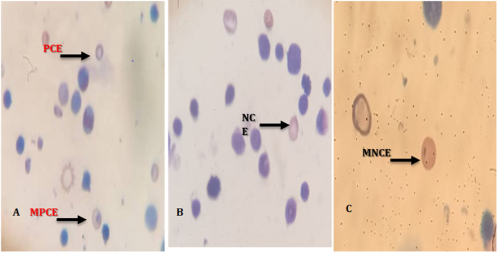
Micronucleus assay can identify spindle poisons Thomson and Perry [13] that give rise to the manifestation of large micronuclei (MN) Yamamoto, et al. [14]. In the present study, there is an appearance of MN that ranged from small to large size in the rats treated with CCl4. The findings of the study revealed that CCl4 had a major impact on the induction of MN. The results are in harmony with the findings from the studies of Abdou, et al. [15] and El-Shorbagy [16] which stated that CCl4 increased the occurrence of MN in the bone marrow of male mice. The significant increase in MN formation could be ascribed to part of a deletion arising from a DNA break or poor repair mechanism of the DNA double- strand [17, 18]. This could have cytotoxic effects and disrupt normal bone marrow cell proliferation. A Comet assay carried out by Mosallam [18] revealed the genotoxic potential of CCl4 in rats. This further strengthens our finding about the genotoxic tendency of CCl4. Administration of protocatechuic acid was able to decrease the frequency of micronucleated cells observed in CCl4-treated rats. This shows the significant influence of protocatechuic acid in reducing the cytogenetic effects of CCl4 on bone marrow cells. Its antigenotoxic effect has also been demonstrated by Anter, et al. [19, 20]. The mechanism by which protocatechuic acid exerts this antigenotoxic effect may be by antioxidant properties and its ability to scavenge ROS as a polyphenolic compound.
Further studies still need to be done to determine the signalling pathways activated by protocatechuic acid in response to the genotoxic response induced by carbon tetrachloride in this study.
Conflicts of Interest
The authors declare that they have no conflict of interest.
References
-
Usui T, Foster SS, Petrini JH (2009) Maintenance of the DNA-damage checkpoint requires DNA-damage-induced mediator protein oligomerization. Mol Cell 33(2): 147- 159.
-
Petković J, Žegura B, Stevanović M, Drnovšek N, Uskoković D, et al. (2011) DNA damage and alterations in expression of DNA damage responsive genes induced by TiO2 nanoparticles in human hepatoma HepG2 cells. Nanotoxicology 5(3): 341-353.
-
Sayed EA, Badr G, Hassan KAH, Waly H, Ozdemir B, et al. (2021) Induction of liver fibrosis by CCl4 mediates pathological alterations in the spleen and lymph nodes: The potential therapeutic role of propolis. Saudi J Biol Sci 28(2): 1272-1282.
-
Fahmy MA, Diab KA, Abdel-Samie NS, Omara EA, Hassan ZM (2018) Carbon tetrachloride induced hepato/ renal toxicity in experimental mice: antioxidant potential of Egyptian Salvia officinalis L essential oil. Environ Sci Pollut Res Int 25(28): 27858-27876.
-
El Rabey HA, Rezk S, Sakran MI, Mohammed GM, Bahattab O, et al. (2021) Green coffee methanolic extract and silymarin protect against CCl4-induced hepatotoxicity in albino male rats. BMC complement Medical and Therapies 21(1): 1- 11.
-
Dong S, Chen QL, Song YN, Sun Y, Wei B, et al. (2016) Mechanisms of CCl4-induced liver fibrosis with combined transcriptomic and proteomic analysis. J Toxicol Sci 41: 561-572.
-
Alkreathy HM, Khan RA, Khan MR, Sahreen S (2014) CCl4 induced genotoxicity and DNA oxidative damages in rats: Hepatoprotective effect of Sonchus arvensis. BMC Complementary Altern Med 21: 414-452.
-
Doherty AT, Ellard S, Parry EM, Parry J (1996) An investigation into the activation and deactivation of chlorinated hydrocarbons to genotoxins in metabolically competent human cells. Mutagenesis 11(3): 247-274.
-
Ingawale DK, Mandlik SK, Naik SR (2014) Models of hepatotoxicity and the underlying cellular, biochemical and immunological mechanism (s): a critical discussion. Environ Toxicol Pharmacol 37(1): 118-133.
-
Tirkey N, Pilkhwal S, Kuhad A, Chopra K (2005) Hesperidin, a citrus bioflavonoid, decreases the oxidative stress produced by carbon tetrachloride in rat liver and kidney. BMC pharmacol 31(5): 2.
-
Adeyanju AA, Molehin OR, Asejeje FO, Oyenuga V, Etokakpan RU (2022) Protocatechuic acid through modulation of signaling pathways and oxidative stress exerts protective effects in rat model of carbon tetrachloride-induced renal and reproductive toxicities. Comp Clin Pathol 31: 465-474.
-
Masella F, Santangelo C, D’Archivio M, Li Volti G, Giovannini C, et al. (2012) Protocatechuic acid and human disease prevention: biological activities and molecular mechanisms. Curr Med Chem 19(18): 2901- 17.
-
Thomson EJ, Perry PE (1988) The identification of micronucleated chromosomes: a possible assay for aneuploidy. Mutagenesis 3(5): 415-418.
-
Yamamoto KI, Yasumoto K (1980) A comparison of diameters of micronuclei induced by clastogens and by spindle poisons. Mutation Research/ Fundamental and Molecular Mechanisms of Mutagenesis 71(1): 127-131.
-
Abdou HS, Salah SH, Booles HF, Abdel Rahim EA (2012) Effect of pomegranate pretreatment on genotoxicity and hepatotoxicity induced by carbon tetrachloride (CCl4) in male rats. Journal of Medicinal Plants Research 6(17): 3370-3380.
-
El-Shorbagy HM (2017) Molecular and anti-oxidant effects of wheat germ oil on CCl4-induced renal injury in mice. Journal of Applied Pharmaceutical Science 7(5): 94-102.
-
Lin B, Qi X, Fang L, Zhao L, Zhang R, et al. (2021) In vivo acute toxicity and mutagenic analysis of crude saponins from Chenopodium quinoa Willd husks. RSC Advances 11(8): 4829-4841.
-
Mosallam S (2020) Efficacy of ascorbic acid against toxicity initiated by mono-sodium glutamate on Albino mice. Journal of Pharmacy and Biological Sciences 15(1): 1-8.
-
Anter J, Romero-Jiménez M, Fernández-Bedmar Z, Villatoro-Pulido M, Analla M, et al. (2011) Antigenotoxicity, cytotoxicity, and apoptosis induction by apigenin, bisabolol, and protocatechuic acid. J Med Food 14(3): 276-283.
-
Adeyanju AA, Asejeje FO, Molehin OR, Owoeye O, Olatoye EO, et al. (2021) Protective role of protocatechuic acid in carbon tetrachloride-induced oxidative stress via modulation of proinflammatory cytokines levels in brain and liver of Wistar rats. Journal Basic Clin Physiol Pharmacol 33(2): 143-154.
- Pattern of Gonadal Hormones in Oral Testosterone-Supplimented Male Wistar Rats with Diabetes-Induced Hypogonadism
- Re-Evaluation of the Genotoxicity of Currently Used Food Dyes in Mouse Multiple Organs Via Continuous Administration by Drinking Using the Comet Assay
- Pharmacogenetics of Type 2 Diabetes Mellitus: Linking Genetic Variability to Drug Efficacy and its Cardiovascular Outcomes
- Exploratory Proteomic Profiling of SARS-CoV-2 Infected THP-1 Macrophages Reveals Alterations in Inflammatory Response and Cellular Metabolism
- Study of Genotoxicity of Hepatocarcinogens in Multiple Organs in Mice by Feeding and Drinking Using the Comet Assay
- Spirulina Polypeptides Inhibit the Growth of Human Lung Tumor (H460) Cells