Androgenic Anabolic Steroids Perturb Hepatocellular Tissue Damage Mediated by Oxidative Stress in the Presence of Endogenous Antioxidant
Objective of the study: Anabolic androgenic steroids (AASs) unlawfully misused for their anabolic effects has deleterious effects of major concerns in public health even in the presence of endogenously produced antioxidants. This study is to evaluate the interaction between AAS metabolites and endogenously produced superoxide dismutase (SOD) activity in AAS treatment and AAS abstinence. Materials and Methods: Twenty (20) adult male Wistar used were divided into four (4) groups, A and B served as the positive and negative control given normal saline and olive oil respectively, C and D was given 120mg/kg/bodyweight oral AAS for 21 days while D served as the 7 days withdrawal group after AAS treatment by evaluating histopathological changes in the liver histology, mucin/ glycogen granules, network of reticulum fibers, liver function enzymes; alanine aminotransferase (ALT) and aspartate aminotransferase (AST), lipid peroxidation enzyme malondialdehyde (MDA) and antioxidant enzyme. Results and Discussion: AAS treatment perturbed hepatocellular membrane integrity seen in the increase MDA, AST and ALT activity which caused disruption of the hepatocytes cell integrity attributed to a decline in endogenous SOD. However, AAS withdrawal group gradually reversed AAS mediated heaptocellular pathogenesis associated with progressive elevation of endogenous SOD, decline in MDA and activities of liver function test. Conclusion: AAS mediates hepatic injury through lipid peroxidation and a decline in endogenous antioxidant enzyme, withdrawal of AAS slows down the oxidative damage process when the endogenous antioxidant production increases and mobs off ROS generated by AAS ingested thereby protecting against liver injury and hepato-cellular break down.
Introduction
Androgenic anabolic steroids (AAS) are basically synthetic analogue of testosterone which binds to androgen receptors (ARs) thereby mediating muscle enhancement by promoting protein synthesis [1, 2]. They are mainly consumed illegally by sportmen and non-athletes for esthetic purposes Park M, et al. [3, 4] and this has several adverse side effects Gagliano-Jucá T, et al. [5] such as kidney dysfunction Barakat LA, et al. [6], cardiovascular dysfunction Tousson E, et al. [7] liver dysfunction Ziaolhagh SJ, et al. [8] and testicular disruption [9].
The abuse of AAS is of public concern because the global life expectancy of young adults recorded to have used AAS without medical prescription is between 1.5- 4% Sagoe D, et al. [10, 11] as the drug can potentiate brain and cardiac dysfunction [12].
Of all the class of anabolic androgenic steroids, testosterone undecanoate, danazol, and oxandrolone, are the commonly consumed for it helps to increase lean muscle protein synthesis and decline body fat mass [13].
The liver is easily prone to toxic side effects of AAS, since it is the main site for drug metabolism and steroid clearance [14]. According to Albano, et al. [15] hepatoxicity due to drug intoxication is characterized by an increased infiltration of the liver parenchyma by neutrophils, lymphocytes, and eosinophil’s Niedfeldt MW [14, 16] resulting in alteration of the normal histoarchitecture of the hepatic tissue reportedly presented like ground glass appearance of the hepatocytes, and liver damage [17].
The pathogenesis of AAS liver injury is characterized by an elevation in liver function enzymes, severe vascular injury to the liver [14]. This damaging effect is proposed to be associated with toxicant mediating oxidative stress that activates androgen receptors which leads to alterations in the mitochondrial enzymes leading to mitochondrial degeneration in the hepatic cells [18]. The oral forms are resistant to immediate degradation and hepatotoxic Castellanos MM, et al. [19] but their parenteral administration also leads to hepatic dysfunction [20].
Testosterone and other class of AASs has the ability to cross the blood into cell membrane via connected intra- cytoplasmic androgenic receptors., to their target site which is the cell nucleus to alter DNA function, cellular metabolism and androgenic functions cellular activities [21, 22].
A report hypothesized/ suggested that the activation of androgen receptor (AR) in the hepatic cells is proposed to be linked with the generation of reactive oxygen species (ROS) that results in mitochondrial mediated hepatic degeneration [14, 18] Oxidative tissue damage is associated to cellular toxicity [23].
In recent years, great report has been linked to the role of endogenous and exogenous antioxidant for their role as a prophylactic or therapeutic agent against side effects of drug abuse [24, 25, 26, 27]. The disruption of redox signaling and antioxidants balance cannot be overemphasized [25].
Therefore, this research is being carried out to understand the interplay between AAS and endogenous antioxidant by assessment of histopathological changes in the liver (using H and E, PAS for mucin/ glycogen granules and reticulin stain for reticular fibers), lipid peroxidation for oxidative tissue damage, endogenous antioxidant activity of SOD and liver function enzymes (alanine aminotransferase (ALT) and aspartate aminotransferase (AST) activity.
Materials and Methods
Experimental Animal Procurement and Breeding
The twenty (20) adult male Wistar rats used for this study were reared in the Animal Holding Facility Department of Anatomy, Bingham University, Karu, Nasarawa State Nigeria. The Animal acclimatized for two weeks in aerated metallic cages under a standard environmental laboratory condition (12hr: 12hrs, dark and light cycles, room temperature) and fed pelleted rats’ feed (Vital feeds Limited, Mararaba, Nasarawa State Nigeria) and water, ad libitum. All experimental procedures aligned with guidelines stipulated by the Guide for the Care and Use of Laboratory Animals published by the US National Institutes of Health [28] and the National Research Council’s Guide for the Care and Use of Laboratory Animals [29] as approved by the Institutional Committee for Evaluation of Animal Use in Research.
Experimental Duration
The experiment was carried out for 28 days.
Experimental Animal Groups and Procedure
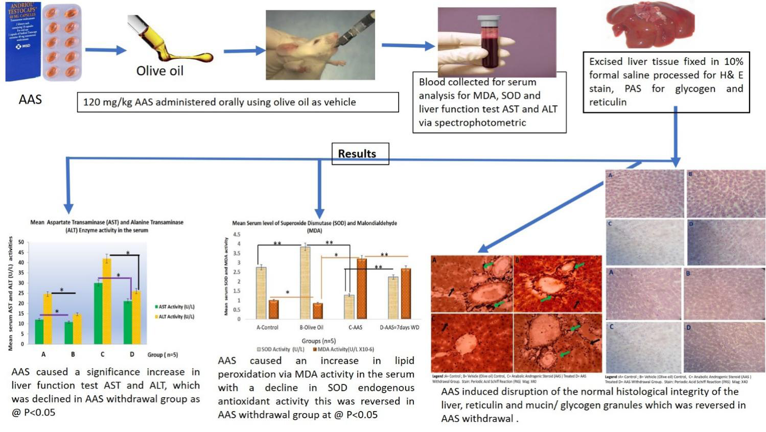
The twenty-adult male Wistar rat used were group into four (4) which are Group A serves as the control, B- given the oral Olive oil (used as a vehicle for drug administration) for 21 days, the group C – given oral 120mg/kg of Anabolic Androgenic Steroid (AAS) for 21days and group D – A recovery group for 7 days following 21 days of oral 120mg/kg AAS treatment.
**Drug of Study**
Testosterone Undecanoate (Andriol Testocaps, Netherlands) at a dose of 120mg/kg bodyweight was given orally using olive oil as vehicle as this was reported to enhance drug bioavailability and increased the rate of absorption for oral delivery because AAS has lipid-based formulations [30].
**Experimental Animal Euthanasia**
They were randomly euthanized by decapitation twenty-four hours after the last drug. Their final body weight was taken on the day of the last dose administration. The humane method was adopted using cervical dislocation was adopted.
Blood Sample and Liver Tissue Collection
The rats in the different experimental groups were fasted overnight, and blood samples obtained from retro-orbital plexus. The blood was centrifuge at 5,000 r.p.m for 10minutes and sera obtained aliquoted into sample cuvettes placed on ice ready for spectrophotometric analysis using commercial kits for each enzyme of study according to methods stipulated in the kit manual. The liver tissue was excised wet wight taken and rapidly fixed in 10% formol saline (buffered) for histopathological analysis.
**Estimation of AST and ALT Activity**
The liver function test was carried out using alanine aminotransferase (ALT) and aspartate aminotransferase (AST) enzyme assay kit. Their activity was measured at 520nm wavelength using a spectrophotometer. AST and ALT are used as biomarker to detect liver injury.0.5ml of Buffered Substrate (R1) was placed in a test tube and incubated in a water bath at a temperature of 37°C for 3 minutes. 0.1ml of serum was added to the test solution while distilled water was added to the standard blank and working pyruvate to the standard solution. All solutions were mixed and incubated at 37°C for 60 minutes. 2,4-DNPH (R2), was added to all solutions after incubating. The mixtures were left to stand for 20 minutes at room temperature. 5ml of 0.4M NaOH was added to solutions, mixed and left to stand at room temperature for 5 minutes. The absorbance was read at 520nm and the enzyme activity was derived.
**Estimation of Antioxidant Enzyme-Superoxide Dismutase (SOD) Activity**
Superoxide dismutase (SOD) is the major endogenous antioxidant enzyme that provides first-line protection against oxidative stress mediated by generation of reactive oxygen species (ROS). Superoxide dismutase (SOD) was assayed with a spectrophotometer as stipulated by procedure in the commercial kit’s manufacturer (Northwest Life Sciences Specialties, Washington, United States).
**Estimation of Lipid Peroxidation Enzyme-Malondialdehyde Activity**
The malonaldehyde a biomarker to measure extent of lipid peroxidation. Small amounts of malonaldehyde (MDA) are produced during lipid peroxidation and these are able to react with thiobarbituric acid (TBA) to generate a pink-coloured complex which in an acidic solution absorbs light at 532nm.0.4ml of test sample was mixed with 1.6ml of TriskCl buffer to which 0.5ml of 30% TCA was added. Then 0.5ml of TBA was added and placed in a water bath to incubate for 45minutes at 80°C. The incubation produced a pink-coloured reaction mixture which was then cooled in ice and centrifuged at 4,000 r.p.m for 15 minutes. The absorbance of the clear pink supernatant was then read at 532nm. The plasma levels of MDA for lipid peroxidation were measured using the MDA commercial assay kit via a spectrophotometer according to the procedure supplied by the manufacturer (Abcam, Cambridge, MA, USA).
Calculation
$$MDA\left(\frac{\text{Units}}{\text{mg protein}}\right) = \text{absorbance} \times \text{volume of mixture}$$
$$E_{532} \times \text{Volume of sample} \times \text{mg protein}$$
Where $E_{532}$ is molar absorptivity at 532nm = 1.56×10^5
**Hepatic Tissue Histopathological Analysis**
The formalin-fixed liver tissue samples were processed using an automated tissue processor (LEICA TP) through been dehydrated in ascending ethanol grades, cleared in xylene, and impregnated and embedded in paraffin as described by Bancroft, et al. [31]; Suvarna, et al. [32] and Memudu, et al. [33] methods. The paraffin embedded liver tissue blocks were cut into sections of 5 µm thick each and then stained with H & E (hematoxylin and eosin) for histological analysis, PAS stain for mucin granules/ glycogen and Reticulin stain for reticular fiber Bancroft, et al. [31] and then cover slipped with Dibutylphthalate Polystyrene Xylene (DPX) mounting media (Sigma, USA).
Tissue Photomicrography
Sections of the stained liver tissues were visualized using an Olympus light microscope and micrographs captured with an attached digital microscopic camera (MV 500 Cameroscope).
Statistical Analysis
Statistical analysis was done using software GraphPad Prism 8.0 (GraphPad, USA). One-way analysis of variance (ANOVA) and Tukey post hoc test was used to for multiple comparison. Differences at p ≤ 0.05 were considered significant (*). The Shapiro-Wilk test was used to check all data for normality. All the values are expressed as mean ± SEM. The results from biochemical enzyme study were examined by Tukey post hoc test (for multiple comparisons) p values (*p < 0.05).
Results
AAS increased a Body Weight Gain
AAS increased final body weight in the treated group as compared with the control, Olive oil and AAS withdrawal groups. There was no significant change in the body weights of the control and Olive treated groups (Figures 1 & 2).
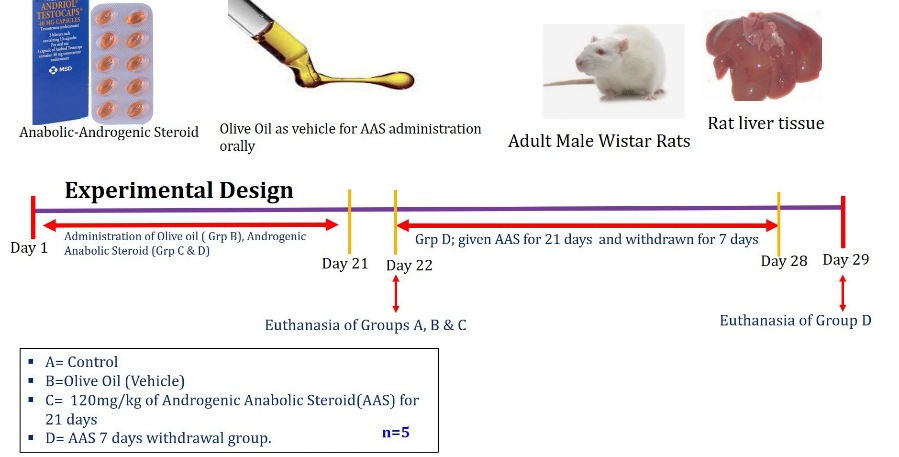
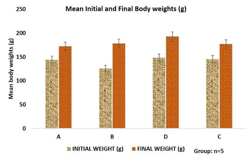
Figure 2: Graphical representation of the mean initial and final body weight of experimental animals. Data are expressed as the mean ± SD. Data analyzed using one way Analysis of Variance (ANOVA). Data (*) taken as significant statistically when p ≤ 0.05. Legend: A=Control, B=Olive Oil Control, B= 120mg/kg b.w.t/ daily AAS for 21days and D= the 7days withdrawal groups following 21 days of 120mg/kg AAS treatment.
AAS Treatment Elevated Absolute Weight of the Liver Tissue
This study shows that AAS administration increased the absolute weight of the liver when compared with the control, olive oil and AAS withdrawal group at p ≤ 0.05. However, there was no significance difference in the absolute weight of the liver in the control and olive oil groups at p ≤ 0.05 (Figure 3). The AAS withdrawal group has a significance decline in absolute liver weight as compared with the AAS treated group, but there was still a significant increase as compared with the groups A and B.
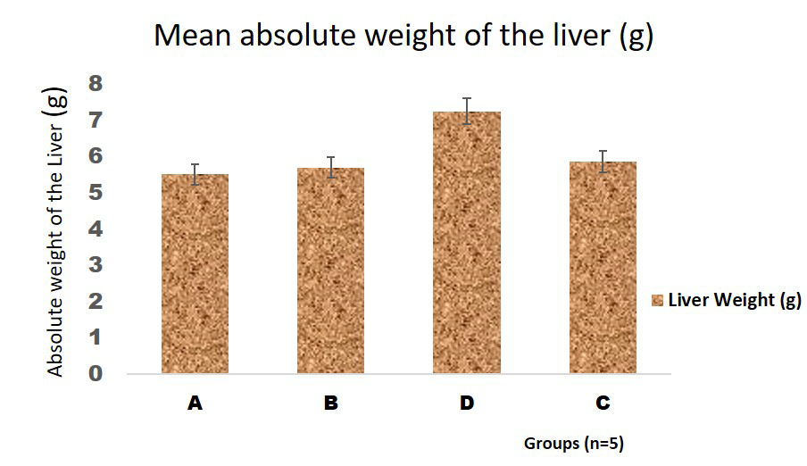
Figure 3: Graphical representation of the mean absolute weight of the liver of adult male Wistar rats used in this study. The data obtained were analyzed using one way Analysis of Variance (ANOVA) and obtained expressed as the mean ± SD. Data (*) taken as significant statistically when p ≤ 0.05. Legend: A=Control, B=Olive Oil Control, B= 120mg/kg b.w.t/ daily AAS for 21days and D= the 7days withdrawal group following 21 days of 120mg/kg AAS treatment.
AAS Induced Elevation of Serum Level of Aspartate Transaminase (AST) and Alanine Transaminase (ALT) Enzyme
Aspartate Transaminase (AST) and Alanine Transaminase (ALT) indicators for liver function and hepatic injury. In study, these enzymes were quantified to evaluate that status of liver function due to assault of AAS against the liver in the midst of endogenous antioxidant enzymes. It was deduced that AAS caused a significant increase in serum levels of AST and ALT enzymes relative to the control, olive oil and AAS withdrawal group (Figure 4). There was a reduction in AAS withdrawal study group relative to the AAS treated groups at p ≤ 0.05. It was observed that olive treated group had a decline in AST and ALT as compared with the control group. However, there was no significant difference in ALT in the AAS withdrawal and the control group.
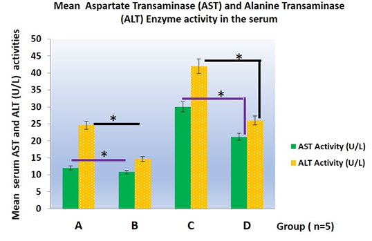
Figure 4: Graphical representation of the mean absolute weight of the liver of adult male Wistar rats used in this study. The data obtained were analyzed using one way Analysis of Variance (ANOVA) and obtained expressed as the mean ± SD. Data (*) taken as significant statistically when p ≤ 0.05. Legend: A=Control, B=Olive Oil Control, B= 120mg/kg b.w.t/ daily AAS for 21days and D= the 7days withdrawal groups following 21 days of 120mg/kg AAS treatment.
AAS Induced Oxidative Tissue Damaged Demonstrated by Enhancing Lipid Peroxidation and Reducing Endogenous Antioxidant Superoxide Dismutase (SOD)
Lipid peroxidation indicator is done by the assessment of biological maker malondialdehyde (MDA) and antioxidant enzyme superoxide dismutase (SOD) is the main endogenous antioxidant enzyme which acts against ROS mediated oxidative stress. This report demonstrated that AAS enables an increase in MDA activity that implies that there is production of ROS due to oxidative stress as compared with the control, olive oil and AAS withdrawal groups. Olive treated and AAS withdrawal groups shows a decline in MDA activity when compared with AAS treated group. AAS attenuated endogenous SOD activity in comparison to the control, olive oil and AAS withdrawal groups. However, AAS withdrawal group had a mild elevation in SOD activity that correlates with a reduction in MDA activity (Figure 5).
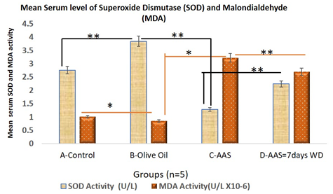
Figure 5: Graphical representation of the mean serum activity of malondialdehyde (MDA) and superoxide dismutase (SOD) which demonstrates changes in liver oxidative stress and lipid peroxidation in AAS treated experimental animals used in this study. The data obtained were analyzed using one way Analysis of Variance (ANOVA) and obtained expressed as the mean ± SD. Data (*) taken as significant statistically when p ≤ 0.05. Legend: A=Control, B=Olive Oil Control, B= 120mg/kg b.w.t/ daily AAS for 21days and D= the 7days withdrawal groups following 21 days of 120mg/kg AAS treatment.
Histological Result of the Liver
The histological presentation of the hepatic tissue appears normal in the livers of the control and olive oil study groups (Figures 6A, 6B & 6D). Which is characterized by well stained eosinophilic nucleus and basophilic cytoplasm, with some sinusoidal spaces and properly aligned hepatic plates of the hepatocytes. The AAS treated liver demonstrated histopathological characterization of liver injury presented as palely stained cytoplasm, ground glass appearance of the hepatocytes, some cellular swelling seen in hepatocytes with centrally located nuclei with cytoplasmic vacuolations and focal mononuclear cellular infiltrations within the hepatic tissues (Figure 6C). However, the AAS withdrawal group showed mild regenerative process in the liver tissue as compared with AAS treated study group characterized by a decline focal mononuclear cell infiltration, reduced ground glass appearance of hepatocytes, less vacuolation in the hepatocytes cytoplasmic areas and improved basophilic staining of the cytoplasm (Figure 6D).
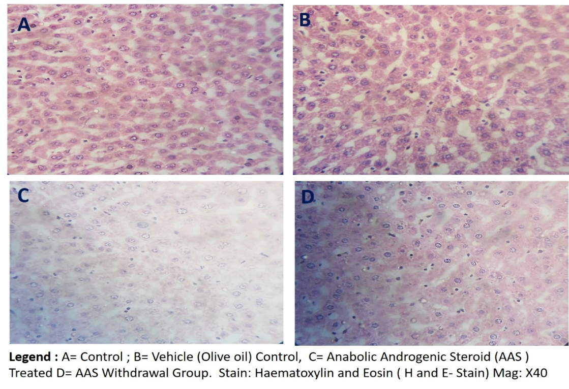
Figure 6: Representative photomicrograph of the H&E-stained hepatic tissue sections of the experimental Wistar rats of study, showing normal histological pictures in the vehicle control (A) and olive oil-treated (B) rats as compared with the AAS treated (C) rats. Legend: A=Control, B=Olive Oil Control, B= 120mg/kg b.w.t/ daily AAS for 21days and D= the 7days withdrawal groups following 21 days of 120mg/kg AAS treatment. Magnification x400, Scale bar 20 microns (20µm).
Histopathological Changes in Glycogen/ granules in the Hepatic Tissue Associated with AAS Administration
The glycogen granules were preserved in the cytoplasm of the hepatocytes in the livers of the control and olive oil study groups (Figures 7A, 7B & 7D) demonstrated in the positive PAS stain as compared with the AAS treated (Figure 7C) characteristic by loss of glycogen granules. However, we observed a progressive restoration of glycogen granules as seen in the moderately positive PAS stain (Figure 7D).
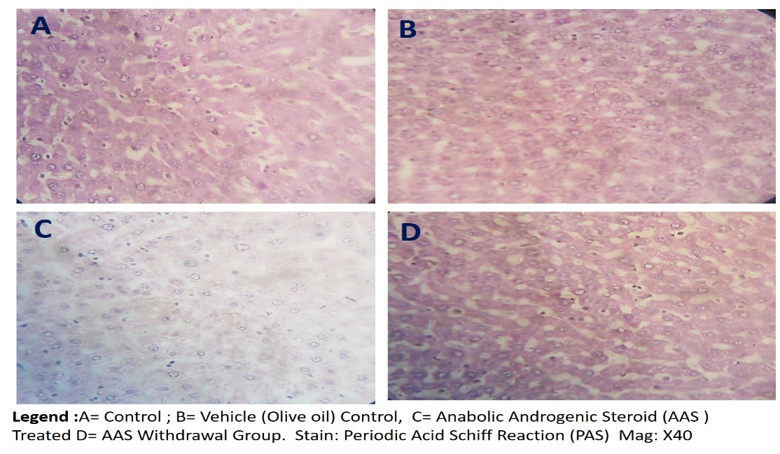
Figure 7: Representative photomicrograph of the periodic acid Schiff’s (PAS) reaction stain for glycogen granules/ mucins in the hepatic tissue sections of the experimental Wistar rats of study, showing normal histological pictures in control, the vehicle control (A) and olive oil-treated (B) rats as compared with the AAS treated (C) rats. Legend: A=Control, B=Olive Oil Control, B= 120mg/kg b.w.t/ daily AAS for 21days and D= the 7days withdrawal groups following 21 days of 120mg/kg AAS treatment. Magnification x400, Scale bar 20 microns (20µm).
AAS Mediates Distortion of the Network of Reticulin Fibers that Supports the Hepatic Plates
The reticulin fibers appears protected in the hepatic plates and around the portal triad/ central vein in the hepatic tissue of the livers of the control and olive oil study groups (Figure 8A, 8B & 8D) demonstrated in darkish brown (green arrows) presentation of the reticulin fibers. However, the reticulin fiber are not adequately presented in AAS treated liver tissue hepatic portal plates (black arrows) as seen in (Figure 8C). There was appreciable presentation of the reticulin fibers the AAS withdrawal (Figure 7D) as compared to the AAS study group (Figure 9).
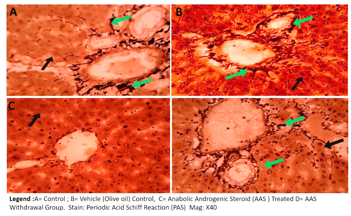
Figure 8: Representative photomicrograph of the histochemical stain for reticulin fibers in the hepatic tissue sections of the experimental Wistar rats of study, showing normal histological pictures in control, the vehicle control (A) and olive oil-treated (B) rats as compared with the AAS treated (C) rats. Legend: A=Control, B=Olive Oil Control, B= 120mg/kg b.w.t/ daily AAS for 21days and D= the 7days withdrawal groups following 21 days of 120mg/kg AAS treatment. Green arrows: reticulin fibers, black arrow= hepatocytes/ hepatic plates Magnification x400, Scale bar 20 microns (20µm).
Discussion
To date, there is dearth information on the therapeutic agents that could lessen AAS associated complications Behairy A, et al. [34] so also the information on the interplay between toxic effects of AAS and endogenously produced antioxidants. Hence, this present work tested the interactions between endogenous antioxidant and AAS for cell survival, to educate AAS users on the role of AAS and endogenously produced antioxidant enzymes in health and disease conditions.
In this current study, AAS resulted in an improvement in body weight, this correlates with it reports on body enhancements or use in body building Sagoe D, et al. [1] through its ability to induced an increase in muscle protein synthesis Kaufman MJ, et al. [13] when its crosses the blood and bind with the androgenic receptors on the skeletal muscle cells to activate anabolic function Pope Jr HG, et al. [22] associated with binding to DNA which results in replication and protein synthesis within the muscles Barceloux D, et al. [21] The have been contradictory findings on AAS influence on increasing body weight.
Bueno, et al. [35] reported an increase, while Traish, et al. [36] reported a decline in body weight linked to AAS intake. These conflicting reports may be due to the dose of AAS and duration of administration as seen in a report by Gbar, et al. [37] that testosterone undecanoate increases body weight and muscle strength when given in for a long duration. The withdrawal study group showed that abstinence from AAS intake results in a reduction in body mass.
Liver is involved in the drug metabolism and detoxification Behairy A, et al. [34]. The weight of the liver as evaluated in histopathological cases related to drug induced liver injury. This study revealed that the wet weight of the liver increased in AAS treatment when compared with the control, Olive oil vehicle and AAS withdrawal group. This increase in liver mass is linked to toxicological report on liver, that hepatic injury can results in an increase in liver weight as demonstrated in this present study. The AAS withdrawal group has a reduction in liver weight. The study group for AAS withdrawal presents with a mild decrease in body mass. The half-life of testosterone undecanote (TU) has been reported to be 1 – 12 days [30]. This shows that effectiveness of a decline in TU activity in the blood after withdrawal from drug administration.
Histopathological evaluation of the liver involves the assay for aminotransferases (AST and ALT) mostly use as specific indicators of hepatocellular necrosis or liver injury [27] They are used as diagnostic tools or biological maker for hepatotoxic liver injury Frankenfeld SP, et al. [38] There was a significant increase in ALT and AST levels in AAS treated rats, this elevation seen correlates with reports made by Neamat- Allah [39] and Behairy, et al. [34]. This increase in serum AST and ALT activities is proposed to be due to hepatic cell membrane distortion Saggu S, et al. [40] and cell membrane dysfunction that results in their release into the blood stream from the degenerating hepatic cells Behairy A, et al. [34], which is an indicator for degeneration of the lipid membrane bilayer of the hepatocytes. Olive oil vehicle has a decline in AST and ALT activity in our study, which is linked to olive oil a polyunsaturated fatty acid, being a strong antioxidant. Covas MI [41] has reported that polyunsaturated fats decline the risk of liver disease or hepatocellular necrosis due to its ability to decrease lipoproteins sensitivity to peroxidation in the liver cells. The decline in AST and ALT level in the olive oil study group aligns with Tuck, et al. [42] reports. The withdrawal of AAS results in a decline in serum level of AST and ALT, this implies a withdrawal of toxicant results in a decrease as the effectiveness of drug in the withdrawal group, indicating recovery in hepatic failure [43].
This present report demonstrates histopathological evaluation of the liver, to ascertain interaction between AAS metabolites and endogenous antioxidant SOD, aside from liver function test, serum activities of MDA and SOD were assayed. This is because, the liver is one of the organs most exposed to oxidative tissue damage because of their metabolic functions associated with detoxification [14] and liver disease or injury is attributed to oxidative tissue damage Behairy A, et al. [34] of which antioxidants are reported to avert the cascade reactions in of oxidative tissue injury in hepatic tissue [26].
The malonaldehyde (MDA) enzyme is a biological marker to evaluate the degree of lipid peroxidation in biological tissues, during oxidative tissue damage. AAS treatment caused an elevation in serum MDA level that supports Petrovic A, et al. [18, 34] findings. This is because AAS has the ability to permeate the blood into the hepatocytes by binding with androgenic receptors Niedfeldt MW [14] gain access into the nucleus to act on the DNA and activate cascade reaction linked with the production of ROS that alters normal mitochondrial function Petrovic A [18] that activates oxidative stress in the hepatic tissues through AAS mediated oxidative stress Solimini R, et al. [17, 44] mentioned that activation of the ARs in liver cells may increase ROS leading to hepatic cell degeneration seen in AAS induced hepatotoxicity, also similar report in testis which expresses Behairy A, et al. [34] that mediated oxidative tissue damage.
There was no significance increase in MDA activity in the olive oil treatment group since it is a polyunsaturated fat that inhibit lipid peroxidation in cells41. Olive oil is a good antioxidant that potentiate mobbing off of generated ROS that di [45].
The AAS withdrawal study group caused a marked reduction in MDA activity associated with a decrease in lipid peroxidation due to withdrawal from AAS treatment, this is linked to the absence of circulating AAS metabolites in the blood stream to bind to ARs to activate oxidative tissue damage and liver injury via lipid peroxidation Behairy A, et al. [34] Superoxide dismutase (SOD) protects against oxidative tissue damage. Our findings reveal that AAS results in a reduction in endogenous SOD synthesis, which implies a decline in its production and effectiveness in clearing ROS which resulted in the reported elevation of MDA with corresponding disruptions of liver cell integrity seen in the elevation of AST and ALT, in this present study. Hence, AAS prevents synthesis of endogenous SOD El-Rahman GIA, et al. [26], resulting in disruption of redox signaling and the role of SOD to detoxify intracellular reactive oxygen species (ROS) [25]. SOD activity increased in Olive oil group and AAS withdrawal group, this is because olive oil as a fatty acid prevents generation of ROS and induce endogenous synthesis of SOD while AAS withdrawal help to revival physiologic function of hepatocytes to metabolize and synthesis endogenous enzymes. This support reports by Mohamed WA, et al. [25, 26, 27] on the roles of endogenous and exogenous antioxidant as prophylactic or therapeutic agents against oxidative tissue damage.
The histopathological demonstrated of liver tissue using H and E, PAS and Reticulin stain showed that AAS induces liver damaged histological demonstration using H and E stain shows presence of an increase in infiltration of hepatic tissue by neutrophils, lymphocytes, and eosinophils and ground glass appearance of hepatocytes which correlates with Bond, et al. [16] and Niedfeldt MW [14] reports. This change is supported by elevation of AST, ALT and MDA with decline in SOD. Furthermore, there was loss of mucin granules and glycogen granules depicting loss of liver function to temporary store glycogen. Reticulin fibers were also loss. These histopathological features support early reports Albano GD, et al. [2, 17] associated with AR expression and it’s in the hepatic cells, acting on the DNA and activating some chain reaction linked with generation of ROS that target the mitochondria Petrovic A, et al. [18, 34] leading to generation of ROS that disrupts structural integrity of DNA, proteins and lipids in the liver Petrovic A, et al. [18] thereby altering liver function and structural integrity. However, the olive oil and AAS withdrawal has preserved liver tissue linked to antioxidant enzymes activities as previously discussed associated with preserved liver tissue cellular integrity, decline in AST, ALT, MDA and increase in SOD. The linked between oxidative stress and DNA dysfunction has been well reported in AAS through androgen receptors (AR) binding Petrovic A, et al. [18, 21, 34]. In fact, oxidative stress is implicated in the pathogenesis of wide range of disease the affect the liver heart and other tissues and this is associated with an increase in ROS availability is also linked to DNA damage34. These present results show that AAS caused significant rise in serum ALT, AST, and MDA levels while decreasing the serum level of antioxidant enzyme-SOD and distinct histopathological perturbations of the liver tissues of AAS treated rats, this supports report made by Behairy A [34]. The mechanism by which AAS withdrawal group reduces AAS induced hepatic dysfunction is based on the fact the abstinence from AAS resulted in a gradual buildup of the endogenous antioxidant enzyme demonstrated by an elevation of serum SOD activity which mobs off ROS generated by AAS thereby reducing lipid peroxidation seen in a decline in serum MDA activity Behairy A, et al. [34, 33], this report of decline in toxicity and normalizing of liver function enzymes following withdrawal from AAS intake supports reports made by Petrovic, et al. [18]. Serum activity of SOD, decreased in AAS animals as compared with the control, olive oil and AAS withdrawal groups [34].
Conclusions
AAS mediates hepatocellular tissue damage attributed to a perturbed redox system that results in a reduction in endogenous antioxidant (SOD) production linked to loss of hepatocellular function and membrane integrity demonstrated in an elevation of AST and ALT associated with disruption of the normal histoarchitecture of the liver (hepatocellular necrosis); distortion in the hepatocytes, decrease in glycogen granules and loss of reticulin for membrane integrity, these characteristics were reversed in AAS withdrawal or abstinence giving room for recovery associated with cellular regeneration and endogenous synthesis of SOD for hepato- protective role against circulating ROS.
Conflict of Interest
There is no conflict of Interest
Acknowledgments
None: Author Contribution: Conceptualization, A.E.M; methodology, A.E.M and G.M.S ; software, validation, formal analysis : A.E.M; investigation, resources, data curation, writing—original draft preparation, A.E.M and G.M.S; writing—review and editing, A.E.M and G.M.S; visualization, A.E.M and G.M.S; supervision, A.E.M; All authors have read and agreed to the published version of the manuscript.
Funding
This research did not receive any specific grant from funding agencies in the public, commercial, or not-for-profit sectors. Authors solely funded this research.
References
-
Sagoe D, McVeigh J, Bjornebekk A, Essilfie MS, Andreassen CS, et al. (2015) Polypharmacy among anabolic- androgenic steroid users: a descriptive metasynthesis. Substance Abuse Treatment, Prevention, and Policy 10: 12.
-
Albano GD, Sessa F, Messina A, Monda V, Bertozzi G, et al. (2017) AAS and organs damage: A focus on Nandrolone effects. Acta Med Mediterr 6: 939-946.
-
Park M, Sim J, Jeon Y, Yeon S, Lee J, et al. (2019) Determination of Boldenone in Postmortem Specimens Including Blood and Urine Samples Using LC-MS/MS. J Pharm Biomed Anal 169: 111-115.
-
Parente Filho SLA, Gomes PE, Forte GA, Lima LL, Silva Júnior GB, et al. (2020) Kidney Disease Associated with Androgenic-Anabolic Steroids and Vitamin Supplements Abuse: Be Aware!. Nefrología (English Edition) 40(1): 26-31.
-
Gagliano-Jucá T, Basaria S (2019) Abuse of Anabolic Steroids: A Dangerous Indulgence. Curr Opin Endocr Metab Res 9: 96-101.
-
Barakat LA, Tousson E, Ibrahim W, Abd El-Hakeem A (2015) Role of Propolis in Improving Hepatic and Renal Damage in Boldenone Undecylenate in Male Rats. Am J Biol Chem 3: 8.
-
Tousson E, El-Moghazy M, Massoud A, El-Atrash A, Sweef O, et al. (2016) Physiological and Biochemical Changes after Boldenone Injection in Adult Rabbits. Toxicol Ind Health 32(1): 177-182.
-
Ziaolhagh SJ, Khojasteh L, Ziaolhagh SS, Yahyaei B (2018) The Effect of Boldenone Anabolic Steroid, and Endurance and Resistance Training on Liver Damage Markers in Rats. Feyz J Kashan Univ Med Sci 22(2): 143-152.
-
Behairy A, El-Sharkawy NI, Saber TM, Soliman MM, Metwally MMM, et al. (2020) The Modulatory Role of Vitamin C in Boldenone Undecylenate Induced Testicular Oxidative Damage and Androgen Receptor Dysregulation in Adult Male Rats. Antioxidants (Basel) 9(11): 1053.
-
Sagoe D, Pallesen S (2018) Androgen abuse epidemiology. Current Opinion in Endocrinology, Diabetes Obesity 25: 185-194.
-
Sandvik MR, Bakken A, Loland S (2018) Anabolic– androgenic steroid use and correlates in Norwegian adolescents. European Journal of Sport Science 18(16): 903-910.
-
Bjørnebekk A, Walhovd KB, Jørstad ML, Due-Tønnessen P, Hullstein IR, et al. (2017) Structural brain imaging of long-term anabolic-androgenic steroid users and nonusing weightlifters. Biological Psychiatry 82(4): 294- 302.
-
Kaufman MJ, Kanayama G, Hudson JI, Pope HG (2019) Supraphysiologic-dose anabolic–androgenic steroid use: A risk factor for dementia?. Neurosci Biobehav Rev 100: 180-207.
-
Niedfeldt MW (2018) Anabolic steroid effect on the liver. Curr Sport med Rep 17: 92-102.
-
Albano GD, Amico F, Cocimano G, Liberto A, Maglietta F, et al. (2021) Adverse Effects of Anabolic-Androgenic Steroids: A Literature Review. Healthcare (Basel) 9(1): 97.
-
Bond P, Llewellyn W, Van Mol P (2016) Anabolic androgenic steroid-induced hepatotoxicity. Med Hypotheses 93: 150-153.
-
Solimini R, Rotolo MC, Mastrobattista L, Mortali C, Minutillo A, et al. (2017) Hepatotoxicity Associated with Illicit Use of Anabolic Androgenic Steroids in Doping. Eur Rev Med Pharmacol Sci 21(1S): 7-16.
-
Petrovic A, Vukadin S, Sikora R, Bojanic K, Smolic R, et al. (2022) Anabolic androgenic steroid-induced liver injury: An update. World J Gastroenterol 28(26): 3071- 3080.
-
Castellanos MM, Martínez EG, Prieto AS, Ramos PR, Terente MP (2018) Bile cast nephropathy associated with severe liver dysfunction caused by anabolic steroids. Nefrologia 38(2): 221-223.
-
Rasmussen JJ, Schou M, Madsen PL, Selmer Ch, Johansen ML, et al. (2018) Increased blood pressure and aortic stiffness among abusers of anabolic androgenic steroids: potential effect of suppressed natriuretic peptides in plasma?. Journal of Hypertention 36(2): 277-285.
-
Barceloux DG, Palmer RB (2013) Anabolic-androgenic Steroids. Disease-a-Month 59(6): 226-248.
-
Pope HG Jr, Kanayama G, Athey A, Ryan E, Hudson JI, et al. (2014) The lifetime prevalence of anabolic-androgenic steroid use and dependence in Americans: current best estimates. Am J Addict 23(4): 371-377.
-
Profumo E, Buttari B, Tinaburri L, D’arcangelo D, Sorice M, et al. (2018) Oxidative Stress Induces HSP90 Upregulation on the Surface of Primary Human Endothelial Cells: Role of the Antioxidant 7,8-Dihydroxy- 4-Methylcoumarin in Preventing HSP90 Exposure to the Immune System. Oxidative Med Cell Longevity 10: 2373167.
-
Castro JP, Fernando R, Reeg S, Meinl W, Almeida H, et al. (2019) Non-enzymatic Cleavage of Hsp90 by Oxidative Stress Leads to Actin Aggregate Formation: A Novel Gain-Of-Function Mechanism. Redox Biol 21: 101108.
-
Mohamed WA, Abd-Elhakim YM, Ismail SAA (2019) Involvement of the Anti-inflammatory, Anti-apoptotic, and Anti-secretory Activity of Bee Venom in its Therapeutic Effects on Acetylsalicylic Acid-Induced Gastric Ulceration in Rats. Toxicology 419: 11-23.
-
El-Rahman GIA, Behairy A, Elseddawy NM, Batiha GES, Hozzein WN, et al. (2020) Saussurea Lappa Ethanolic Extract Attenuates Triamcinolone Acetonide-Induced Pulmonary and Splenic Tissue Damage in Rats via Modulation of Oxidative Stress, Inflammation, and Apoptosis. Antioxidants (Basel) 9(5): 396.
-
Abd-Elhakim YM, Abdel-Motal SM, Malhat SM, Mostafa HI, Moselhy AAA, et al. (2021) Curcumin Mitigates Neurotoxic and Neurobehavioral Changes of Gentamicin and Sodium Salicylate in Rats by Adjusting Oxidative Stress and Apoptosis. Life Sci 265: 118824.
-
(1996) National Institutes of Health.
-
NRC (1986) National Research Council’s Guide for the Care and Use of Laboratory Animals
-
Kalepu S, Manthina M, Padavala V (2013) Oral lipid-based drug delivery systems-an overview. Acta Pharmaceutica Sinica B 3(6): 361-372.
-
Bancroft JD, Gamble M (2008) Theory and practice of histological techniques. In: 6th (Edn.), Churchill Livingstone, London.
-
Suvarna SK, Layton C, Bancroft JD (2019) Bancroft’s theory and practice of histological techniques: Theory and practice of histological techniques. In: 8th (Edn.), Churchill Livingstone, London.
-
Memudu AE, Pantong S, Osahon I (2020) Histomorphological evaluations on the frontal cortex extrapyramidal cell layer following administration of N-Acetyl cysteine in an aluminum-induced neurodegeneration rat model. Metab Brain Dis 35(5): 829-839.
-
Behairy A, Mohamed WAM, Ebraheim LLM, Soliman MM, Abd-Elhakim YM, et al. (2021) Boldenone Undecylenate- Mediated Hepatorenal Impairment by Oxidative Damage and Dysregulation of Heat Shock Protein 90 and Androgen Receptors Expressions: Vitamin C Preventive Role. Front Pharmacol 12: 651497.
-
Bueno A, Carvalho FB, Gutierres JM, Lhamas CL, Brusco I, et al. (2017) Impacts of Dose and Time of Boldenone and Stanazolol Exposure in Inflammatory Markers, Oxidative and Nitrosative Stress and Histopathological Changes in the Rat Testes. Theriogenology 90: 101-108.
-
Traish MA, Abdullah B, Yu G (2011) Andregen deficiency and mitochondrial dysfunction: implications for fatique muscle dysfunction, insulin resistance, diabetes, and cardiovascular disease. Horm Mol Biol clin Invest 8: 432- 444.
-
Gabr F, El-Maaty A, Amal M, Aotifa AM, Hassan TA (2009) Effects of Growth Promoter Boldenone Undecylenate on Weaned Male Lambs. Nat Sci 7: 61-69.
-
Frankenfeld SP, Oliveira LP, Ortenzi VH, Rego-Monteiro IC, Chaves EA, et al. (2014) The Anabolic Androgenic Steroid Nandrolone Decanoate Disrupts Redox Homeostasis in Liver, Heart and Kidney of Male Wistar Rats. PLoS ONE 9(9): e102699.
-
Neamat-Allah A (2014) Effect of Boldenone Undecylenate on Haematological and Biochemical Parameters in Veal Calves. Global Veterinaria 13(6): 1092-1096.
-
Saggu S, Kumar R (2007) Modulatory Effect of Seabuckthorn Leaf Extract on Oxidative Stress Parameters in Rats during Exposure to Cold, Hypoxia and Restraint (C-H-R) Stress and Post Stress Recovery. J Pharm Pharmacol 59(12): 1739-1745.
-
Covas MI (2007) Olive oil and the cardiovascular system. pharmacol Res 55(3): 175-186.
-
Tuck KL, Hahball P (2002) Major phenolic compound in olive metabolism and health effects. J Nut Biochem 13(11): 636-644.
-
Rosalk S, Mcintyre N (1999) Biochemical investigations in the management of liver disease. Hepatobiliary Diseases pp: 39-71.
-
Patanè FG, Liberto A, Maria Maglitto AN, Malandrino P, Esposito M, et al. (2020) Nandrolone Decanoate: Use, Abuse and Side Effects. Medicina 56(11): 606.
-
Waiz SA, Raies-Ul-Haq M, Waiz HA, Gupta S, Pathak AK (2015) Preliminary Study on the Protective Effect of Vitamin C on Monosodium Glutamate-Induced Hepatotoxicity in Rats. Comp. Clin Pathol 24: 1063-1068.
- Pattern of Gonadal Hormones in Oral Testosterone-Supplimented Male Wistar Rats with Diabetes-Induced Hypogonadism
- Re-Evaluation of the Genotoxicity of Currently Used Food Dyes in Mouse Multiple Organs Via Continuous Administration by Drinking Using the Comet Assay
- Pharmacogenetics of Type 2 Diabetes Mellitus: Linking Genetic Variability to Drug Efficacy and its Cardiovascular Outcomes
- Exploratory Proteomic Profiling of SARS-CoV-2 Infected THP-1 Macrophages Reveals Alterations in Inflammatory Response and Cellular Metabolism
- Study of Genotoxicity of Hepatocarcinogens in Multiple Organs in Mice by Feeding and Drinking Using the Comet Assay
- Spirulina Polypeptides Inhibit the Growth of Human Lung Tumor (H460) Cells