Angiogenesis and its Inhibitors – Areas in Pharmacological Research
It’s surprising to know that, if all the blood vessels in the body were lined up end-to-end, they would form a line that could circle the earth twice. These vessels which carry our life’s blood snake throughout our body visible through our skin only as a faint bluish track. Vessels play a central role in many
Introduction
It’s surprising to know that, if all the blood vessels in the body were lined up end-to-end, they would form a line that could circle the earth twice. These vessels which carry our life’s blood snake throughout our body visible through our skin only as a faint bluish track. Vessels play a central role in many physiological and pathological processes in our body. Angiogenesis, the process of building new blood vessels and controlling the propagation of blood vessels are fundamental to human health, as they play key roles in wound healing and tissue growth. When dysregulated, new blood vessels formation contributes to numerous ischaemic, malignant, inflammatory, infectious and immune disorders. Molecular insights into these processes are offering new therapeutic opportunities. Angiogenesis is defined as formation of new blood vessels from pre-existing vessels. Normally this process helps to repair injured vessels, and is necessary in menstruation and pregnancy. Angiogenesis is controlled by tightly regulated series of biologic “on (promoters /stimulators) and off (inhibitors)” switches. The Scottish anatomist and surgeon John Hunter provided the first recorded scientific insights into the field of angiogenesis. He recognized that overall regulation of angiogenesis follows a basic law of nature founded by Aristotle that says “form follows function” - a fundamental law of nature in which the anatomical structure (form) of a system adapts to accommodate changes in the function of the system. The modern era of angiogenesis began with the works of Surgeon Dr Judah Folkman who hypothesized that tumour growth is dependent upon angio-genes in his theory, published in the New England Journal of Medicine, in 1971 [1, 2]. Angiogenesis is the physiological process through which new blood vessels form from pre-existing vessels [3, 4, 5]. In precise usage this is distinct from vasculogenesis, which is the de novo formation of endothelial cells from mesoderm cell precursors, and from neovascularization, although discussions are not always precise (especially in older texts) [6]. The first vessels in the developing embryo form through vasculogenesis, after which angiogenesis is responsible for most, if not all, blood vessel growth during development and in disease [7]. Angiogenesis is a normal and vital process in growth and development, as well as in wound healing and in the formation of granulation tissue. However, it is also a fundamental step in the transition of tumors from a benign state to a malignant one, leading to the use of angiogenesis inhibitors in the treatment of cancer. The essential role of angiogenesis in tumor growth was first proposed in 1971 by Judah Folkman, who described tumors as "hot and bloody," illustrating that, at least for many tumor types, flush perfusion and even hyperemia are characteristic [8].
Types of Angiogenesis
Sprouting Angiogenesis
Sprouting angiogenesis was the first identified form of angiogenesis. It occurs in several well-characterized stages. First, biological signals known as angiogenic growth factors activate receptors on endothelial cells present in pre-existing blood vessels. Second, the activated endothelial cells begin to release enzymes called proteases that degrade the basement membrane to allow endothelial cells to escape from the original (parent) vessel walls. The endothelial cells then proliferate into the surrounding matrix and form solid sprouts connecting neighboring vessels. As sprouts extend toward the source of the angiogenic stimulus, endothelial cells migrate in tandem, using adhesion molecules called integrins. These sprouts then form loops to become a full-fledged vessel lumen as cells migrate to the site of angiogenesis. Sprouting occurs at a rate of several millimeters per day, and enables new vessels to grow across gaps in the vasculature. It is markedly different from splitting angiogenesis because it forms entirely new vessels as opposed to splitting existing vessels.
Intussusceptive of Angiogenesis
By intussusception, also known as splitting angiogenesis, a new blood vessel is created by splitting of an existing blood vessel in two. Intussusception was first observed in neonatal rats. In this type of vessel formation, the capillary wall extends into the lumen to split a single vessel in two. There are four phases of intussusceptive angiogenesis. First, the two opposing capillary walls establish a zone of contact. Second, the endothelial cell junctions are reorganized and the vessel bilayer is perforated to allow growth factors and cells to penetrate into the lumen. Third, a core is formed between the 2 new vessels at the zone of contact that is filled with pericytes and myofibroblasts. These cells begin laying collagen fibers into the core to provide an extracellular matrix for growth of the vessel lumen. Finally, the core is fleshed out with no alterations to the basic structure. Intussusception is important because it is a reorganization of existing cells. It allows a vast increase in the number of capillaries without a corresponding
| Stimulator | Mechanism | ||||||
| Fgf | Promotes Proliferation & Differentiation Of Endothelial Cells, Smooth Muscle Cells, And Fibroblasts | ||||||
| Vegf | Affects Permeability | ||||||
| VEGFR And NRP-1 | Integrate Survival Signals | ||||||
| Ang 1 and Ang 2 | Stabilize Vessels | ||||||
| PDGF (BB- Homodimer) And PDGFR | Recruit Smooth Muscle Cells | ||||||
| TGF- Β, Endoglin And TGF- Β Receptors | ↑Extracellular Matrix Production | ||||||
| CCl 2 | Recruits Lymphocytes To Sites Of Inflammation | ||||||
| Histamine | |||||||
| Integrins Α β , Α β ( v 3 v 5 ?[9]) And Α β 5 1 | Bind Matrix Macromolecules And Proteina ses | ||||||
| VE- Cadherin And CD31 | Endothelial Junctional Molecules | ||||||
| Ephrin | Determine Formation Of Arteries Or Veins | ||||||
| Plasminogen Activators | Remodels Extracellular Matrix, Releases And Activates Growth Factors | ||||||
| Plasminogen Activator Inhibitor-1 | Stabilizes Nearby Vessels | ||||||
| Enos And COX-2 | |||||||
| Ac133 | Regulates Angioblast Different iation | ||||||
| Id1/Id3 | Regulates Endothelial Transdifferentiati on |
Table 1: Overview of angiogenesis
Role of FGF (Fibroblast Growth Factor) in Angiogenesis
The fibroblast growth factor (FGF) family with its prototype members FGF-1 (acidic FGF) and FGF-2 (basic FGF) consists to date of at least 22 known members [9, 10, 11]. Most are single-chain peptides of 16-18 kDa and display high affinity to heparin and heparan sulfate. In
general, FGFs stimulate a variety of cellular functions by binding to cell surface FGF-receptors in the presence of heparin proteoglycans. The FGF-receptor family is composed of seven members, and all the receptor proteins are single-chain receptor tyrosine kinases that become activated through autophosphorylation induced by a mechanism of FGF-mediated receptor dimerization. Receptor activation gives rise to a signal transduction cascade that leads to gene activation and diverse biological responses, including cell differentiation, proliferation, and matrix dissolution, thus initiating a process of mitogenic activity critical for the growth of endothelial cells, fibroblasts, and smooth muscle cells. FGF-1, unique among all 22 members of the FGF family, can bind to all seven FGF-receptor subtypes, making it the broadest-acting member of the FGF family, and a potent mitogen for the diverse cell types needed to mount an angiogenic response in damaged (hypoxic) tissues, where upregulation of FGF-receptors occurs [12]. FGF-1 stimulates the proliferation and differentiation of all cell types necessary for building an arterial vessel, including endothelial cells and smooth muscle cells; this fact distinguishes FGF-1 from other pro-angiogenic growth factors, such as vascular endothelial growth factor (VEGF), which primarily drives the formation of new capillaries [13, 14]. Besides FGF-1, one of the most important functions of fibroblast growth factor-2 (FGF-2 or bFGF) is the promotion of endothelial cell proliferation and the physical organization of endothelial cells into tube-like structures, thus promoting angiogenesis. FGF-2 is a more potent angiogenic factor than VEGF or PDGF (platelet- derived growth factor); however, it is less potent than FGF-1. As well as stimulating blood vessel growth, aFGF (FGF-1) and bFGF (FGF-2) are important players in wound healing. They stimulate the proliferation of fibroblasts and endothelial cells that give rise to angiogenesis and developing granulation tissue; both increase blood supply and fill up a wound space/cavity early in the wound-healing process.
Role Vascular Endothelial Growth Factor (VEGF) in Angiogenesis
Vascular endothelial growth factor (VEGF) has been demonstrated to be a major contributor to angiogenesis, increasing the number of capillaries in a given network. Initial in vitro studies demonstrated bovine capillary endothelial cells will proliferate and show signs of tube structures upon stimulation by VEGF and bFGF, although the results were more pronounced with VEGF [15]. Upregulation of VEGF is a major component of the physiological response to exercise and its role in angiogenesis is suspected to be a possible treatment in vascular injuries [16, 17, 18, 19]. In vitro studies clearly demonstrate that VEGF is a potent stimulator of angiogenesis because, in the presence of this growth factor, plated endothelial cells will proliferate and migrate, eventually forming tube structures resembling capillaries [8]. VEGF causes a massive signaling cascade in endothelial cells. Binding to VEGF receptor-2 (VEGFR-2) starts a tyrosine kinase signaling cascade that stimulates the production of factors that variously stimulate vessel permeability (eNOS, producing NO), proliferation/survival (bFGF), migration (ICAMs/VCAMs/MMPs) and finally differentiation into mature blood vessels. Mechanically, VEGF is upregulated with muscle contractions as a result of increased blood flow to affected areas. The increased flow also causes a large increase in the mRNA production of VEGF receptors 1 and 2. The increase in receptor production means muscle contractions could cause upregulation of the signaling cascade relating to angiogenesis. As part of the angiogenic signaling cascade, NO is widely considered to be a major contributor to the angiogenic response because inhibition of NO significantly reduces the effects of angiogenic growth factors. However, inhibition of NO during exercise does not inhibit angiogenesis, indicating there are other factors involved in the angiogenic response [8].
Role of Angiopoietins in Angiogenesis
The angiopoietins, Ang1 and Ang2, are required for the formation of mature blood vessels, as demonstrated by mouse knock out studies [20]. Ang1 and Ang2 are protein growth factors which act by binding their receptors, Tie-1 and Tie-2; while this is somewhat controversial, it seems that cell signals are transmitted mostly by Tie-2; though some papers show physiologic signaling via Tie-1 as well. These receptors are tyrosine kinases. Thus, they can initiate cell signaling when ligand binding causes a dimerization that initiates phosphorylation on key tyrosines.
Role of Metalloproteinase (MMP) in Angiogenesis
Another major contributor to angiogenesis is matrix metalloproteinase (MMP). MMPs help degrade the proteins that keep the vessel walls solid. This proteolysis allows the endothelial cells to escape into the interstitial matrix as seen in sprouting angiogenesis. Inhibition of MMPs prevents the formation of new capillaries [21]. These enzymes are highly regulated during the vessel formation process because destruction of the extracellular matrix would decrease the integrity of the microvasculature [8].
Role of Delta-Like Ligand 4 (DII4) In Angiogenesis
Delta-like ligand 4 (DII4) is a protein with a negative regulatory effect on angiogenesis [22, 23]. Dll4 is a transmembrane ligand, for the notch family of receptors.
Role of Class 3 Semaphorins in angiogenesis
Class 3 Semaphorins (SEMA3s) regulate angiogenesis by modulating endothelial cell adhesion, migration, proliferation, survival and the recruitment of pericytes [10]. Furthermore, semaphorins can interfere with VEGF- mediated angiogenesis since both SEMA3s and VEGF-A compete for Neuropilin receptor binding at endothelial cells [24, 25]. The relative expression levels of SEMA3s and VEGF-A may therefore be important for angiogenesis [10].
Chemical inhibition of angiogenesis
Angiogenesis inhibitor can be endogenous or come from outside as drug or a dietary component.
Application of Angiogenesis in Medicine
Angiogenesis may be a target for combating diseases characterized by either poor vascularisation or abnormal vasculature E.g Heart Disease sufferers [26]. Application of specific compounds that may inhibit or induce the creation of new blood vessels in the body may help combat such diseases. The presence of blood vessels where there should be none may affect the mechanical properties of a tissue, increasing the likelihood of failure. The absence of blood vessels in a repairing or otherwise metabolically active tissue may inhibit repair or other essential functions. Several diseases, such as ischemic chronic wounds, are the result of failure or insufficient blood vessel formation and may be treated by a local expansion of blood vessels, thus bringing new nutrients to the site, facilitating repair. Other diseases, such as age- related macular degeneration, may be created by a local expansion of blood vessels, interfering with normal physiological processes. The modern clinical application of the principle of angiogenesis can be divided into two main areas: anti- angiogenic therapies, which angiogenic research began with, and pro-angiogenic therapies. Whereas anti- angiogenic therapies are being employed to fight cancer and malignancies [27, 28], which require an abundance of oxygen and nutrients to proliferate, pro-angiogenic therapies are being explored as options to treat cardiovascular diseases, the number one cause of death in the Western world. One of the first applications of pro- angiogenic methods in humans was a German trial using fibroblast growth factor 1 (FGF-1) for the treatment of coronary artery disease [13, 29, 30]. Also, regarding the mechanism of action, pro- angiogenic methods can be differentiated into three main categories: gene-therapy, targeting genes of interest for amplification or inhibition; protein-therapy, which primarily manipulates angiogenic growth factors like FGF-1 or vascular endothelial growth factor, VEGF; and cell-based therapies, which involve the implantation of specific cell types. There are still serious, unsolved problems related to gene therapy. Difficulties include effective integration of the therapeutic genes into the genome of target cells, reducing the risk of an undesired immune response, potential toxicity, immunogenicity, inflammatory responses, and oncogenesis related to the viral vectors used in implanting genes and the sheer complexity of the genetic basis of angiogenesis. The most commonly occurring disorders in humans, such as heart disease, high blood pressure, diabetes and Alzheimer's disease, are most likely caused by the combined effects of variations in many genes, and, thus, injecting a single gene may not be significantly beneficial in such diseases. In contrast, pro-angiogenic protein therapy uses well- defined, precisely structured proteins, with previously defined optimal doses of the individual protein for disease states, and with well-known biological effects [1]. On the other hand, an obstacle of protein therapy is the mode of delivery. Oral, intravenous, intra-arterial, or intramuscular routes of protein administration are not always as effective, as the therapeutic protein may be metabolized or cleared before it can enter the target tissue. Cell-based pro-angiogenic therapies are still early stages of research, with many open questions regarding best cell types and dosages to use.
Tumor Angiogenesis
Cancer cells are cells that have lost their ability to divide in a controlled fashion. A malignant tumor consists of a population of rapidly dividing and growing cancer cells that progressively accrues mutations. However, tumors need a dedicated blood supply to provide the oxygen and other essential nutrients they require in order to grow beyond a certain size (generally 1–2 mm3).
Without angiogenesis a tumor cannot grow beyond a limited size [31, 32]. Tumors induce blood vessel growth (angiogenesis) by secreting various growth factors (e.g. VEGF) and proteins. Growth factors such as bFGF and VEGF can induce capillary growth into the tumor, which some researchers suspect supply required nutrients, allowing for tumor expansion. Unlike normal blood vessels, tumor blood vessels are dilated with an irregular shape [33]. In 2007, it was discovered that cancerous cells stop producing the anti-VEGF enzyme PKG. In normal cells (but not in cancerous ones), PKG apparently limits beta-catenin, which solicits angiogenesis [34]. Other clinicians believe angiogenesis really serves as a waste pathway, taking away the biological end products secreted by rapidly dividing cancer cells. In either case, angiogenesis is a necessary and required step for transition from a small harmless cluster of cells, often said to be about the size of the metal ball at the end of a ball-point pen, to a large tumor. Angiogenesis is also required for the spread of a tumor, or metastasis. Single cancer cells can break away from an established solid tumor, enter the blood vessel, and be carried to a distant site, where they can implant and begin the growth of a secondary tumor. Evidence now suggests the blood vessel in a given solid tumor may, in fact, be mosaic vessels, composed of endothelial cells and tumor cells. This mosaicity allows for substantial shedding of tumor cells into the vasculature, possibly contributing to the appearance of circulating tumor cells in the peripheral blood of patients with malignancies [35]. The subsequent growth of such metastases will also require a supply of nutrients and oxygen and a waste disposal pathway. Endothelial cells have long been considered genetically more stable than cancer cells. This genomic stability confers an advantage to targeting endothelial cells using antiangiogenic therapy, compared to chemotherapy directed at cancer cells, which rapidly mutate and acquire 'drug resistance' to treatment. For this reason, endothelial cells are thought to be an ideal target for therapies directed against them [36].
Formation of Tumor Blood Vessels in Angiogenesis
The mechanism of blood vessel formation by angiogenesis is initiated by the spontaneous dividing of tumor cells due to a mutation. Angiogenic stimulators are then released by the tumor cells. These then travel to already established, nearby blood vessels and activates their endothelial cell receptors. This induces a release of proteolytic enzymes from the vasculature. These enzymes target a particular point on the blood vessel and cause a pore to form. This is the point where the new blood vessel will grow from. The reason tumor cells need a blood supply is because they cannot grow any more than 2-3 millimeters in diameter without an established blood supply which is equivalent to about 50-100 cells [37].
Role of Angiogenesis in Cardiovascular Disease
Angiogenesis represents an excellent therapeutic target for the treatment of cardiovascular disease. It is a potent, physiological process that underlies the natural manner in which our bodies respond to a diminution of blood supply to vital organs, namely the production of new collateral vessels to overcome the ischemic insult [13]. A large number of preclinical studies have been performed with protein-, gene- and cell-based therapies in animal models of cardiac ischemia, as well as models of peripheral artery disease. Reproducible and credible successes in these early animal studies led to high enthusiasm that this new therapeutic approach could be rapidly translated to a clinical benefit for millions of patients in the Western world suffering from these disorders. A decade of clinical testing both gene- and protein-based therapies designed to stimulate angiogenesis in under perfused tissues and organs, however, has led from one disappointment to another. Although all of these preclinical readouts, which offered great promise for the transition of angiogenesis therapy from animals to humans, were in one fashion or another, incorporated into early stage clinical trials, the FDA has, to date (2007), insisted that the primary endpoint for approval of an angiogenic agent must be an improvement in exercise performance of treated patients [38]. These failures suggested that either these are the wrong molecular targets to induce neovascularization, that they can only be effectively used if formulated and administered correctly, or that their presentation in the context of the overall cellular microenvironment may play a vital role in their utility. It may be necessary to present these proteins in a way that mimics natural signaling events, including the concentration, spatial and temporal profiles, and their simultaneous or sequential presentation with other appropriate factors [39].
Exercise and Angiogenesis
Angiogenesis is generally associated with aerobic exercise and endurance exercise. While arteriogenesis produces network changes that allow for a large increase in the amount of total flow in a network, angiogenesis causes changes that allow for greater nutrient delivery over a long period of time. Capillaries are designed to provide maximum nutrient delivery efficiency, so an increase in the number of capillaries allows the network to deliver more nutrients in the same amount of time. A greater number of capillaries also allows for greater oxygen exchange in the network. This is vitally important to endurance training, because it allows a person to continue training for an extended period of time. However, no experimental evidence suggests that increased capillarity is required in endurance exercise to increase the maximum oxygen delivery [8].
Macular Degeneration and Angiogenesis
Over expression of VEGF causes increased permeability in blood vessels in addition to stimulating angiogenesis. In wet macular degeneration, VEGF causes proliferation of capillaries into the retina. Since the increase in angiogenesis also causes edema, blood and other retinal fluids leak into the retina, causing loss of vision. Anti- angiogenic drugs targeting the VEGF pathways are now used successfully to treat this type of macular degeneration.
Types of Angiogenesis Process
Sprouting: characterized by sprouts composed of endothelial cells, which usually grow toward an angiogenic stimulus Intussusceptive: formation of blood vessels by a splitting process in which elements of interstitial tissues invade existing vessels.
The Angiogenesis Process
The overall process of angiogenesis is initiated by hypoxia through hypoxia inducible factors which induces angiogenic stimulators and endothelial cell activation. Hypoxia is an important stimulus for expansion of vascular bed. Activated Endothelial cells secrete proteases (Matrix metalloproteases and plasminogen activator) which degrade extracellular tissue to facilitate endothelial penetration. A stringent control of proteolytic activity is ensured by the release of proteolytic inhibitors like tissue inhibitor of matrix metallo-proteins and Plasminogen activator inhibitor. Increase in concentration of various growth factors occurs due to extracellular matrix degradation which stimulates endothelial cell migration and proliferation. The endothelial cells follow a leader endothelial cell and migrate through the degraded matrix and form small sprouts. The rate of sprout elongation increases with endothelial cell proliferation. These processes are also mediated through cell adhesion molecules. In the final phase maturation of the neo-vasculature occurs. New basement membrane is formed. Extracellular proteolysis is locally inhibited during this process to facilitate deposition and assembly of extracellular membrane components. Polarity of the endothelial cells (EC) is established by cell adhesion molecules in order to form a lumen. After sufficient angiogenesis the angiogenic factors are downregulated or concentration of inhibitors increases (Flow chart 1).
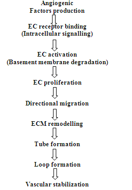
Flow chart -1: The process of angiogenesis
Endogenous Angiogenesis Modulators (EAM)
The process of angiogenesis is a fine dynamic balance between positive and negative regulators that mediate both physiological and pathological angiogenesis. Some factors are both pro and antiangiogenic functions. Some of the important angiogenic promoters are: Vascular Endothelial Growth Factors (VEGF), Fibroblast Growth Factors (FGF), Platelet Derived Growth Factors (PDGF), Angopoietin, Endothelins, Angiotensin 2, Erythropoeitin etc. Some of the angiogenic inhibitors are: Somatostatin, Angiostatin, Endostatin, Plasminogen Activator Inhibitor
Bioequivalence & Bioavailability International Journal
(PAI), platelet factor 4, Prolactin, Antithrombin III, Vasostatin etc.
Role of VEGF
VEGF, mainly VEGF –A, is one of the most potent proangiogenic factor. This family includes structurally related growth factors VEGF B, C ,D, E, F. They are expressed in various tissues including brain, kidney and liver. It mediates its action through two receptors, VEGFR 1 & VEGFR 2. It plays a crucial role in physiological angiogenesis, several human cancers, diabetic retinopathy, RA, atherosclerosis.
Pathological Angiogenesis
• Both angiogenesis insufficiency and excess can lead to various disorders. Insufficient angiogenesis leads to many disorders including ischemic tissue injury, cardiac failure, delayed healing of gastric ulcers, recurrent aphtous ulcers, pre- eclampsia, age related diseases, purpura, pulmonary fibrosis, ALS, alzheimer etc.
• Excessive angiogenesis is pathological in cancer, diabetic retinopathy, atherosclerosis, rheumatoid arthritis, chrons disease, diabetes, psoriasis, endometriosis etc.
Angiogenesis in Pharmacology
This would be beneficial in many conditions likely to be improved by clinical manipulation of angiogenesis. Angiogenesis can be induced by angiogenic proteins or endothelial progenitor cells synthesizing angiogenic growth factors injected directly into site to stimulate blood vessel growth. The right genes could also be activated to induce signalling cascade that would lead to angiogenesis. The major concern is regarding safety (for e.g. VEGF forms leaky and tortuous vessels and triggering of dormant tumours and atherosclerosis) of these procedures.
Becaplermin (Regranex) is approved (1997 by FDA) as topical recombinant PDGF preparation for diabetic neuropathic lower extremity ulcers. Drugs for Myocardial infarction, peripheral ischemia, cerebral ischemia and other diseases of insufficient angiogenesis are in experimental stages.
Antiangiogenic Therapy
Several diseases benefit from inhibition of excessive angiogenesis. The drugs may be of two types:
• Agents primarily developed for anti – angiogenesis property or
• Those developed or used for other biologic effects but also have anti-angiogenic activity e.g. celecoxib, rosiglitazone, interferon alpha, everolimus, vorinostat, zolendronic acid
• Some of the approved drugs are:
• Monoclonal antibody therapy: Bevacizumab, Cetuximab, Panitumumab, Trastuzumab, Ranizumab
• Tyrosine kinase inhibitors - Sorafenib, Sunitinib, Thalidomide
• Matrix Metalloprotein (MMP)-Inhibiting Drugs – Marimastat, Batimastat
Metronomic Therapy and Angiogenesis
Metronomic chemotherapy refers to the close, rhythmic administration of low doses of cytotoxic drugs, with minimal or no drug-free breaks, over prolonged periods. Metronome refers to a musical instrument that produces regular, metrical ticks representing fixed, regular aural pulse.
The main characteristics of metronomic chemotherapy are:
• Frequent (dose-dense) administration of chemotherapy without any interruptions
• Using a biological optimized dose instead MTD
• No application of hematopoietic growth factors
• Preference for oral drugs
• Low incidence of treatment related side-effects
• Potential for delayed development of resistance.
To summarise, while angiogenesis as a hallmark of tumour development and metastasis is now a validated target for cancer treatment, the overall benefits of anti-angiogenic drugs from the perspective of impacting survival have left much to desire, endorsing a need for developing more effective therapeutic regimens.
Quantification of Angiogenesis
Quantifying vasculature parameters such as microvascular density has various complications due to preferential staining or limited representation of tissues by histological sections. Recent research has shown complete 3D reconstruction of tumor vascular structure and quantification of vessel structures in whole tumors in animal models [40, 41].
Conclusion
This review clearly shows that there are lots of research areas to exploit the diagnostic and therapeutic importance of angiogenesis as well as its inhibition.
Hence, we opine that this mini review will through light in the minds of young researchers to develop this area of research for the benefit of the recipients.
References
-
Blsht M, Dhasmana DC, Blst SS (2010) Angiogenesis: future of pharmacological modulation. Indian Journal of pharmacology 42(1): 2-8.
-
Jain RK, Peter F (2001) Carmeliet "Vessels of Death and Life". Scientific American 285(6): 26-33.
-
Santulli G (2013) Angiogenesis insights from a systematic overview. New York: Nova Science.
-
Birbrair A, Zhang T, Wang ZM, Messi ML, Mintz A, et al. (2015) Pericytes at the intersection between tissue regeneration and pathology. Clinical Science 128(2): 81-93.
-
Birbrair A, Zhang T, Wang ZM, Messi ML, Olson JD, et al. (2014) Type-2 pericytes participate in normal and tumoral angiogenesis. American Journal of Physiology Cell Physiology 307(1): C25-38.
-
Risau W, Flamme I (1995) Vasculogenesis. Annual Review of Cell and Developmental Biology 11: 73-91.
-
Flamme I, Frölich T, Risau W (1997) Molecular mechanisms of vasculogenesis and embryonic angiogenesis. Journal of Cellular Physiology 173(2): 206-210.
-
Penn SJ (2008) Retinal and Choroidal Angiogenesis. Springer pp: 119.
-
Burri PH, Hlushchuk R, Djonov V (2004) Intussusceptive angiogenesis: its emergence, its characteristics, and its significance. Developmental Dynamics 231(3): 474-488.
-
Prior BM, Yang HT, Terjung RL (2004) What makes vessels grow with exercise training?. Journal of Applied Physiology 97(3): 1119-1128.
-
Sheppard D (2002) Endothelial integrins and angiogenesis: not so simple anymore. The Journal of Clinical Investigation 110(7): 913-914.
-
Mecollari V, Nieuwenhuis B, Verhaagen J (2014) A perspective on the role of class III semaphorin signaling in central nervous system trauma. Frontiers in Cellular Neuroscience 8: 328.
-
Ornitz DM, Itoh N (2001) Fibroblast growth factors. Genome Biology 2(3): reviews3005.1-reviews 3005.12.
-
Blaber M, DiSalvo J, Thomas KA (1996) X-ray crystal structure of human acidic fibroblast growth factor. Biochemistry 35(7): 2086-2094.
-
Stegmann TJ (1998) FGF-1: a human growth factor in the induction of neoangiogenesis. Expert Opinion on Investigational Drugs 7(12): 2011-2015.
-
Khurana R, Simons M (2003) Insights from angiogenesis trials using fibroblast growth factor for advanced arteriosclerotic disease. Trends in Cardiovascular Medicine 13(3): 116-122.
-
Goto F, Goto K, Weindel K, Folkman J (1993) Synergistic effects of vascular endothelial growth factor and basic fibroblast growth factor on the proliferation and cord formation of bovine capillary endothelial cells within collagen gels. Laboratory Investigation 69(5): 508-517.
-
Ding YH, Luan XD, Li J, Rafols JA, Guthinkonda M, et al. (2004) Exercise-induced overexpression of angiogenic factors and reduction of ischemia/reperfusion injury in stroke. Current Neurovascular Research 1(5): 411-420.
-
Gavin TP, Robinson CB, Yeager RC, England JA, Nifong LW, et al. (2004) Angiogenic growth factor response to acute systemic exercise in human skeletal muscle. Journal of Applied Physiology 96(1): 19-24.
-
Kraus RM, Stallings HW, Yeager RC, Gavin TP (2004) Circulating plasma VEGF response to exercise in sedentary and endurance-trained men. Journal of Applied Physiology 96(4): 1445-1450.
-
Lloyd PG, Prior BM, Yang HT, Terjung RL (2003) Angiogenic growth factor expression in rat skeletal muscle in response to exercise training. American Journal of Physiology. Heart and Circulatory Physiology 284 (5): H1668- H1678.
-
Thurston G (2003) Role of Angiopoietins and Tie receptor tyrosine kinases in angiogenesis and lymphangiogenesis. Cell Tissue Res 314(1): 61-68.
-
Haas TL, Milkiewicz M, Davis SJ, Zhou AL, Egginton S, et al. (2000) Matrix metalloproteinase activity is required for activity-induced angiogenesis in rat skeletal muscle. American Journal of Physiology. Heart and Circulatory Physiology 279(4): H1540- H1547.
-
Lobov IB, Renard RA, Papadopoulos N, Gale NW, Thurston G, et al. (2007) Delta-like ligand 4 (Dll4) is induced by VEGF as a negative regulator of angiogenic sprouting. Proceedings of the National Academy of Sciences of the United States of America 104(9): 3219-3224.
-
Hellström M, Phng LK, Hofmann JJ, Wallgard E, Coultas L, et al. (2007) Dll4 signalling through Notch1 regulates formation of tip cells during angiogenesis. Nature 445(7129): 776-780.
-
Soker S, Takashima S, Miao HQ, Neufeld G, Klagsbrun M (1998) Neuropilin-1 is expressed by endothelial and tumor cells as an isoform-specific receptor for vascular endothelial growth factor. Cell 92(6): 735- 745.
-
Herzog B, Pellet-Many C, Britton G, Hartzoulakis B, Zachary IC (2011) VEGF binding to NRP1 is essential for VEGF stimulation of endothelial cell migration, complex formation between NRP1 and VEGFR2, and signaling via FAK Tyr407 phosphorylation. Mol Biol Cell 22(15): 2766-2776.
-
Ferrara N, Kerbel RS (2005) Angiogenesis as a therapeutic target. Nature. 438(7070): 967-974.
-
Folkman J, Klagsbrun M (1987) Angiogenic factors. Science 235(4787): 442-447.
-
Folkman J (1996) Fighting cancer by attacking its blood supply. Scientific American. 275(3): 150-154.
-
Stegmann TJ, Hoppert T, Schneider A, Gemeinhardt S, Köcher M, et al. (2000) Induction of myocardial neoangiogenesis by human growth factors. A new therapeutic approach in coronary heart disease. Herz 25(6): 589-599.
-
Folkman J (1998) Angiogenic therapy of the human heart. Circulation. 97(7): 628-629.
-
McDougall SR, Anderson AR, Chaplain MA (2006) Mathematical modelling of dynamic adaptive tumour- induced angiogenesis: clinical implications and therapeutic targeting strategies. Journal of Theoretical Biology 241(3): 564-589.
-
Spill F, Guerrero P, Alarcon T, Maini PK, Byrne HM (2015) Mesoscopic and continuum modelling of angiogenesis. J Math Biol 70 (3): 485-532.
-
Gonzalez-Perez RR, Rueda BR (2013) Tumor angiogenesis regulators (first ed.). Boca Raton: Taylor & Francis. Pp: 462.
-
Allard WJ, Matera J, Miller MC, Repollet M, Connelly MC, et al. (2004) Tumor cells circulate in the peripheral blood of all major carcinomas but not in healthy subjects or patients with nonmalignant diseases. Clin Cancer Res 10(20): 6897-6904.
-
Bagri A, Kouros-Mehr H, Leong KG, Plowman GD (2010) Use of anti-VEGF adjuvant therapy in cancer: challenges and rationale. Trends in Molecular Medicine 16(3): 122-132.
-
Nishida N, Yano H, Nishida T, Kamura T, Kojiro M (2006) Angiogenesis in cancer. Vasc Health Risk Manag 2(3): 213-219.
-
Hariawala MD, Sellke FW (1997) Angiogenesis and the heart: therapeutic implications. J R Soc Med 90(6): 307-311.
-
Cao L, Mooney DJ (2007) Spatiotemporal control over growth factor signaling for therapeutic neovascularization. Adv Drug Deliv Rev 59(13): 1340- 1350.
-
Chia-Chi Chien, Ivan MK, Cheng LW, Hwu Yeukuang, Chen NY, et al. (2013) Complete microscale profiling of tumor microangiogenesis. Biotechnology Advances 31(3): 396-401.
- Effects of 5-HTP and Melatonin on the Sleep Cycle of Medical Students
- Adsorption of Bisphenol A on NH4OH- Modified Rice Husk and Sugar Cane Bagasse Biochar
- Comparative Assessment of the Reinforcement Efficiency of Palm Fruit Fibre and Coconut Fibre in High Density Polyethylene (HDPE) Matrix Composite
- Importance of Bio Compounds Naturally Present in Food with Functionality in Animal Metabolism
- Sub-Acute Study on the Cardiotoxic Effects of Monosodium Glutamate Ingestion in Albino Rat
- Weight Management and Its Natural Solutions: A Review