Effect of Ethanol Extract of Beta Vulgaris on Liver Enzymes of Monosodium Glutamate-Induced Hepatotoxicity in Wistar Rats
Objective: Certain drugs may cause liver injury when administered even within the therapeutic ranges. Monosodium glutamate (MSG) has been studied and identified to have some harmful effect on the liver which may lead to hepatotoxicity. The Effect ethanol extract of Beta vulgaris (EEBV) on liver enzymes of MSG-induced hepatotoxicity in wistar rats was evaluated. Method: The total number of 30 wistar rats was used in this study. The 30 wistar rats were categorized into 5 major groups, A, B, C, D, and E. Each groups have 6 wistar rats and specifically given a distinct treatment but the same standard diet. Group A served as Normal control and received no treatment; group B received MSG (4mg/kg) without treatment, group C received MSG (4mg/kg) + EEBV (200mg/kg), group D received MSG (4mg/kg) + EEBV (400mg/kg) and group E received MSG (400mg/kg) + Hydrocortisone (100mg/kg). Throughout the experiment, the body weight of the rats was recorded every week until week 6.Blood samples and liver tissues were collected for biochemical and histopathology analyses for hepatic injury using standard methods. The statistical analysis was done using ANOVA and the multiple comparison was done using SPSS. Difference were considered significant at p<0.05 level of significance. Result: Findings from the study, monosodium glutamate induced a significant hepatotoxicity, which was significantly ameliorated by low and high doses of ethanol extract of Beta vulgaris . Conclusion: Beta vulgaris is a potent plant for treatment of hepatotoxicity against monosodium glutamate.
Introduction
Monosodium glutamate (MSG), also known as sodium glutamate, is the sodium salt of glutamic acid. MSG is found naturally in some foods including tomatoes and cheese in this glutamic acid form. MSG is used in cooking to add umami flavour to food, similar to how naturally occurring glutamate does in stews and meat soups. According to the Food and Drug Administration in 2021, MSG is used in cooking as a flavour enhancer with an umami taste that accentuates the meaty, savoury flavour of food, much like naturally occurring glutamate does in stews and meat soups. Recently, research into the toxicity potential of MSG has increased. MSG has been shown to be toxic to the heart of experimental rats [1], and the liver, being the primary organ for xenobiotic or ingested chemical metabolism, may not be exempted to toxicity.
Hepatotoxicity refers to liver dysfunction or liver damage that is associated with an overload of drugs or xenobiotics [2]. The chemical that cause liver injury are called hepatotoxins or hepatotoxicants. Hepatotoxicants are exogenous compounds of clinical relevance and may include overdoses of certain medicinal drugs, industrial chemicals, natural chemicals like microcystins, herbal remedies and dietary supplements. Certain drugs may cause liver injury when introduced even within the therapeutic ranges. Hepatotoxicity may result not only from direct toxicity of the primary compound but also from a reactive metabolite or from an immunologically-mediated response affecting hepatocytes, biliary epithelial cells and/or liver vasculature [3].
There are some natural plants which play a very important role in the development of the body while some helps to mitigate the incidence of body toxicity. Beta vulgaris are among the natural plants which plays a very crucial role in the kidney and liver. Though, it’s relevance has not been seen in our contemporary society and some people do not really know the importance of taking an extract of Beta Vulgaris as it regards to the general wellbeing.
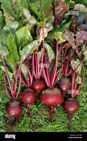
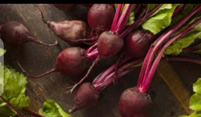
Beta vulgaris (beet) is a species of flowering plant in the subfamily Betoideae of the family Amaranthaceae. Economically, it is the most important crop of the large order Caryophyllales. It has several cultivar groups: the sugar beet, of greatest importance to produce table sugar; the root vegetable known as the beetroot or garden beet; the leaf vegetable known as chard or spinach beet; and mangelwurzel, which is a fodder crop. Three subspecies are typically recognised [4]. All cultivars fall into the subspecies Beta vulgaris subsp. vulgaris (Figures 1 & 2).
Beets have been used in folk medicines all over the world for different reasons. From the past to the present, the consumption of vegetables and fruits has gained great importance to people in terms of their overall health. A lot of research has indicated that increasing consumption of plant foods like beetroot reduce the risk of obesity, diabetes mellitus, and cardiovascular disease [5]. The current study aims to evaluate the protective effects of ethanol extract of Beta vulgaris administration on the hepatotoxicity of wistar rats when induced with monosodium glutamate (MSG).
Material and Method
Chemical
The chemical used was monosodium glutamate (C5H9NO4 .Na) purity 99% (Figure 3); it was sold in glocery store in Enugu under the license of Ajinomoto co.inc of Nigeria. A stock solution was prepared by dissolving 200g and 400g of MSG crystals in 100 ml of distilled water. The dose schedule was so adjusted that the amount of MSG administration per animal was as per their respective weight. The applied doses were selected according to Tyl and Friedman [6].
Experimental Animal
A total of thirty (30) Wistar rats were used in this study, aged 10 weeks and weighing (200 ± 20 g). All animals were procured from the Panaceas Laboratory building at College Road Enugu State. Housing conditions were standardized at 22 ± 2°C with a humidity ratio of 50 %. During the days of the experiments, the rats had free access to their diets and drinking water.
Ethical Approval
An Ethical clearance of this study was obtained from Research Ethics Committee, Directorate of Research and Publications, College of Medicine, University of Nigeria Enugu Campus.
Preparation of Beta vulgaris Extract
Freshly cultivated beetroot stems were purchased from a certified local supplier in Ogbete market Enugu state, and the ethanolic extract was prepared as described previously by Albasher, et al. [7]. Briefly, all stems were washed with water, the skin was removed, and the pulp was chopped into smaller slices (1 mm). Then, all stem parts were dried at 40°C and blended to yield a powder soaked in ethanol 1:10 (w/v) for 48 h at 4°C. The extract was filtered, and the solvent was removed under a vacuum. The extract was then lyophilized and kept at -20°C until use.
Administration of Monosodium glutamate (MSG)
A stock solution was prepared by dissolving 200g and 400g of MSG crystals in 100 ml of distilled water to give a stock solution of 6000mg/ml. The dose schedule was so adjusted that the amount of MSG administration per animal was as per their respective weight. The applied doses were selected according to Ikebunwa [1]. The appropriate dose for kg was measured out for oral administration via an oro- gastric tube using a 2ml syringe.
Experimental Design
The 30 Wistar rats were categorized into 5 major groups, A, B, C, D, and E. Each group had 6 Wistar rats and specifically given a distinct treatment but the same standard diet. Throughout the experiment, the body weight of the rats was recorded every week. A representative diagram of the experimental design and classification of the various groups is shown below: Group A: Served as Normal control and received no treatment Group B: Received MSG (4mg/kg) without treatment Group C: Received MSG (4mg/kg) + EEBV (200mg/kg) Group D: Received MSG (4mg/kg) + EEBV (400mg/kg) Group E: Received MSG (400mg/kg) + Hydrocortisone (100mg/kg).
Biochemical Analysis
Assessment of Liver Function: Serum was used for the assay of liver enzymes [aspartate aminotransferase (AST), alanine aminotransferase (ALT), alkaline phosphatase (ALP)], acid phosphatase (ACP), using standard laboratory kits from Randox Laboratories Ltd. Measurement of bilirubin (Total and Direct): Colorimetric method as described by Jendrassik, et al. [8]. Measurement of ALT and AST: Determination of ALT and AST were by colorimetric method as described by Reitman, et al. [9]. Measurement of ALP: Determination of ALP was by colorimetric method as described by Kind, et al. [10].
Histopathological Analysis
The excised liver tissues were fixed in 10% formal saline for 24 hours before being processed for light microscopy using the standard paraffin wax embedding technique. The paraffin-embedded liver tissues were cut at 5 microns with a rotary microtome (Leitz 1520 Rotary Microtome, Leica Biosystems, Nussloch, Germany). Baker, et al. [11] described the hematoxylin and eosin procedure for staining tissue sections. Histological sections were examined under an Olympus TM light microscope.
Statistical Analysis
Data obtained from present study were expressed as mean ± SEM. They were analyzed by one-way analysis of variance (ANOVA) based on the modified method of Scheffe [12]. Multiple comparisons of means were made using the least significant difference (LSD) test with the statistical package for social sciences (SPSS) for Windows version 11.0 package. Differences were considered significant at p<0.05 level of significance.
Results
Biochemical Results
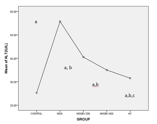
The mean comparison of mean serum ALT levels of all the treatment groups was statistically significant higher when compared with control (25.25±02.21, p=0.01). Serum ALT levels was significantly higher in MSG group than that of other treatment groups (55.75±2.32,p=0.01). The Serum ALT levels of hydrocortisone group was significantly lower when compared with MSG +BV200 (40.50±2.50, P=0.04) Figure 4.
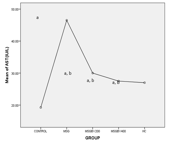
The mean comparison of mean serum AST levels of all the treatment groups was statistically significantly higher when compared with NC (19.25±0.85, P=0.01). Serum AST levels of other treatment groups were significantly lower when compared with MSG group (46.50±3.01,P=0.01). When compared with MSG+BV200, the Serum AST levels of MSG+BV400 (27.50±2.18, P=0.91) was not significant (Figure 5).
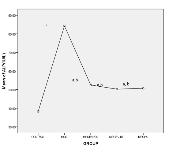
The mean comparison of mean serum ALP levels of all the treatment groups was statistically significantly higher when compared with NC (38.25±2.95, P=0.01). Serum ALP levels of other treatment groups were significantly lower when compared with MSG group (84.25±3.25,P=0.01). When compared with MSGBV200, the Serum ALP levels of MSGBV400 (50.25±1.89, P=0.98) and MSGHC (50.75+ 1.65, P=1.00) was not significant (Figure 6).
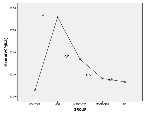
The mean comparison of mean serum ACP levels of all the treatment groups was statistically significantly higher when compared with NC (52.75±2.06, P=0.01). Serum ACP levels of other treatment groups were significantly lower when compared with MSG group (85.75±3.07,P=0.01). When compared with MSGBV200, the Serum ALP levels of MSGBV400 (58±1.47, P=0.25) and MSGHC (56+ 3.59, P=0.14) was not significant (Figure 7).
Histopathological Results
Microscopical examination of the liver isolated from the rat at sacrifice revealed no histopathological alteration in the control rats (Plate 1). In MSG only group, For plate 2, sections of the liver presented in this group showed severe multifocal areas of hepatocellular necrosis (Plate 2). In MSG + EEBV (200 mg/kg), sections of the liver presented in this group showed mild multifocal areas of hepatocellular necrosis (Plate 3). In MSG + EEBV (400 mg/kg), sections of the liver presented in this group showed mild multifocal areas of hepatocellular necrosis (Plate 4). In MSG + Hydrocortisone, sections of the liver presented in this group showed the normal hepatic histomorphology (Plate 5, Table 1).
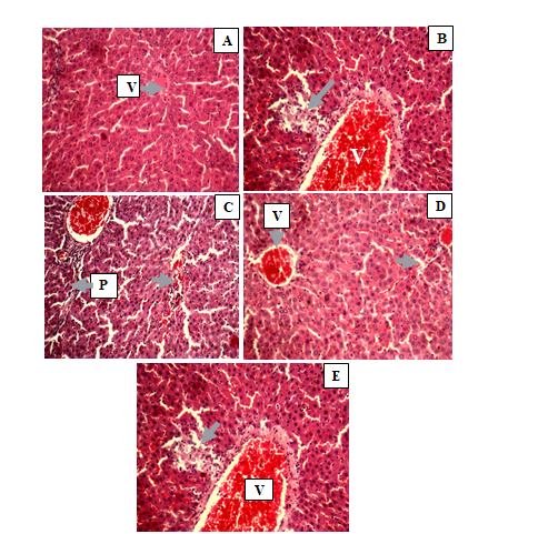
Plate 1(A): Histology of the Liver of rat in control group (group 1). Sections of the liver presented in this group showed the normal hepatic histomorphology. Central vein (V) (B): Histology of the liver in group 2 (MSG only). Sections of the liver presented in this group showed severe multifocal areas of hepatocellular necrosis (arrow). Central vein (V) (C): Showing the Histology of the liver in group 3 (MSG + low EEBV). Sections of the liver presented in this group showed mild multifocal areas of hepatocellular necrosis (arrow). Central vein (V); Portal area (P) (D): Plate showing the Histology of the liver in group 4 (MSG + high EEBV). Sections of the liver presented in this group showed mild multifocal areas of hepatocellular necrosis (arrow). Central vein (V) (E): Plate showing the Histology of the liver in group 5 (MSG +hydrocortsone). Sections of the liver presented in this group showed the normal hepatic histomorphology. Central vein (V); Portal area (P) H&E x200.
| Week 1 | Week 3 | Week 6 | Change in body weight (%) | |
|---|---|---|---|---|
| (g) | (g) | Change in body weight (%) | (g) | |
| A | 182.25±13.86 | 196.50±6.81 | 211.25±10.66 | ↑15.9 |
| B | 157.25±12.26 | 169.67±26.10 | 199.00±31.11 | ↑26.55 |
| C | 174.50±5.00 | 200.00±12.29^{c}$ | $207.00±13.85 | ↑18.62 |
| D | 170.00±11.92 | 191.33±3.79 | 217.67±19.66 | ↑28.04 |
| E | 166.75±24.86 | 195.67±15.95 | 185.00±19.92 | ↑11.39 |
| p-values | 0.2062 | 0.0135 | 0.2814 |
Table 1: Effect of B. V on MSG induced Nephro and Hepatotoxicity on body weight.
Values were expressed as mean± standard deviation (n=5). *P<0.05 indicated that there was a statistical significant (P<0.05) diffup B using One way Analysis of variance with Tukey HSD for multiple comparison among experimental groups.
Discussion
Studies on monosodium glutamate has shown that monosodium glutamate causes a progressive increase in body weight. This weight gain effect of monosodium glutamate may be due to its high content of glutamate (78%) or may be likened to its sweetened flavor. When MSG is added to food, it provides a flavoring function similar to the naturally occurring free glutamate which differ from the four classic tastes of sweet, sour, salty and bitter. As food additive, MSG is described and listed on food labels as a “Flavouring” or “hydrolysed vegetable protein”. Through its stimulation of the orosensory receptors and by improving the palatability of meals, MSG influences the appetite positively, and induces weight gain [13].
The results from this research indicated a progressive increase in body weights of MSG group showing percentage change in body weight of 26.55% when compared to other treatment groups, this could be it’s ability to induce leptin resistance, possibly influencing energy balance thereby leading to weight gain.
When compared to the groups treated with beet root at doses of 200mg/kg and 400mg/kg, the group that received MSG + BV200 showed a percentage change of 18.62% and those treated with MSG + BV400 showed a percentage change of 28.04% Showing a dose dependent change. These changes in body weight may be due to its high fiber content or due to some phytochemical components like dietary phenolic compounds which studies has shown its ability to prevent obesity and have detoxification effect. The progressive increase shown in week one to week 6 may be due to the specie of beet root found in Africa that may be lacking the necessary phytochemical component.
The studies done on monosodium glutamate have shown that it causes an increase in the serum level of liver enzymes including ALT, ALP, ACP and AST. These liver enzymes especially the ALT is known to be the most sensitive bio maker of liver damage. This damage is likened to be due to ammonium overload caused by intake of monosodium glutamate leading to constant release of this serum liver enzymes [14].
The results following this research shows a significant increase in the serum level of all the liver enzymes of the group labelled MSG only (Group B) when compared to the control group and other treatment groups. This could be attributed to deleterious effect of MSG on hepatocytes. The results of the histochemical studies revealed that with increasing dose of monosodium glutamate consumption, there were varying degrees of dilatations of the central vein of the liver which contained lysed red blood cells in the treatment group compared to the control sections of the liver. The necrosis observed is in consonance with the findings recorded in previous work on MSG [15].
When compared with the treatment groups treated with (MSG 4mg/kg + EEBV 200mg/kg) and (MSG 4mg/kg + EEBV 400mg/kg) respectively there is a significant decrease in (MSG 4mg/kg + EEBV 200mg/kg) group and (MSG 4mg/kg + EEBV 400mg/kg), this decrease in the serum level of liver enzymes could be attributed to the hepatoprotective effect of beetroots. This hepatoprotective effect of beet root may be due to the phytochemical componoents like belatain that is antioxidative in function and reduces oxidative stress.
The results obtained from this study suggest that oral administration of beetroot extract can ameliorate rat liver injury through possibly the interruption of apoptosis machinery or secondary necrosis. Beetroot juice has potential hepatoprotective effects on the liver in a dose- dependent manner contrary to Ogur, et al. [16] who stated that beetroot consumption increases the level of serum nitric oxides which causes hepatotoxicity of the liver and increase in liver enzymes.
Summary of Findings
- Monosodium glutamate increases the mean serum level of hepatic enzymes (Alanine transferase, Aspartate transaminase, Alanine phosphatase and Acid Phosphatase).
- Beta vulgaris extract decreases the mean serum level of hepatic enzymes (Alanine transferase, Aspartate transaminase, Alanine phosphatase and Acid Phosphatase).
- Monosodium glutamate intake causes progressive increase in body weight.
- Beta Vulgaris Administration causes a significant change in body weight.
Conclusion
The results obtained from this study suggest that oral administration of 200 or 400 mg/kg of beetroot extract can ameliorate rat liver injury through possibly the interruption of apoptosis machinery or secondary necrosis. Further studies will be required to isolate its active ingredients and determine the mechanism of action.
References
-
Ikebunwa OA (2023) Evaluation of the cardiac effect of monosodium glutamate (Ajinomoto) in albino rats. Open Access Research Journal of Life Sciences 5(2): 57-62.
-
Uchendu IK. (2018) Effect of aqueous extract of bitter leaf (Vernonia amygdalina) against acetaminophen-induced liver damage in rats. Bioequivalence and Bioavailability International Journal 2(1): 000122.
-
Gulati K, Reshi MR, Rai N, Ray A (2018) Hepatotoxicity: Its mechanisms, experimental evaluation and protective strategies. Am J Pharmacol 1 (1): 1004.
-
Neelwarne B, Halagur SB (2012) Red beet: an overview. In: Neelwarne B (Ed.), Red beet biotechnology: Food and pharmaceutical applications 14: 1-43.
-
Clifford T, Howatson G, West DJ, Stevenson EJ (2015) The potential benefits of red beetroot supplementation in health and disease. Nutrients 7(4): 2801-2822.
-
Tyl R, Friedman M (2003) Effects of acrylamide on rodents reproductive performance. Reprod Toxicol 17(1): 1-13.
-
Albasher G, Almeer R, Al-Otibi FO, Al-Kubaisi N, Mahmoud AM (2019) Ameliorative effect of Beta vulgaris root extract on chlorpyrifos-induced oxidative stress, inflammation and liver injury in rats. Biomolecules 9(7): 261.
-
Malloy HT, Evelyn KA (1937) The determination of bilirubin with the photoelectric colorimetric method. J Biol Chem 119: 481-490.
-
Reitman S, Frankel SA (1957) A colorimetric method for the determination of serum glutamic oxalacetic and glutamic pyruvic transaminases. Am J Clin Pathol 28(1): 56-63.
-
Kind PRH, King EJ (1954) Colorimetric method for determination of serum alkaline phosphatise. J Clin Path 7: 322.
-
Baker FJ, Silverton RE, Pallister CJ (1998) _Introduction_ _to Laboratory Technology._ In: Baker, et al. (Eds.), _Introduction_ _to_ _Laboratory_ _Technology._ 7th(Edn.), Butterworth-Heinemann, Woburn, MA, USA, pp: 448.
-
Scheffe H (1952) An analysis of variance for paired comparisons. _J Am Statistics Assoc_ 47: 381-400.
-
Kazmi Z, Fatima I, Perveen S, Malik SS (2017) Monosodium glutamate: Review on clinical reports. International Journal of food properties 20(Supp2): 1807-1815.
-
Zanfirescu A, Ungurianu A, Tsatsakis AM, Nițulescu GM, Kouretas D, et al. (2019) A review of the alleged health hazards of monosodium glutamate. Comprehensive reviews in food science and food safety 18(4): 1111- 1134.
-
Eweka AO, Om’Iniabohs FAE (2007) Histological studies of the effects of monosodium glutamate on the small intestine of adult Wistar rat. Electron J Biomed 2: 14-18.
-
Ogur R, Coskun O, Korkmaz A, Oter S, Yaren H, et al. (2005) High nitrate intake impairs liver functions and morphology in rats; protective effects of α-tocopherol. Environmental toxicology and Pharmacology 20(1): 161-166.
- Effects of 5-HTP and Melatonin on the Sleep Cycle of Medical Students
- Adsorption of Bisphenol A on NH4OH- Modified Rice Husk and Sugar Cane Bagasse Biochar
- Comparative Assessment of the Reinforcement Efficiency of Palm Fruit Fibre and Coconut Fibre in High Density Polyethylene (HDPE) Matrix Composite
- Importance of Bio Compounds Naturally Present in Food with Functionality in Animal Metabolism
- Sub-Acute Study on the Cardiotoxic Effects of Monosodium Glutamate Ingestion in Albino Rat
- Weight Management and Its Natural Solutions: A Review