Bilateral Giant Opthalmic Carotid Artery Aneurysms Presenting as Unilateral Diplopia: A Case Report
Introduction: The etiology of giant aneurysms is multifactorial. It becomes symptomatic in 9/100.000, most frequently in the fifth and sixth decades of the life. Intracavernous carotid artery aneurysms (ICAAs) represent less than 1% of intracranial aneurysms and show a slight female predominance. Case-Report: We report an interesting case of bilateral ICAA in 42-year-old female presented as acute diplopia and emphasize the need of a thorough systemic evaluation in young patients with diplopia and treated with endovascular coil embolization. A 42-year-old woman was admitted to the neurology department suffering from the unilateral diplopia. Neurological examination showed the left sixth cranial nerve palsy and had no further general motor or sensory symptoms. Digital Subtraction Angiogram (DSA) showed bilateral ophthalmic artery aneurysm. The patient was treated with endovascular loose packing coil embolization and Derivo 3.5x30mm flow diverter stent. Conclusion: It should be kept in mind that intracavernous carotid artery aneurysms may be bilateral in patients with unilateral cranial nerve symptoms.
Introduction
The aneurysms account for 0.2-7.9% of all cerebrovascular lesions. The ophthalmic artery aneurysms constitute 5% of the aneurysms [1]. It becomes symptomatic in 9/100.000, most frequently in Bilateral Giant Opthalmic Carotid Artery Aneurysms Presenting as Unilateral Diplopia: A Case Report the fifth and sixth decades of the life [2]. Intracavernous carotid artery aneurysms (ICAAs) represent less than 1% of intracranial aneurysms and show a slight female predominance [2, 3]. Bilateral intracavernous carotid artery aneurysms with unilateral cranial nerve symptoms are extremely rare and difficult to treat.
J Human Anat
We report an unusual case of bilateral ICAA opthalmic aneurysm in the 42year-old female patient presented as acute unilateral diplopia and treated with endovascular loose packing coil embolization and Derivo 3.5x30mm flow diverter stent.
Case Presentation
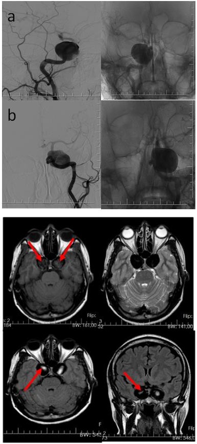
A 42-year-old woman was admitted to the neurology department suffering from the unilateral diplopia. Neurological examination showed the left sixth cranial nerve palsy and had no further general motor or sensory symptoms. In view of the clinical findings, magnetic resonance imaging of the brain was undertaken. Coronal, axial T1 and T2 weighted images demonstrated bilateral heterogeneously enhanced masses indicating in the bilateral ICCA (Figure 1). Digital Subtraction Angiogram (DSA) showed bilateral ophthalmic artery aneurysms (Figure 2).
Premedication with double antiplatelet and steroid treatment was started before the endovascular procedure. The patient was treated endovascularly under general anesthesia with one-month interval. Endovascular loose packing coil embolization and Derivo 3.5x30mm flow diverter stent was placed to the segment of the damaged ICA. Contrast stagnation was observed at the aneurysm sac soon after the flow diverter placement.
Lafci Fahrioglu S, et al. Bilateral Giant Opthalmic Carotid Artery Aneurysms Presenting as Unilateral Diplopia: A Case Report. J Human Anat 2020, 4(1): 000143.
Figures 2a & 2b: Digital angiography (DSA) imaging of the right (a) and the left (b) sides- ICAAs.
Discussion
The etiology of giant aneurysms is multifactorial, and many structural and hemodynamic stress factors have been previously discussed. However, pathogenesis of the cavernous aneurysm is not yet defined, and idiopathic intracavernous aneurysms are the most common [3]. Many bilateral intracavernous aneurysm cases have definite causative factors suggesting weakness of the carotid arterial wall. They can occur after radiation therapy or in association with connective tissue disorders such as fibromuscular dysplasia and Paget’s Disease [4].
Our patient presented with diplopia which is the most common presenting symptom [5]. One study reported 206 CCAs cases (65%) presenting symptom of diplopia, followed by retro-orbital pain (34%). Moreover, the most common finding at presentation was a cavernous sinus syndrome that involved a combination of the third, fourth, and sixth cranial nerves (18% of 206 CCAs) [6].
Without any intervention, improvement of diplopia and concurrent reduction of pain have both been reported. One study showed that, if left untreated, either diplopia or pain improved in 56% of CCAs. Based on that report, selection bias—related to severity of symptoms— Copyright© Lafci Fahrioglu S, et al.
has impeded comparison of outcomes and complications between untreated and treated patients [5, 7]. The likelihood of an aneurysm becoming symptomatic is directly related to its size. Optimal management of symptomatic giant carotid aneurysms remains controversial. However, present treatment options favor bypass or embolization to direct surgery with very good results [8].
Conclusion
To summarize we report an interesting case of bilateral ICAA in 42-year-old female presented as acute diplopia and emphasize the need of a thorough systemic evaluation in young patients with diplopia and treated with endovascular coil embolization.
References
-
Purvin VA (2009) Neuro-ophthalmic aspects of aneurysms. Int Ophthalmol Clin 49(3): 119-132.
-
Terzidou C, Dalianis G, Zacharaki F (2012) Ocular Symptomatology, Management, and Clinical Outcome of a Giant Intracranial Aneurysm. Case Reports Medicine 2012: 4.
-
Linskey ME, Sekhar LN, Hirsch W, Yonas H, Horton JA (1990) Aneurysms of the intracavernous carotid Lafci Fahrioglu S, et al. Bilateral Giant Opthalmic Carotid Artery Aneurysms Presenting as Unilateral Diplopia: A Case Report. J Human Anat 2020, 4(1): 000143. artery: clinical presentation, radiographic features, and pathogenesis. Neurosurgery 26(1): 71-79.
-
Sekhar LN, Ramanathan D, Hallam DK, Ghodke BV, Kim LJ (2011) What is the correct approach to aneurysm management in 2011?. World Neurosurg 75(3-4): 409-411.
-
Rehman T, Ali R, Taylor C, Yonas H (2010) Bilateral giant cavernous carotid artery aneurysms in a child with juvenile Paget's disease. World Neurosurg 73(6): 691-693.
-
Dinca EB, Brehar F, Giovani A, Ciurea AV (2017) Challenges in a case of ophthalmic artery aneurysm associated with abnormal internal carotid arteries. Asian J Neurosurg 12(1): 106-108.
-
Vanikieti K, Poonyathalang A, Jindahra P, Cheecharoen P, Chokthaweesak W (2018) Occipital lobe infarction: a rare presentation of bilateral giant cavernous carotid aneurysms: a case report. BMC Ophthalmol 18(1): 25.
-
Stiebel-Kalish H, Kalish Y, Bar-On RH, Setton A, Niimi Y, et al. (2005) Presentation, natural history, and management of carotid cavernous aneurysms. Neurosurg 57(5): 850-857. Copyright© Lafci Fahrioglu S, et al.
- Pattern of Breast Lesions in Ovu Inland, Delta State, South Southern Nigeria
- Morphometric Analysis of the Human Femur: Exploring Platymetric and Robusticity Indices Among the Nigerian Population
- Anatomical Variation of Arteria Lusoria: Clinical Implications for Dysphagia Lusoria and Surgical Risk
- Morphometric Study of the Vertebral Body and Pedicle of Typical Cervical Vertebrae Using Radiological Image
- Epigenetic Mechanisms Driving Human Evolutionary Changes
- Neuroprotective Effects of Ginkgo Biloba Extract on Bilateral Common Carotid Artery Ischaemic Stroke Induced in Wistar Rat