Determination of Aflatoxins in Pig Feed. Using the Thin Layer Chromatography Technique
Aflatoxins are the most important mycotoxins in terms of food safety, due to their frequency and toxicity, they are characterized by being secondary metabolites synthesized by fungi. In order to determine the presence of aflatoxins in pork feed, five samples from different commercial companies were analyzed. The methodology used prior to the sample extraction process was the thin layer chromatography technique, observed using long-wave ultraviolet light. It could be seen in samples 1,3,4,5, the presence of Aflatoxins, toxicogenic strains of Aspergillus parasiticus, which produce aflatoxins, G1 and G2, is characterized by the fluorescence coloration, in sample 2 the presence was observed of aflatoxins toxicogenic strains of Aspergillus flavus. Toxigenic strains of Aspergillus flavus generally produce only aflatoxins B1 and B2, while toxicogenic strains of Aspergillus parasiticus produce aflatoxins B1, B2, G1 and G2. AFB1 aflatoxins have been considered obvious carcinogens in experimental animals. It is important to carry out systematic research with low-cost analytical techniques to understand the mechanisms of production and action of aflatoxins in foods, in order to prevent, monitor and control these types of toxins.
Introduction
Aflatoxins belong to the mycotoxin family, which are chemicals produced by toxigenic strains of fungi, mainly Aspergillus flavus and Aspergillus parasiticus. They are of great interest in toxicological research, mainly due to their high incidence in foods and because their consumption in low, medium or high doses causes acute or chronic toxic effects [1, 2]. Ingestion of aflatoxins can cause a disease known as aflatoxicosis, which can cause death in humans and animals. These mycotoxins are usually designated with letters, which refer to a physical characteristic of the type of compound, for example, B1 and B2 fluoresce blue and G1 and G2 fluoresce green when exposed to long-wave ultraviolet radiation.
Toxigenic strains of Aspergillus flavus generally produce only aflatoxin B1 and B2, while toxicogenic strains of Aspergillus parasiticu s produce aflatoxins B1, B2, G1 and G2 [3, 4]. According to this classification, the International Agency for Research on Cancer (IARC) considers aflatoxin AFB1 as an obvious carcinogen in experimental animals, making it the most important aflatoxin in public health (International
Agency For Research On Cancer (IARC) [5]. They have also been implicated in the pathogenesis of protein-energy malnutrition (PEM), and it has been established that even for different types of animals, the acute oral lethal dose (LD50, in mg/kg) is different. It has been suggested that feeding broiler chickens with foods containing low concentrations of AFB1 can cause hepatotoxicity and induce chronic aflatoxicosis. Likewise, they have suggested that there are differences in the health impacts of aflatoxins between different human races caused by chronic exposure to these toxins [3].
The climatic environment conducive to the production of mycotoxins is in a tropical climate, where the optimal conditions for the growth of aflatoxigenic fungi are met, mainly with the conditions of relative humidity and temperature; that end up contaminating highly consumed foods if these conditions are not taken into account for their preservation. Even though it has been detected as natural contaminants in a large number of agricultural products and its presence has been confirmed in practically all areas of the world and, to a greater or lesser extent, in almost all staple foods. Foods considered most susceptible to fungal contamination with consequent aflatoxin production typically include corn, peanuts, pistachios, walnuts, cotton seeds, and dried coconut meat (copra) [6]. Aflatoxins have also been found in oilseeds such as sunflower and soybeans, in unrefined vegetable oils, in other nuts such as almonds and hazelnuts, in species such as paprika, chili, pepper, etc., in fruits dried such as dried figs, raisins, coffee, cocoa, cereals, their derived products and in feed [7].
It is important to know the pathogenic action of aflatoxins in synergy with other mycotoxins such as zearalenone, fumonisin or deoxynivalenol (DON), which cause food poisoning that is increasingly common in pig farms. This incidence and risk factors correspond to the poor conservation of food bags, without control of humidity, temperature in storage places, coupled with the fact that they are already contaminated, the use of automatic feed storage and distribution equipment, the difficulties for cleaning and disinfecting storage areas make the pathology more vulnerable to these substances [3, 8]. Porcine aflatoxicosis is one of the most common mycotoxicosis, it is not transmissible, it mainly affects pregnant sows and piglets, its chronic effect is seen with decreased animal growth and acute by sudden death, hepatitis, its toxicity is characterized by hepatotoxic actions, carcinogenic, teratogenic and mutagenic [9].
Although the toxic response and clinical symptoms depend on the age of the animals, the productive phase, the level of immunity of the farm, ingestion time and concentration of aflatoxins in the ration alimenticie. The immunosuppressive effect that occurs in contaminated animals facilitates the spread of diseases on the farm. The toxic effects can be synergistic, enhanced with other mycotoxins from different fungi [10].
Panagou and Cols, in 2015, indicated that the optimal temperature and water activity (aw) for the proliferation of A. flavus is 20-30 º C and ± 0.99 aw, knowing them as the typical storage fungi Panagou EZ, et al. [11], while Goyarts, et al. 2011 study the influence of prolonged doses (35-70 days) of zearalenone, deoxynivalenol (DON) and aflatoxins in prepubertal sows of 35 kg w/v on histopathologyof the intestinal and uterine mucosa, and on ovarian alterations and sexual immaturity of young reproductive animals [12].
There are new technologies, storage and distribution methods for pig foods and other types of animals, as well as special control measures for cereals and flours are required to minimize and/or prevent the growth and proliferation of fungi. These measures include periodic sampling and analysis of the stored bags, increasing the purchase of cereals and legumes with certain guarantees (humidity values lower than 12.5 and 9%, respectively), adequately ventilating to ensure rates lower than 0.65 aw. and temperatures ≤20 ºC, use of fungistatics, etc [3].
The tolerance levels in oilseed meals and corn grains that contain aflatoxins greater than 20 µg cannot be used for the production of pig feed, much less for human consumption or young animals (pre-starter and starter feeds). However, researchers have proven that the administration of minimum daily doses between 50-280 µg does not produce clinical symptoms or mortality in piglets, although they may cause slight growth delays [13, 14].
According to René, et al. tolerance to aflatoxins is usually greater as the live weight of the animal increases and doses of 20.50 and 100 µg/kg for pigs weighing 35, 60 and 100 kg respectively do not have harmful effects. The LD50 is 0.60 mg/kg of live weight and doses of 1-2 mg/kg produce death in just 18-72 hours with necrosis and hemorrhages in the intestinal mucosa. Weaned piglets are more sensitive than adult pigs, since daily doses of 0.30 mg/kg body weight (approximately 6 mg/kg feed) cause growth retardation, jaundice, necrosis with hepatic hemorrhages and death7 -14 days [15, 16]. Given the contamination of food and raw materials with toxic substances, such as aflatoxins, it is necessary to develop low-cost analytical techniques, with good sensitivity and accessible development, which can be used in training programs for students in various areas. of research especially in the analytical determination of toxins in biological and non-biological matrices. The process of determining mycotoxins in solid and liquid foods is carried out in several phases that, in general terms, include sample preparation (grinding and homogenizing in the case of solid foods and ultrasonication in the case of liquid foods), extraction and subsequent purification to eliminate interferences and finally determination using instrumental techniques [17]. The most widely used method to determine aflatoxins is based on chromatographic techniques, such as: thin-layer chromatography (TLC), high-performance liquid chromatography (HPLC) combined with a fluorescence detector or with new equipment, such as UPLC (Ultra Pressure Liquid Chromatography) that improves its characteristics. There are also immunochemical techniques such as ELISA, which is a method that can be qualitative or quantitative; however, although it is fast and sensitive, it can give false positives [18].
Chromatography is a method of separating different components of a sample by a stationary phase with the help of the mobile phase (liquid or gas), each component of the sample has particular properties that will allow its interaction in a different way between the stationary phase and mobile, in this way each component is delayed in a particular way and if the flow rate, the characteristics of the stationary and mobile phase are adequate, complete separation of all the components of the sample will be achieved [19]. Thin layer chromatography is simple, low-cost and very useful when discarding samples with the identification of these toxins. Using silica gel plates with fluorescence, the presence and types of aflatoxins present can be determined with the specific mobile phase system that can be separated and also with a stationary phase that allows us to observe the fluorescence that reflects the type of aflatoxins [20].
Analytical Procedure for Aflatoxin Extraction
Most methods used for the determination of mycotoxins need reliable extraction and purification methods. These steps are vital to a successful protocol as they are time- consuming (sample preparation is the main time factor in an analysis and takes approximately two-thirds of the total time) and will affect the final choice of detection procedure. The extraction method used to extract mycotoxins from the biological matrix is dependent on the structure of the toxin. Hydrophobic toxins such as aflatoxins are extracted using organic solvents. The used organic solvents are methanol, cloruro de sodio, acetonitrile, acetone, dichloromethane hexano, cloroformo, and a mixture of them [21]. Five (05) samples of pork feed from different commercial companies labeled as Sample A, Sample B, Samples C, Samples D, Samples E were analyzed.
50 g of each ground concentrated feed were weighed, 200 ml of a methanol-water solution (170 ml-30 ml) was added to each sample, then placed on the mechanical shaker for 30 minutes. Subsequently, 40 ml of the solution (supernatant) was taken, placed in a separatory funnel with 40 ml of 10% sodium chloride, mixed and then 25 ml of N-Hexane was added and extracted for 10 minutes. It was allowed to settle to separate the two phases, the lower phase was placed in another separating funnel and the upper phase was discarded. The aflatoxins are extracted from the lower phase, in double extraction with 25 ml of chloroform each, they were shaken for 10 minutes, allowed to rest and the phases were separated, obtaining the organic phase (lower) by placing it in a container with a lid; then 25 ml of chloroform is added again, it is extracted again for 10 minutes, it is allowed to rest and the other organic phase is obtained again, placing it in the same container as the previous one, with a final volume. of 45 ml, it was brought to concentration, until dry in a water bath.
Preparation of the Purification Column
A 20cc syringe was used as a column, crystallized cotton was placed at the bottom of the column, 1g of silica gel 60 for column chromatography (70-230mesh) and 1g of anhydrous sodium sulfate were added to the top.
Aflatoxin Purification Procedure using Vacuum with a Kitasate
The column was washed with 3ml of N-Hexane and 3ml of dichloromethane using vacuum, then the evaporated samples were taken up with 3ml of dichloromethane and added to the column without vacuum. The container containing the sample was washed with 2 portions of 1 ml each of dichloromethane and added to the column. The column was washed again with 3 ml of N-Hexane, 3 ml of ethyl ether and 3 ml of dichloromethane using vacuum. The vacuum was turned off and the aflatoxins were extracted with 3 portions of 3 ml of a chloroform - acetone mixture each (9 ml of chloroform + 1 ml of acetone), collecting it in a test tube, then it was dried in a water bath and resumed with 0.5 ml of chloroform to seed on the F254 silica gel chromatography plate. Subsequently, it was taken to the chromatography tank prepared with the specific system for aflatoxins (90ml of chloroform and 10ml of acetone), when the mobile phase reached 15cm, the plate was removed, allowed to dry and the paths of the samples were observed in the ultraviolet light (UVL) lamp 20.
Results
| Consistency | Solubility Water | Fluorescence LUV | |
|---|---|---|---|
| 1 | Light brown solid food fragments, dried | Solubilizes with precipitate | Green Fluorescence |
| 2 | Dark brown solid food fragments, dried | Solubilizes with precipitate | Blue Fluorescence |
| 3 | Dried light brown solid food fragments | Solubilizes with precipitate | Green Fluorescence |
| 4 | Dried dark brown solid food fragments | Solubilizes with precipitate | No Fluorescence |
| 5 | Dried dark brown solid food fragments | Solubilizes with precipitate | No Fluorescence |
Table 1: Physical characteristics and observations of the samples.
Morales and Barrios, 2024
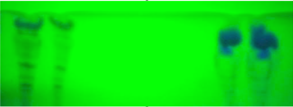
According to the methodology used it is described in Table 1, the samples 1,3,4,5 above, the presence of Aflatoxins was found, toxicogenic strains of Aspergillus parasiticu s, which produce aflatoxins, G1 and G2, characterization that is done by fluorescence coloration, in the Sample 2 found the presence of aflatoxin, toxicogenic strains of Aspergillus flavus that only produce aflatoxin B1 and B2. En la Figure 1 se observa la fluoressencia azul de la muestra 2.
Morales and Barrios, 2024.
Discussion and Conclusions
Ingestion of aflatoxins can cause aflatoxicosis, a disease that can cause death in humans and animals. These mycotoxins are designated with letters, according to their toxicity, which refer to a physical characteristic of the type of compound. The results of this study, based on chromatographic separation, correlate with the classification carried out by encompasses et al in the year 2000, with respect to the color of the fluorescence observed in the positive samples. Evidently, the presence of aflatoxins could be observed with Sample 2, toxicogenic strains of Aspergillus flavus that only produce aflatoxins B1 and B2, considered obvious carcinogens in experimental animals. On the other hand, the purification procedure used in a protocol is the most important step, since the purity of the sample affects the sensitivity of the results. Small amounts of a target molecule can be masked by interfering compounds, which are found not only in the matrix, but also in the chemicals, materials and solvents used in the art. The material used was free of contamination, such as alkaline detergents, which can form salts with the compounds and lead to lower detection rates21. In the determination of aflatoxins in the five samples analyzed, the extraction and separation process was rigorously controlled to avoid interference, contamination or carryover of the analyte.
The negative impact of aflatoxins on human and animal health has been demonstrated, particularly by their carcinogenicity.
Hence the importance of establishing health controls for the storage and transfer of animal feed, cereals, flour, nuts that are sold in bulk. The need to carry out systematic research with low-cost analytical techniques is shown to understand the mechanisms of production and action of the same in foods, raw materials, human and animal organisms, generating increasingly solid scientific bases to prevent, monitor and control these types of toxins.
References
-
Ciegler A (1975) Mycotoxins: occurrence, chemistry, biological activity. Lloydia 38(1): 21-35.
-
Smith J, Hacking A (2003) Fungal toxicity. Filamentous fungi. Londres: Arnold EV pp: 238-64
-
Abarca L, Bragulat R, Castellá G, Accensi F, Cabañes J (2000) Emerging mycotoxin-producing fungi. Rev Iberoam Micol 17: 63-68.
-
Hesseltine C, Sorrenson W, Smith M (2007) Taxonomic studies of the aflatoxin-producing strains in the Aspergillus flavus group. Micología 62(1): 123-132.
-
Chemical Agents and Related Occupations (2012) IARC Monographs on the Evaluation of Carcinogenic Risks to Humans 100: 225-248.
-
Arroyo N, Manzanares J, Pérez H, Gracia L, Campaña A (2014) Control of mycotoxins in foods. Granada, Spain; University of Granada. Bulletin No 7: 17.
-
Soriano-del-Castillo JM (2007) Mycotoxins in foods. In: 1st (Edn.), Díaz de Santos Editions, Madrid, Spain.
-
Deng S, Tian L, Liu F, Jin S, Liang G, et al. (2010) Toxic effects and residue of aflatoxin B1 in Tilapia (Oreochromis miloticus x 38 O. aureus) during longterm dietary exposure. Aquaculture 307(3): 233-240.
-
Durán C, Castillo P (2006) Mycotoxins: secondary metabolites of filamentous fungi. Educación Química pp: 128
-
Fung F, Clark RF (2004) Health effects of mycotoxins: a toxicological overview. J Toxicol Clin Toxicol 42(2): 217- 324.
-
Panagou EZ, Skandamis PN, Nychas GJE (2003) Modelling the combined effect of temperature, pH and aw on the growth rate of Monascus ruber, a heat-resistant fungus isolated from green table lives. J Appl Microbiol 94(1): 146-156.
-
Goyarts T, Danicke S, Brussow K, Valenta H, Ueberschar K, et al. (2011). On transfer of the Fusarium toxins deoxynivalenol (DON) and zearalenone (ZON) from sows to their fatuses during days 35-70 of gestation. Toxicology Letters 171(1-2): 38-49.
-
Sala R, Reguera G, Pérez B, García-Casado G (2008) Mycotoxins and their impact on pig production. Albéitar 1(112): 34-38.
-
Hussein H, Brasel J (2001) Toxicity, metabolism, and impact of mycotoxins on humans and animals. Toxicology 167: 101-134.
-
Neftalí R, Márquez M (2016) Repercussions of Mycotoxins on High Producing Sows. Pig Farmers and Their Environment 111: 3-7.
-
Urrego J, Díaz G (2006) Aflatoxins: mechanisms of toxicity in the etiology of cellular liver cancer. Pharmaceutical Rev (Colombia) 54: 109-110.
-
Herrero L (2012) Development and validation of a method for analyzing aflatoxins in nuts and cereals. Master’s work. Zaragoza University pp: 56.
-
Huang B, Han Z, Cai Z, Wu Y, Ren Y (2010) Simultaneous determination of aflatoxins B1, B2, G1, G2, M1 and M2 in peanuts and their derivative products by ultra-high- performance liquid chromatography– tandem mass spectrometry. Analytica Chimica Acta 662(1): 62-68.
-
Chromatography Guide (2008) Central University of Venezuela. Caracas, Venezuela, pp: 11.
-
Aguirre O, Pérez J, Pujol M (2001) Validation of analytical methods. A.E.F. Industry. AEFI monographs. Barcelona Spain, pp: 23-134.
-
Turner NW, Subrahmanyam S, Piletskyb SA (2009) Analytical methods for determination of mycotoxins: A review. Anal Chim Acta 632(2): 168-180.
- Pattern of Gonadal Hormones in Oral Testosterone-Supplimented Male Wistar Rats with Diabetes-Induced Hypogonadism
- Re-Evaluation of the Genotoxicity of Currently Used Food Dyes in Mouse Multiple Organs Via Continuous Administration by Drinking Using the Comet Assay
- Pharmacogenetics of Type 2 Diabetes Mellitus: Linking Genetic Variability to Drug Efficacy and its Cardiovascular Outcomes
- Exploratory Proteomic Profiling of SARS-CoV-2 Infected THP-1 Macrophages Reveals Alterations in Inflammatory Response and Cellular Metabolism
- Study of Genotoxicity of Hepatocarcinogens in Multiple Organs in Mice by Feeding and Drinking Using the Comet Assay
- Spirulina Polypeptides Inhibit the Growth of Human Lung Tumor (H460) Cells